Staphylococcus epidermidis is a Gram-positive, coagulase-negative coccus that forms part of the normal human skin microbiota and is an important opportunistic pathogen associated particularly with foreign-body-related infections.
Basic Characteristics
Taxonomy
Domain: Bacteria
Phylum: Bacillota (Firmicutes)
Class: Bacilli
Order: Bacillales
Family: Staphylococcaceae
Genus:Staphylococcus
Species:Staphylococcus epidermidis
Microscopy & Gram Stain
Gram-positive cocci
Cells occur singly, in pairs, and in irregular clusters
Oxygen Relationship
Facultatively anaerobic
Rapid Identification Tests
Catalase: positive
Coagulase: negative
Novobiocin: usually susceptible (S. epidermidis group)
Haemolysis on blood agar: usually absent
Ecology and Clinical Relevance
Natural Habitat
Human skin, especially moist body sites
Axillae, inguinal and perineal areas
Anterior nares
Toe webs
Common Clinical Specimens
Blood cultures
Catheter tips
Device-associated samples
Wound swabs
Skin swabs
Clinical Significance
Common colonizer of human skin and mucosal surfaces
Important opportunistic pathogen in immunocompromised patients
Frequently associated with indwelling devices and implanted biomaterials
A major cause of foreign-body-related and healthcare-associated infections
Differential Considerations
Staphylococcus aureus and other coagulase-positive staphylococci
Other coagulase-negative staphylococci such as S. haemolyticus and S. lugdunensis
Important clues include coagulase negativity, usual absence of haemolysis, and typical novobiocin susceptibility
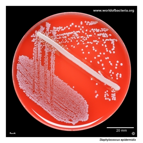
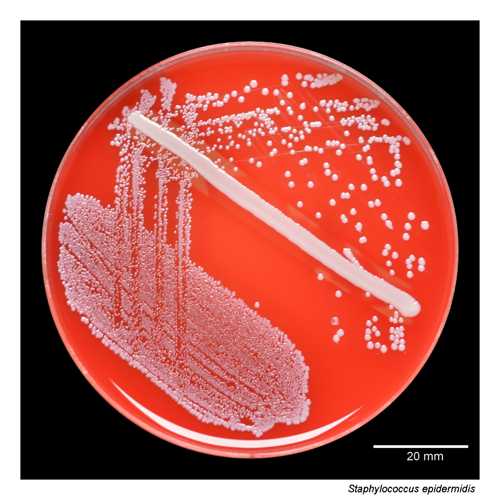
A skin swab collected before blood culture sampling was enriched overnight in nutrient broth at 36 °C and then subcultured onto blood agar. After 24 hours at 36 °C in 5% CO2, Staphylococcus epidermidis formed small grey-white colonies about 2–3 mm in diameter without haemolysis. A streak of S. aureus ATCC 25923 was included on the plate for comparison. Lack of haemolysin production is typical of S. epidermidis, although it is not a fully reliable identification feature.
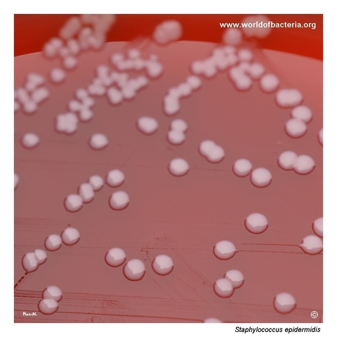
Colonies of Staphylococcus epidermidis growing on sheep blood agar after 24 hours at 36 °C in ambient air. Most strains of S. epidermidis are non-haemolytic on blood agar and may therefore be described as gamma-haemolytic. However, beta-haemolytic isolates do occur in clinical material and can resemble non-pigmented colonies of S. aureus. In clinical practice, S. epidermidis is also notable for its often higher level of antibiotic resistance.
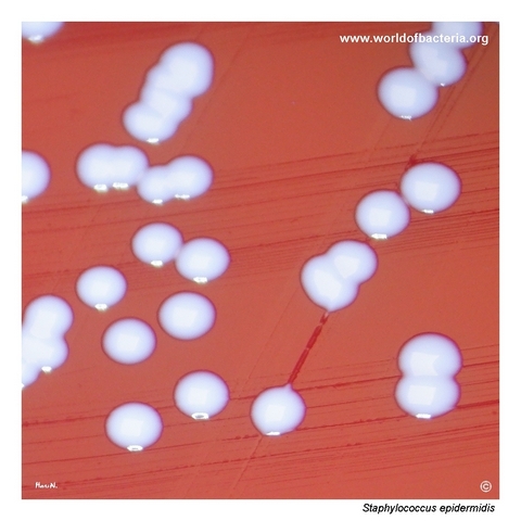
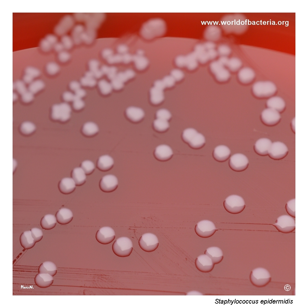
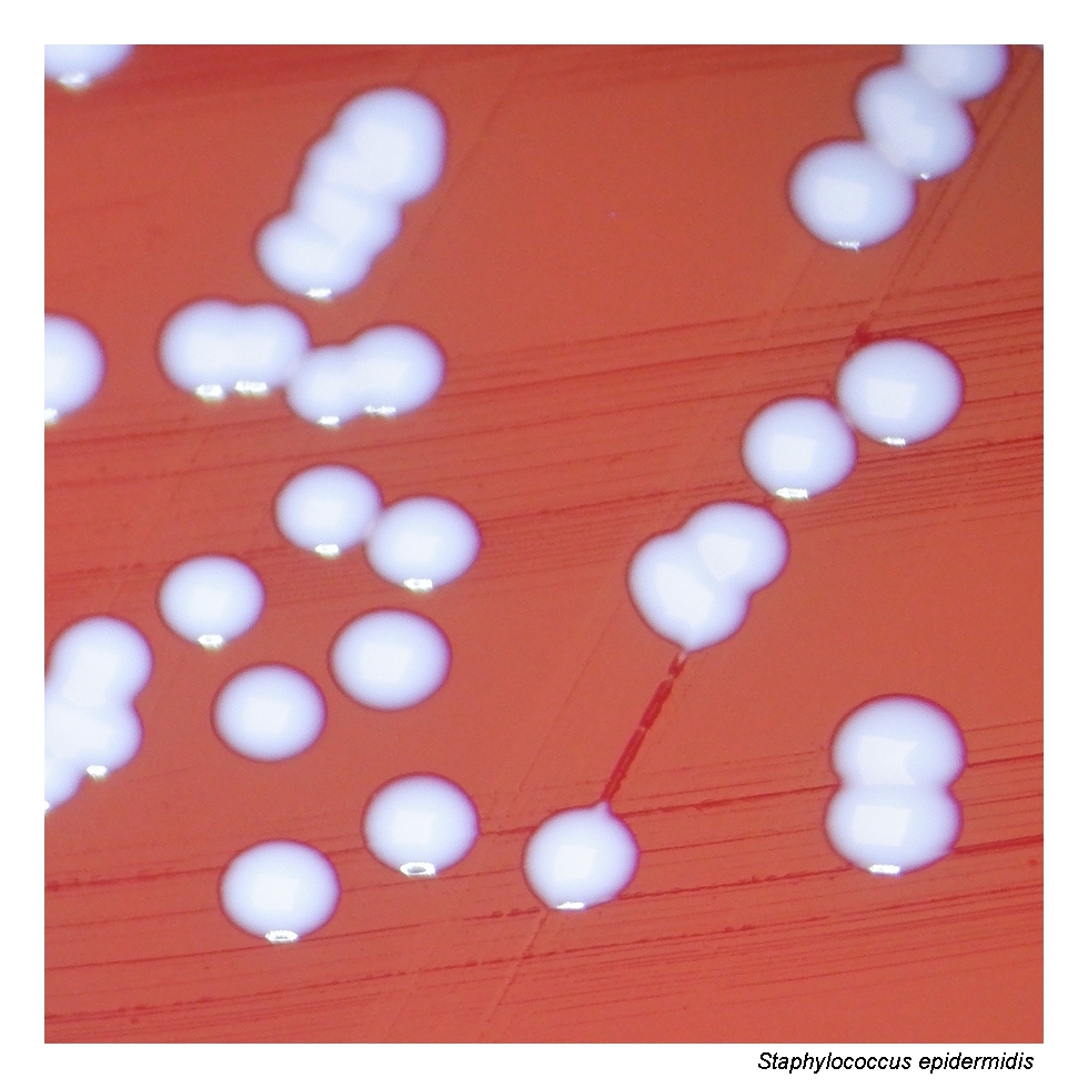
Close-up view of Staphylococcus epidermidis colonies on blood agar after 24 hours at 37 °C. The colonies are smooth, convex, grey-white, and round with entire margins, lacking both beta-haemolysis and yellow pigment production. This colony morphology is typical of S. epidermidis and of many other coagulase-negative staphylococci, a group of species that form part of the normal human skin microbiota but may also act as opportunistic pathogens.
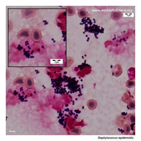
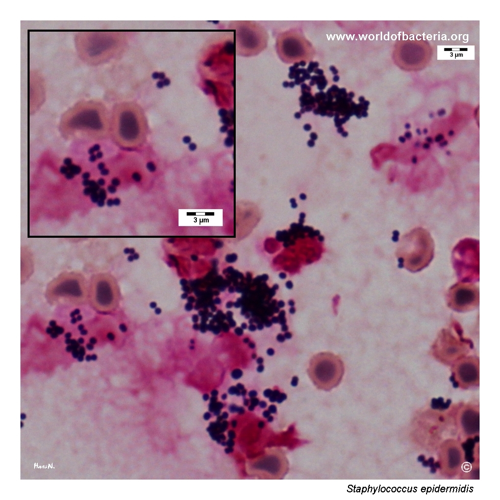
Staphylococcus epidermidis in a positive blood culture. The Gram stain shows Gram-positive cocci occurring singly, in pairs, and in clusters, a morphology indistinguishable from that of other staphylococcal species. In blood cultures, S. epidermidis often represents contamination introduced during venepuncture because of inadequate skin disinfection, although in some patients it may also be a true cause of bloodstream infection.
Diagnostic and Clinical Notes
Staphylococcus epidermidis belongs to the coagulase-negative staphylococci (CoNS) and is one of the most frequently recovered bacterial colonizers of the human body surface. It is particularly prevalent on moist skin sites and in the anterior nares.
Although it is a common commensal organism, S. epidermidis has major clinical relevance as an opportunistic pathogen, especially in immunocompromised patients and in individuals with indwelling or implanted foreign materials.
Among staphylococci, S. epidermidis is often regarded as the second most important pathogen after Staphylococcus aureus because of its role in healthcare-associated and device-related infections.
Its pathogenicity is closely linked to foreign-body-related infections, including infections associated with intravascular catheters, prosthetic joints, cardiac devices, and other polymer-based implants, where biofilm formation can facilitate persistence and reduced antimicrobial susceptibility.
Clinical interpretation always depends on specimen type and context. Recovery from blood cultures may reflect either contamination from the skin flora during venepuncture or true bloodstream infection, and correlation with repeated cultures and clinical findings is therefore essential.
Laboratory Identification
Colony Morphology
On blood agar incubated at 35–37 °C, colonies of Staphylococcus epidermidis are typically small to medium-sized, smooth, convex, grey-white, and round with entire margins after 18–24 hours. Most isolates are non-haemolytic and lack golden-yellow pigmentation.
Microscopy
Gram staining reveals Gram-positive cocci occurring singly, in pairs, and in irregular clusters. The microscopic appearance is not species-specific and resembles that of other staphylococci.
Key Identification Clues
Gram-positive cocci in clusters
Strongly catalase positive
Coagulase negative
Usually non-haemolytic on blood agar
Usually novobiocin susceptible (S. epidermidis group)
Modern Identification Methods
Species-level identification in clinical microbiology laboratories is commonly achieved using MALDI-TOF mass spectrometry. Automated biochemical systems may support routine identification, while molecular methods can be useful for strain characterization, resistance analysis, and epidemiological investigations.
Antibiotic Characteristics
Staphylococcus epidermidis often shows reduced susceptibility to multiple antimicrobial agents, and clinical isolates may be more resistant than many other common skin commensals.
Methicillin resistance is frequent among healthcare-associated isolates and is commonly mediated by the mecA gene, which substantially limits the activity of beta-lactam agents.
Biofilm-associated growth on foreign materials may further complicate therapy because it can impair antimicrobial penetration and contribute to persistent infection.
Note: Antimicrobial susceptibility patterns vary considerably between isolates and institutions; treatment decisions should always be guided by laboratory susceptibility testing and clinical context.