Staphylococcus saprophyticus is a Gram-positive, coagulase-negative coccus best known as an important cause of urinary tract infection, especially acute cystitis in young women.
Basic Characteristics
Taxonomy
Domain: Bacteria
Phylum: Bacillota (Firmicutes)
Class: Bacilli
Order: Bacillales
Family: Staphylococcaceae
Genus:Staphylococcus
Species:Staphylococcus saprophyticus
Microscopy & Gram Stain
Gram-positive cocci
Cells may occur singly, in pairs, short chains, and clusters
Oxygen Relationship
Facultatively anaerobic
Rapid Identification Tests
Catalase: positive
Coagulase: negative
Novobiocin: typically resistant
Haemolysis on blood agar: usually absent
Ecology and Clinical Relevance
Natural Habitat
Human genitourinary and perineal region
May colonize the lower genital tract and adjacent skin
Often discussed in relation to urinary microbiology rather than invasive disease
Common Clinical Specimens
Midstream urine
Catheter urine
Urine from patients with acute cystitis or pyelonephritis
Occasional blood cultures or deeper samples in complicated infection
Clinical Significance
Important cause of uncomplicated urinary tract infection
Classically associated with acute cystitis in young women
Pathogenicity is linked to adhesion to uroepithelium and biofilm formation
Usually much more relevant in urinary specimens than in non-urinary superficial samples
Differential Considerations
Other coagulase-negative staphylococci in urine cultures
Staphylococcus aureus, S. lugdunensis, or S. haemolyticus when colonies are unusually pigmented
Enterococci and other Gram-positive urinary pathogens in direct microscopy
Important clues include urinary tropism, novobiocin resistance, and usually non-haemolytic colonies even when pigmentation is marked
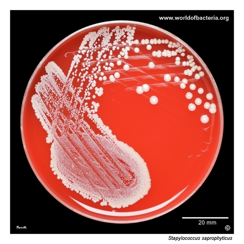
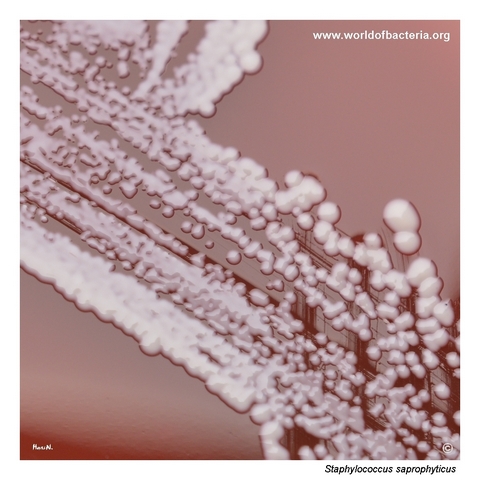
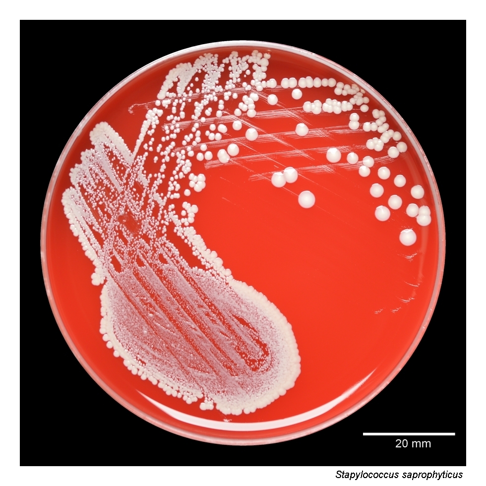
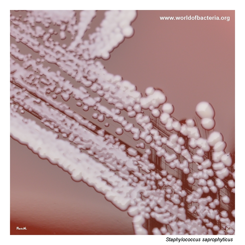
Ten microlitres of midstream urine were inoculated onto blood agar. The patient was a 30-year-old woman with acute cystitis. After 24 hours of incubation at 36 °C in ambient air, the culture was further prolonged for another 24 hours at room temperature. Staphylococcus saprophyticus belongs to the staphylococcal species capable of forming large colonies during extended incubation, sometimes reaching 6 mm or more in diameter after about five days.
These convex, non-pigmented white colonies show a somewhat confluent pattern of growth in the area of primary inoculation and wherever colony counts are high, a feature often seen in Staphylococcus saprophyticus. For most strains, a key virulence factor is the ability to adhere to the uroepithelium and to form bacterial biofilm, which helps explain the organism’s well-known association with urinary tract infections, especially in young women.
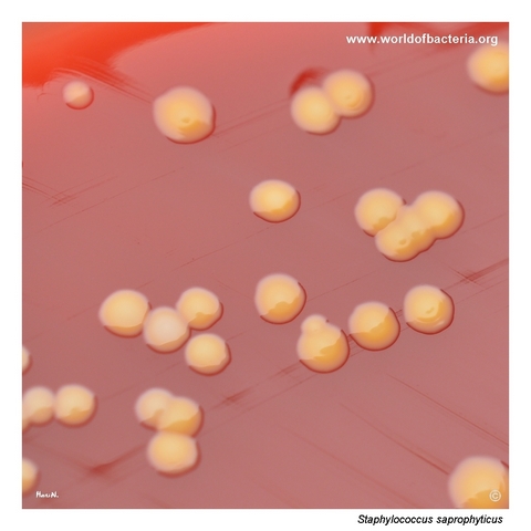
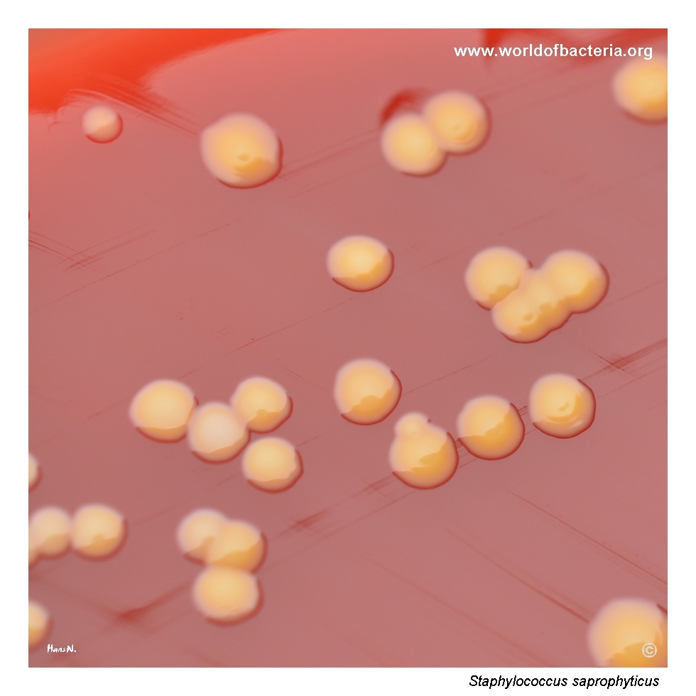
Colonies of Staphylococcus saprophyticus on blood agar after 24 hours of incubation at 36 °C in ambient air. Strongly pigmented isolates such as this one are less common in clinical material and may resemble strains of Staphylococcus aureus, S. lugdunensis, or S. haemolyticus. In S. saprophyticus, however, the conspicuous absence of haemolysis around the colonies remains an important morphological clue during primary culture examination.
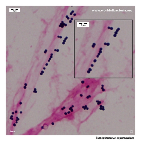
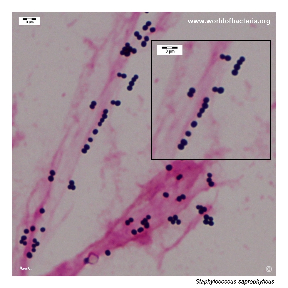
Staphylococcus saprophyticus in a urine specimen, Gram stained. The organism appears as Gram-positive cocci occurring singly, in pairs, short chains, and clusters. Although this microscopic appearance is consistent with the genus Staphylococcus, it is not species-specific. In urinary microbiology, correct interpretation therefore depends on combining Gram stain findings with colony morphology, quantitative culture, and additional identification tests.
Diagnostic and Clinical Notes
Staphylococcus saprophyticus is one of the most important coagulase-negative staphylococci in clinical microbiology because of its well-established role in urinary tract infection. It is classically associated with acute uncomplicated cystitis, especially in young women.
Its pathogenicity in the urinary tract is linked in part to the ability to adhere to uroepithelial cells and to form biofilm, features that support persistence in the urinary environment and help explain its characteristic clinical niche.
In urine culture, the organism may produce relatively large colonies after prolonged incubation, and areas of heavy growth can appear somewhat confluent. These features may be useful practical clues during routine plate examination.
Although most isolates are non-pigmented, occasional strongly pigmented strains do occur and may resemble more visually conspicuous staphylococci. In such cases, the absence of haemolysis remains an important morphological clue that helps distinguish the organism during primary culture.
This isolate was identified by MALDI-TOF mass spectrometry, while biochemical testing supported classification as Staphylococcus saprophyticus subsp. saprophyticus. For routine clinical interpretation, however, species-level identification is usually the more important point.
Laboratory Identification
Colony Morphology
On blood agar incubated at 35–37 °C, colonies of Staphylococcus saprophyticus are typically smooth, convex, and non-haemolytic. Many isolates are white to grey-white, but occasional pigmented strains may occur. With prolonged incubation, colony enlargement can be substantial, and some strains may form notably large colonies.
Microscopy
Gram staining reveals Gram-positive cocci occurring singly, in pairs, short chains, and irregular clusters. The microscopic appearance is compatible with the genus Staphylococcus but is not diagnostic at species level.
Key Identification Clues
Gram-positive cocci in pairs, short chains, and clusters
Strongly catalase positive
Coagulase negative
Typically novobiocin resistant
Usually non-haemolytic on blood agar
Strong clinical association with urinary tract infection, especially in young women
Modern Identification Methods
Species-level identification in clinical microbiology laboratories is commonly achieved using MALDI-TOF mass spectrometry. Subspecies discrimination is not always available by MALDI-TOF alone, and additional biochemical characterization may be used when finer taxonomic resolution is desired.
Antibiotic Characteristics
Staphylococcus saprophyticus is often susceptible to many agents used in urinary tract infection therapy, but susceptibility testing remains important because resistance profiles may vary between isolates and clinical settings.
As with other uropathogens, interpretation should focus on clinically relevant urinary isolates and should take into account the quantitative culture result, patient symptoms, and the likely site of infection.
In routine practice, species confirmation helps distinguish S. saprophyticus from other coagulase-negative staphylococci that may be less clearly pathogenic in urine specimens.
Note: Antimicrobial therapy should always be guided by laboratory susceptibility testing, local recommendations, and clinical context.