Staphylococcus warneri is a Gram-positive, coagulase-negative coccus that commonly forms part of the normal human skin microbiota and is usually of low virulence, although it may occasionally act as an opportunistic pathogen.
Basic Characteristics
Taxonomy
Domain: Bacteria
Phylum: Bacillota (Firmicutes)
Class: Bacilli
Order: Bacillales
Family: Staphylococcaceae
Genus:Staphylococcus
Species:Staphylococcus warneri
Microscopy & Gram Stain
Gram-positive cocci
Cells occur singly, in pairs, and in irregular clusters
Oxygen Relationship
Facultatively anaerobic
Rapid Identification Tests
Catalase: positive
Coagulase: negative
Haemolysis on blood agar: usually absent, but weak haemolysis may occur in some strains
Colony size: usually relatively small
Ecology and Clinical Relevance
Natural Habitat
Human skin microbiota
Commonly recovered from superficial body sites
Often present as part of the background flora in wound and skin specimens
Common Clinical Specimens
Wound swabs
Skin swabs
Blood cultures
Device-associated samples
Occasional deeper clinical materials in opportunistic infection
Clinical Significance
Usually regarded as a low-virulence coagulase-negative staphylococcus
Most often represents colonizing skin flora rather than a major pathogen
May occasionally cause opportunistic infection, especially in predisposed patients
Interpretation depends strongly on specimen type and clinical setting
Differential Considerations
Other coagulase-negative staphylococci recovered from skin or wound material
Micrococcus species in superficially similar pale colonies
More clinically significant CoNS such as S. lugdunensis or S. haemolyticus
Important clues include usually small non-haemolytic colonies, subtle yellowish pigmentation, and the need for proper species-level identification
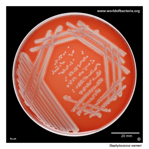
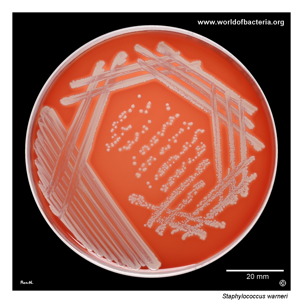
Pure culture of Staphylococcus warneri on blood agar after 24 hours of incubation at 36 °C in ambient air. S. warneri is one of many coagulase-negative staphylococci commonly recovered from wound swabs, usually as part of the surrounding skin microbiota. Most isolates are non-haemolytic, although some strains may show a weak zone of haemolysis close to the colonies, particularly after prolonged incubation.
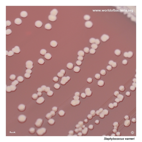
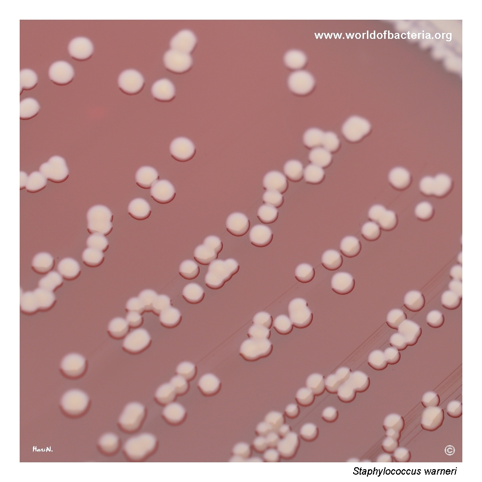
Typical smooth, convex colonies of Staphylococcus warneri, grey-white in colour with a faint yellowish tint. Blood agar, 24 hours, 36 °C, ambient atmosphere. S. warneri usually forms relatively small colonies that do not enlarge markedly even during prolonged incubation, although occasional large-colony strains may occur. Beta-haemolysis may also be observed in some isolates, especially after extended cultivation.
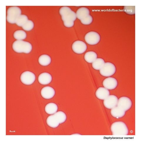
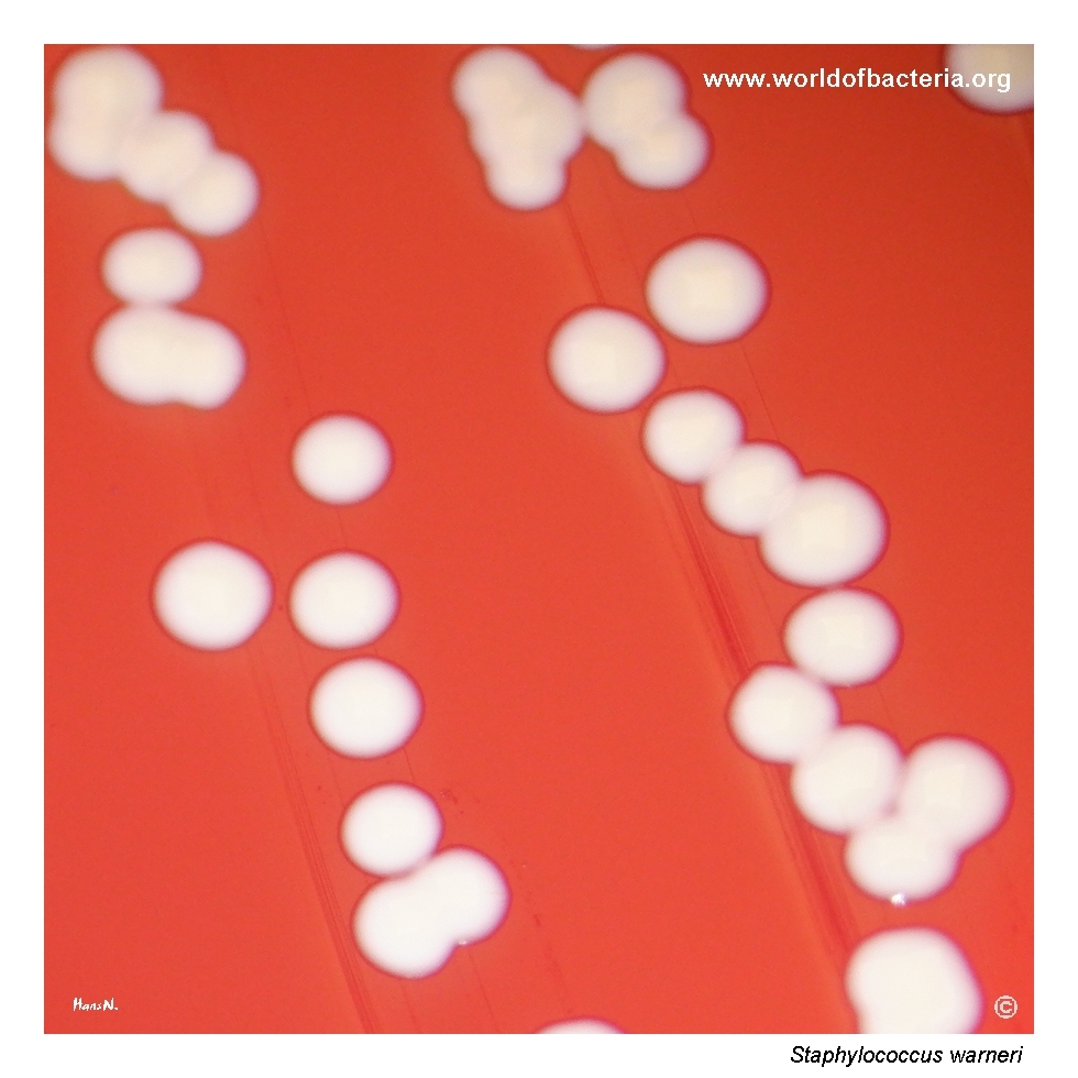
Close-up view of Staphylococcus warneri colonies on blood agar after 24 hours at 36 °C in ambient air. The colonies are smooth, convex, and round with entire margins, measuring approximately 2–3 mm in diameter. A more pronounced yellowish hue is visible in the colony centre, reflecting pigment production. Such subtle pigmentation can help in colony description, although it is not sufficiently specific for identification on its own.
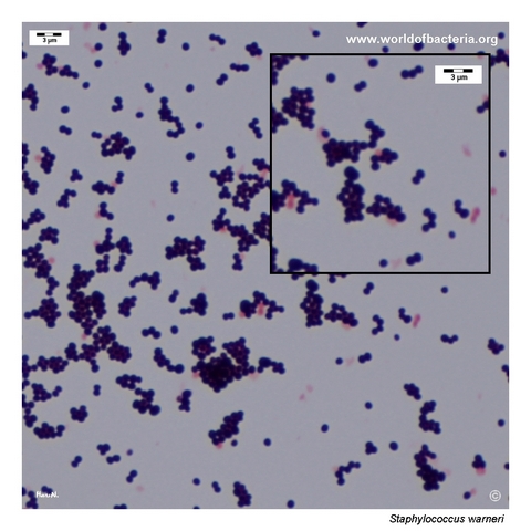
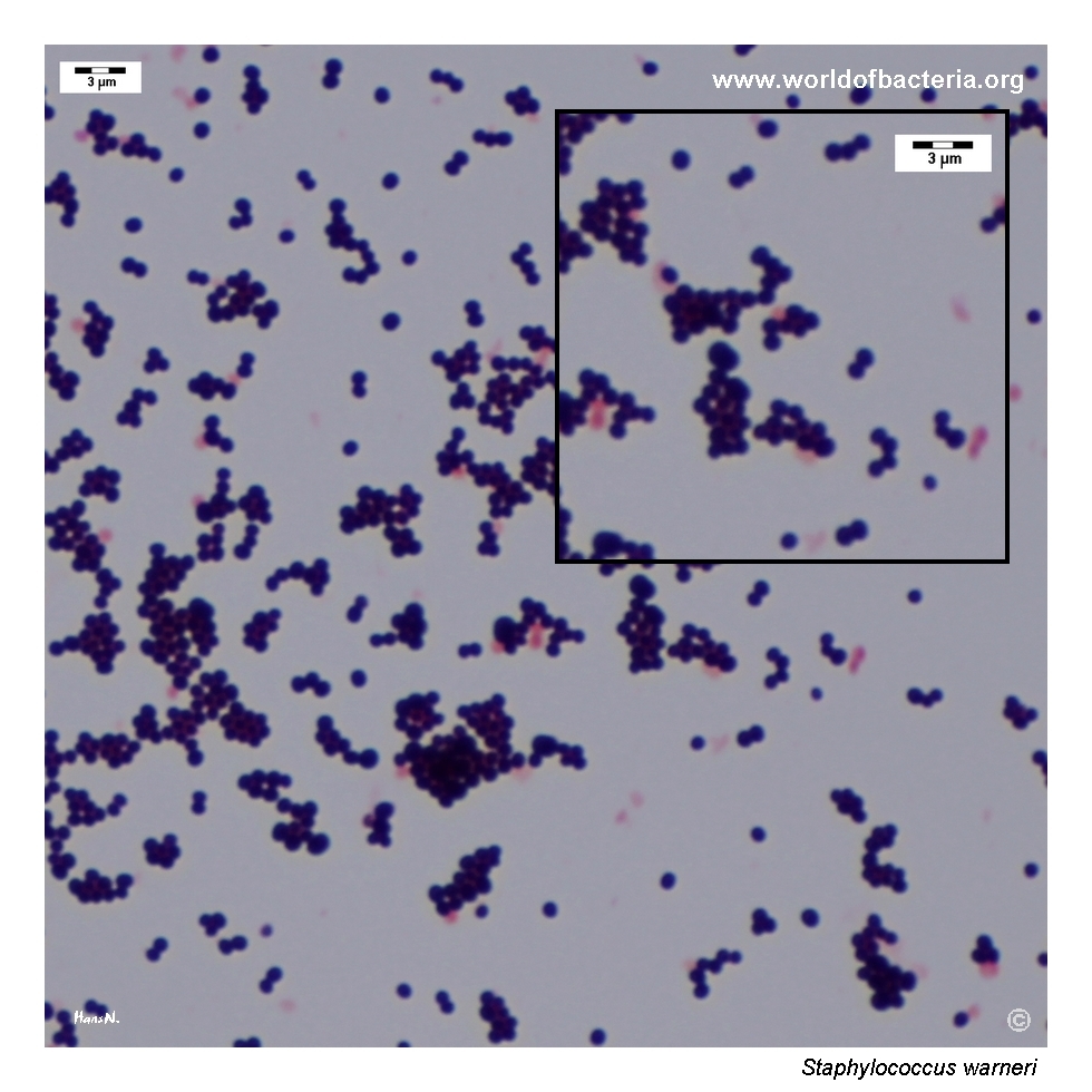
Gram-stained smear prepared from a colony of Staphylococcus warneri grown on blood agar. The organism appears as Gram-positive cocci occurring singly, in pairs, and in clusters, which is the typical microscopic pattern of the genus Staphylococcus. As with other coagulase-negative staphylococci, definitive species identification requires correlation with colony morphology and further laboratory testing.
Diagnostic and Clinical Notes
Staphylococcus warneri is one of the many coagulase-negative staphylococci that commonly colonize human skin and are therefore frequently encountered in routine clinical microbiology. In most superficial specimens, especially wound swabs, its recovery often reflects background skin flora rather than a primary pathogen.
Compared with more aggressive CoNS such as S. lugdunensis, S. warneri is generally regarded as a low-virulence organism. Nevertheless, it may occasionally act as an opportunistic pathogen, particularly in patients with underlying disease, intravascular devices, prosthetic materials, or other predisposing factors.
On blood agar, most isolates are non-haemolytic, but weak haemolysis may be seen in some strains, especially after prolonged incubation. Colonies are usually small, smooth, and slightly pigmented, with a faint yellowish tinge that may be useful descriptively but is not diagnostically specific.
In practice, the main interpretive challenge is deciding whether the isolate is clinically meaningful or merely part of the normal skin microbiota. This decision depends on specimen quality, quantity of growth, culture purity, repetition of isolation, and the overall clinical context.
Because the species is relatively inconspicuous phenotypically, reliable species identification is useful whenever the isolate is recovered from blood, device-related material, or other potentially significant sites.
Laboratory Identification
Colony Morphology
On blood agar incubated at 35–37 °C, colonies of Staphylococcus warneri are typically smooth, convex, round, and grey-white with a faint yellowish tint. Colony size is usually modest, often around 2–3 mm after 24 hours, and many isolates do not enlarge markedly even with longer incubation. Most strains are non-haemolytic, although weak haemolysis may occasionally be observed.
Microscopy
Gram staining reveals Gram-positive cocci occurring singly, in pairs, and in irregular clusters. As in other coagulase-negative staphylococci, this appearance is typical of the genus but is not sufficient for definitive species identification.
Key Identification Clues
Gram-positive cocci in clusters
Strongly catalase positive
Coagulase negative
Usually small, smooth, non-haemolytic colonies
Subtle yellowish pigmentation may be present
Clinical interpretation often requires distinguishing colonization from true infection
Modern Identification Methods
Species-level identification in clinical microbiology laboratories is commonly achieved using MALDI-TOF mass spectrometry. Automated biochemical systems may also be used, but modern identification methods are especially valuable for relatively uncommon or phenotypically subtle coagulase-negative staphylococci such as S. warneri.
Antibiotic Characteristics
Staphylococcus warneri is not usually one of the most aggressive staphylococcal pathogens, but clinically significant isolates should still undergo standard susceptibility testing because resistance patterns may vary among strains.
When the organism is recovered from blood cultures, implanted devices, or repeated clinically relevant specimens, antimicrobial decisions should be based on the confirmed susceptibility result and the likelihood that the isolate represents true infection rather than contamination.
In superficial polymicrobial material, the species often has limited independent significance unless supported by culture predominance or clinical evidence.
Note: Antimicrobial therapy should always be guided by laboratory susceptibility testing, specimen significance, and clinical context.