Micrococcus luteus is a yellow-pigmented, strictly aerobic coccus commonly found on human skin and in the environment, usually of low pathogenic significance but occasionally encountered in clinical microbiology as a contaminant or rare opportunistic isolate.
Basic Characteristics
Taxonomy
Domain: Bacteria
Phylum: Actinomycetota
Class: Actinomycetes
Order: Micrococcales
Family: Micrococcaceae
Genus:Micrococcus
Species:Micrococcus luteus
Microscopy & Gram Stain
Gram-positive cocci
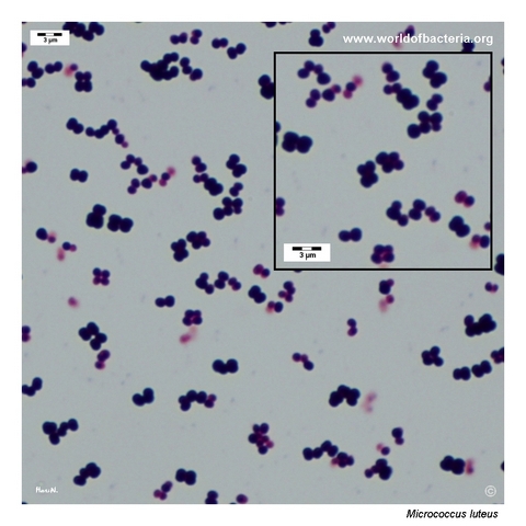
Cells typically occur in tetrads, pairs, and irregular clusters
Tetrad formation is a useful practical clue when distinguishing micrococci from staphylococci on Gram stain
Microscopic morphology supports the genus Micrococcus but does not replace full identification
Oxygen Relationship
Strictly aerobic
Grows well under routine aerobic culture conditions on blood agar
Not expected to behave like a facultatively anaerobic staphylococcus in routine culture
Rapid Identification Tests
Catalase: positive
Oxidase: positive
Pigmentation: colonies are often distinctly yellow
Practical note: a familiar environmental and skin-associated organism that is usually of low pathogenic significance
Identification caveat: modern methods are useful when a clinically significant isolate must be distinguished from look-alike Gram-positive cocci
Ecology and Clinical Relevance
Natural Habitat
Widely distributed on human skin and in the environment, including air, dust, water, and surface contamination
Often encountered in environmental monitoring and in incidental laboratory contamination
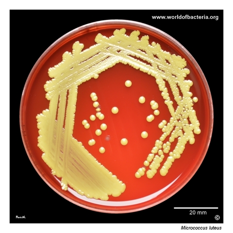
In this page set, the isolate represents accidental contamination of a laboratory culture plate
Common Clinical Specimens
Commonly recovered from skin swabs and environmental monitoring samples
May appear in clinical cultures as a contaminant from skin or surroundings
True infection is uncommon and usually requires a strongly supportive clinical context
Clinical Significance
Usually regarded as a harmless commensal organism or environmental contaminant
Rare opportunistic infections have been reported, mainly in predisposed patients or damaged tissue
In routine microbiology, the main task is often to recognize low pathogenic significance rather than to overinterpret the isolate
Differential Considerations
Kocuria rhizophila and other yellow-pigmented Gram-positive cocci
Staphylococcus aureus and pigmented coagulase-negative staphylococci when only colony colour is considered
Staphylococcus hominis and other non-pigmented skin organisms present in mixed culture
Neisseria meningitidis in this page set serves as a useful contrast organism because its colonies are non-pigmented
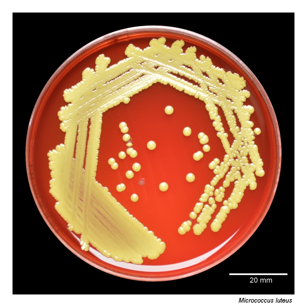
Micrococcus luteus on blood agar. The isolate originated from accidental contamination of a plate in the laboratory. Strains of M. luteus are commonly encountered in skin swabs or during monitoring of environmental sterility. Incubation: 48 hours at 36 °C in 5% CO2.
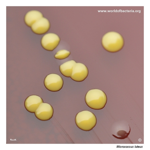
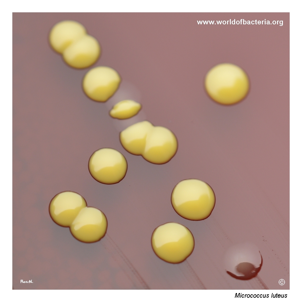
Colonies of Micrococcus luteus are characterized by striking yellow pigmentation. They are usually convex, smooth, and round with entire margins, although rough-surfaced colonies may also occur. The non-pigmented colonies in the image belong to Neisseria meningitidis. Incubation: 48 hours at 36 °C in 5% CO2.
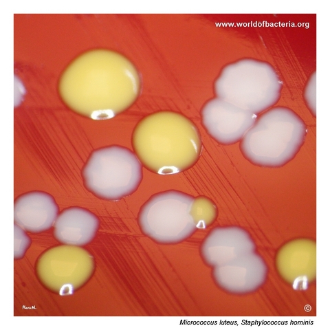
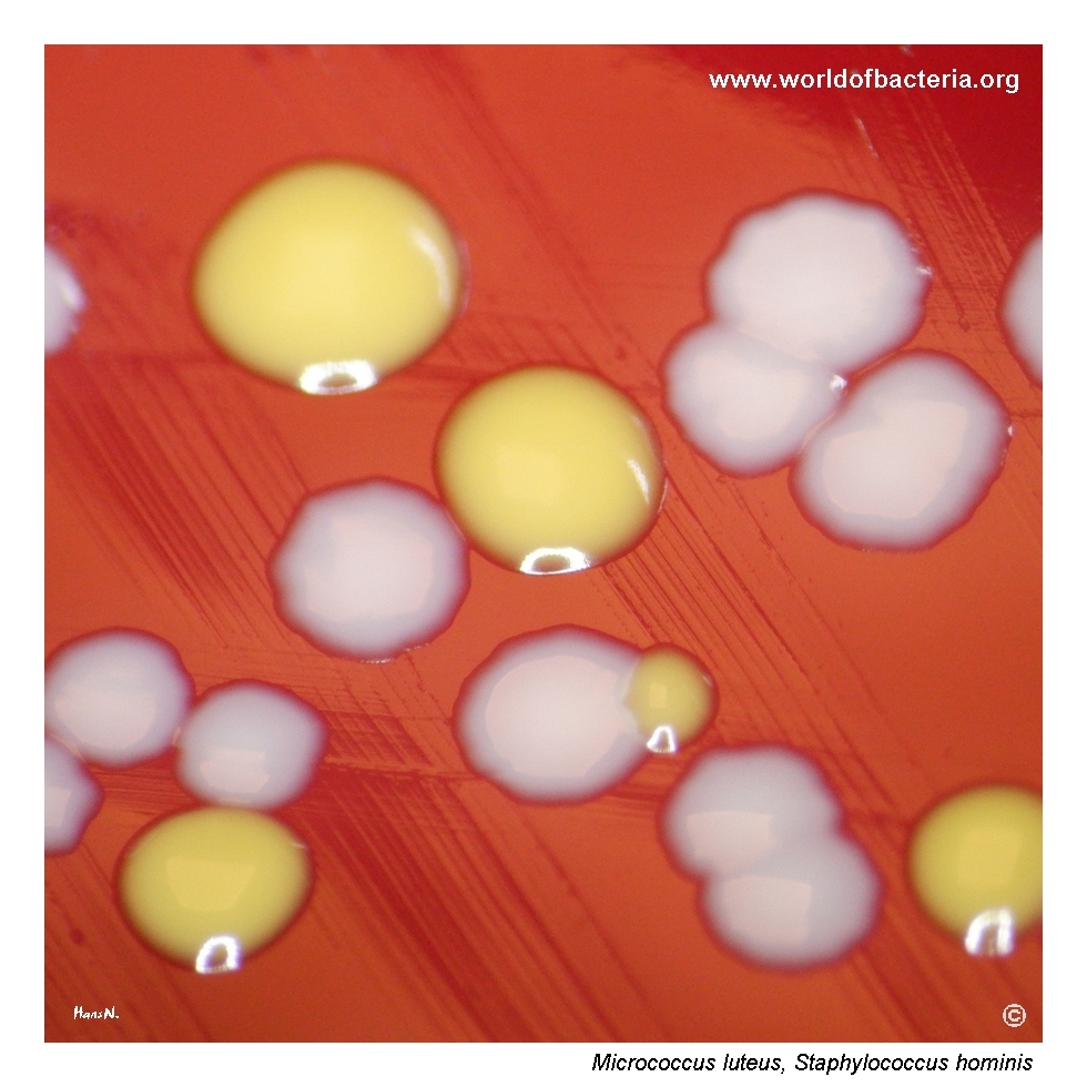
Pigmented colonies of Micrococcus luteus and non-pigmented colonies of Staphylococcus hominis on blood agar. Incubation: 24 hours at 36 °C in ambient atmosphere.
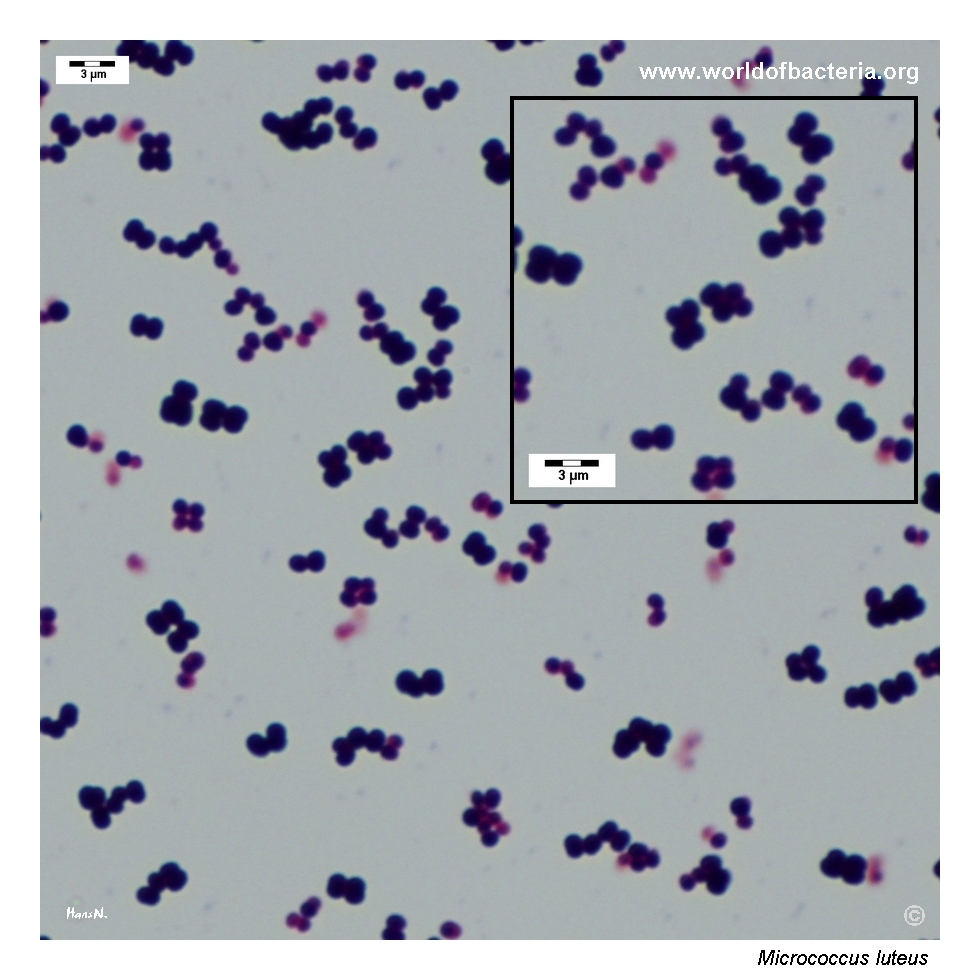
Smear prepared from a colony of Micrococcus luteus grown on blood agar and stained by the Gram method. Gram-positive cocci are arranged mainly in tetrads and pairs.
Diagnostic and Clinical Notes
Micrococcus luteus is a classic teaching organism because it combines two memorable visual features: bright yellow pigmentation and a microscopic tendency to form tetrads. These features make it more distinctive than many low-significance Gram-positive cocci seen in routine cultures.
At the same time, the practical challenge is interpretive rather than descriptive. In most routine settings, recovery of M. luteus reflects skin flora, environmental contamination, or accidental laboratory contamination rather than true disease. That fits well with the source of the isolate shown on this page.
Mixed culture is especially instructive here. The contrast between yellow M. luteus colonies and non-pigmented colonies of other bacteria shows how colony colour can offer a useful first clue during plate reading. However, pigmentation alone is not enough for final identification, because several unrelated organisms may produce yellow colonies.
When clinically important infection is suspected, the organism should not be dismissed solely on appearance. Rare opportunistic infections have been described, but interpretation must be based on specimen type, quantity, repeated isolation, and the condition of the patient rather than on pigment alone.
Laboratory Identification
Colony Morphology
On blood agar, Micrococcus luteus typically forms smooth, convex, round colonies with entire margins and a distinct yellow pigment. Rougher colony variants may also occur. In mixed culture, this colour difference can make the organism visually conspicuous even when it is not clinically important.
Microscopy
Gram staining shows Gram-positive cocci occurring mainly in tetrads and pairs, sometimes with small irregular aggregates. Tetrad formation is one of the most useful practical clues for recognizing a micrococcal morphology and separating it from the more cluster-dominant appearance of staphylococci.
Key Identification Clues
Gram-positive cocci in tetrads and pairs
Strictly aerobic growth pattern
Catalase positive
Oxidase positive
Distinct yellow colony pigmentation
Common association with skin or environmental contamination
Usually low clinical significance unless supported by strong specimen context
Modern Identification Methods
Modern identification is readily achieved by MALDI-TOF mass spectrometry in laboratories with adequate database coverage. Instrumental identification is particularly helpful when yellow-pigmented Gram-positive cocci must be distinguished from look-alike organisms such as Kocuria spp. or pigmented staphylococci, or when the result may affect clinical interpretation.
Antibiotic Characteristics
No simple therapeutic assumption should be made from the appearance of Micrococcus luteus on culture plates. In most routine situations, the more relevant question is whether the isolate is clinically meaningful at all.
Because true infection is uncommon, antimicrobial susceptibility testing is mainly relevant when the organism is recovered from a convincing clinical specimen or repeatedly isolated in a compatible clinical setting.
When treatment is considered necessary, susceptibility should be determined on the individual isolate rather than inferred from its usual reputation as a low-virulence commensal.
Note: Antimicrobial therapy should always be guided by formal laboratory susceptibility testing and the overall clinical picture.