Streptococcus pyogenes is the classical group A streptococcus, best known for its sharply beta-haemolytic colonies on blood agar and for its ability to cause both common mucosal infections and severe invasive disease.
Basic Characteristics
Taxonomy
Domain: Bacteria
Phylum: Bacillota
Class: Bacilli
Order: Lactobacillales
Family: Streptococcaceae
Genus:Streptococcus
Species:Streptococcus pyogenes
Microscopy & Gram Stain
Gram-positive cocci typically arranged in pairs and especially in chains, which is the classic microscopic pattern of beta-haemolytic streptococci
In younger cultures the cells are usually clearly Gram positive, but blood-culture smears may show partial Gram variability when cell-wall maturation is incomplete
Some cells can appear slightly elongated, while others remain more nearly spherical, so morphology should always be interpreted in context
Microscopy strongly supports the diagnosis of a streptococcal organism but does not replace species-level identification
Oxygen Relationship
Facultatively anaerobic streptococcus that grows readily on routine blood agar under capnophilic incubation
In this page set, clearly visible colonies developed after 24 to 48 hours at 36 °C in 5% CO2
The species is therefore not fastidious in routine diagnostic culture, and its pronounced haemolysis often makes it visually prominent
Rapid Identification Tests
Catalase: negative
Haemolysis: typically strong beta-haemolysis on blood agar
Lancefield grouping: group A streptococcus (GAS)
Bacitracin: historically useful as a screening clue, but not sufficiently specific for definitive identification
PYR: typically positive
Practical implication: a classic GAS phenotype should still be confirmed in the full laboratory and clinical context
Ecology and Clinical Relevance
Natural Habitat
Streptococcus pyogenes is a human-associated pathogen and colonizer, most often linked to the pharynx and skin rather than to environmental reservoirs
Its recovery from clinical material is therefore usually more meaningful than the recovery of many commensal streptococci
This page set includes isolates from the vagina, throat, middle-ear-related material, and blood culture
Common Clinical Specimens
Throat swabs
Pus and other purulent material
Blood cultures in invasive disease
Other mucosal specimens, including genital samples, where interpretation depends strongly on the clinical context
Clinical Significance
Streptococcus pyogenes is one of the most important human bacterial pathogens, causing diseases that range from uncomplicated pharyngitis to severe invasive infection
Its classical beta-haemolytic plate phenotype is diagnostically useful, but the species is clinically defined far more by its pathogenic potential than by morphology alone
Historically, GAS was also one of the most feared bacterial pathogens in obstetrics, because puerperal and postpartum sepsis caused by this species had very high mortality before antisepsis, a fact strongly associated with the work of Ignaz Semmelweis on hand hygiene
The blood-culture image in this page set illustrates that GAS in a normally sterile site must always be treated as a serious finding
Differential Considerations
Other large-colony beta-haemolytic streptococci, especially Streptococcus dysgalactiae subsp. equisimilis
Streptococcus agalactiae and other beta-haemolytic streptococci when only the haemolytic plate phenotype is considered
More conspicuous pyogenic pathogens in mixed material
Any invasive blood-culture isolate requiring immediate clinical interpretation
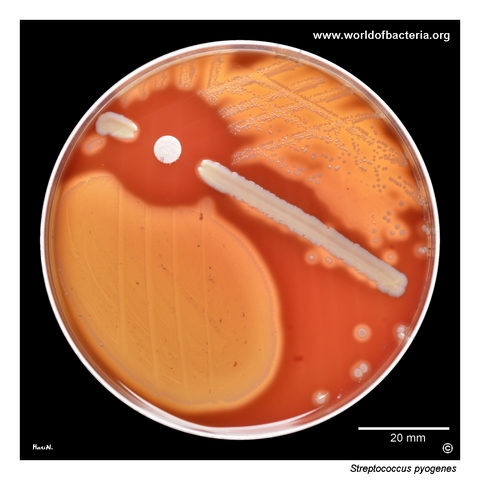
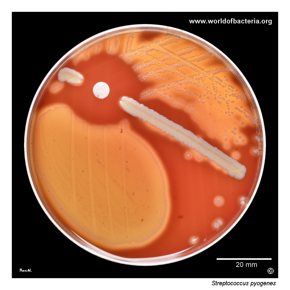
Vaginal swab from a 69-year-old woman with the diagnosis N76.1, subacute and chronic vaginitis. The culture yielded an almost pure growth of Streptococcus pyogenes. A streak of Staphylococcus aureus ATCC 25923 and a bacitracin disk were placed on the plate. Part of the grown colonies was removed during antibiotic susceptibility testing. Culture conditions: 48 hours at 36 °C in 5% CO2. This image illustrates that S. pyogenes may also be recovered in high quantity outside the pharynx. In women, GAS has major historical significance because puerperal and postpartum infections caused by this organism were among the most feared and often fatal bacterial complications of childbirth before the era of antisepsis and antibiotics.
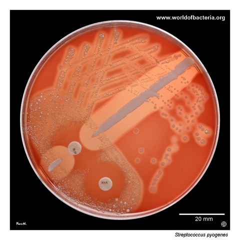
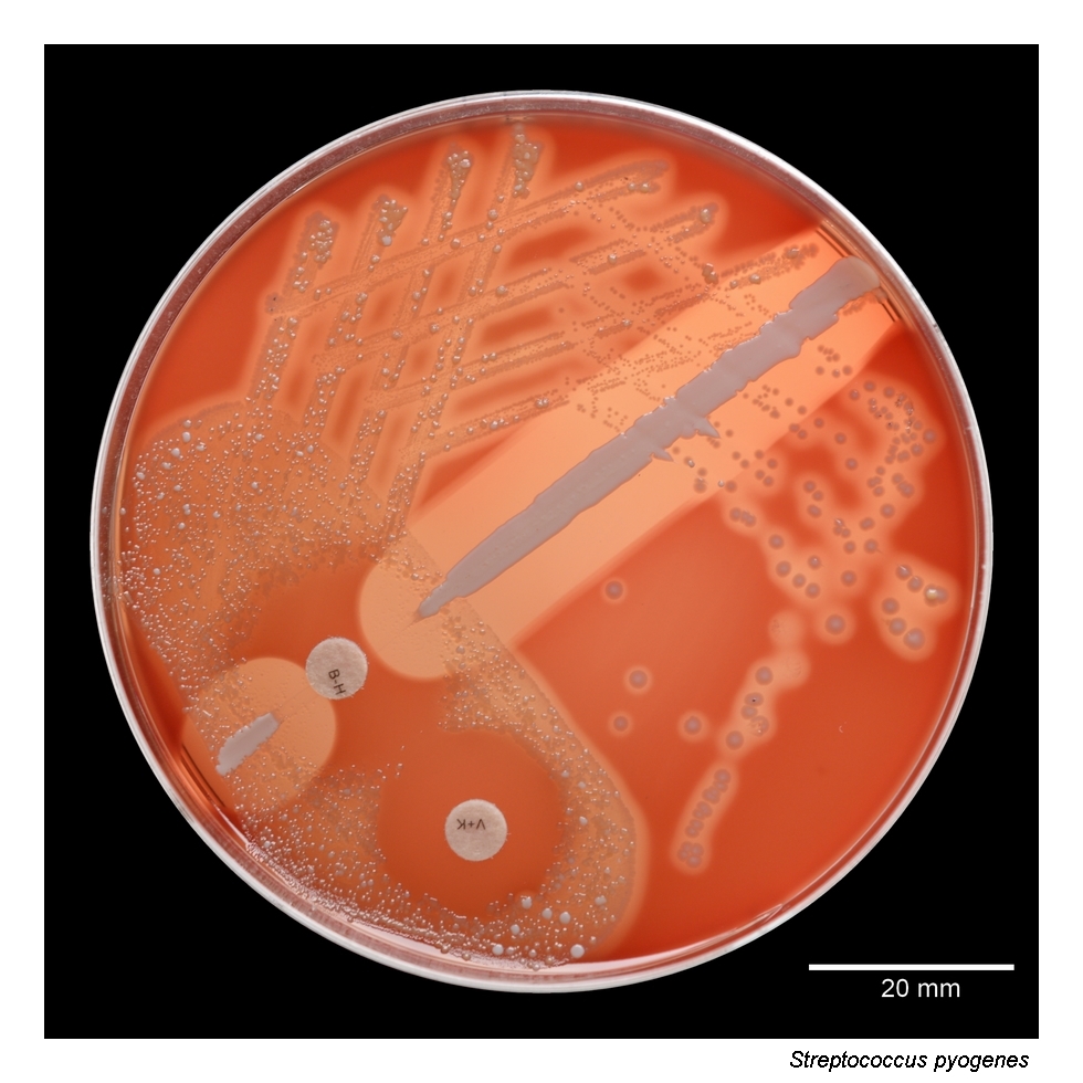
Colonies of Streptococcus pyogenes on blood agar isolated from a throat swab. The colonies measure approximately 2–3 mm in diameter and are surrounded by a clear zone of beta-haemolysis. Culture conditions: 24 hours at 36 °C in 5% CO2. This is the classic plate appearance expected for group A streptococci and remains one of the most important visual clues during routine primary culture reading.
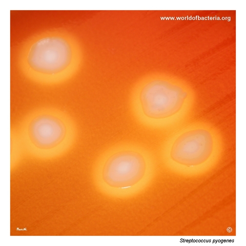
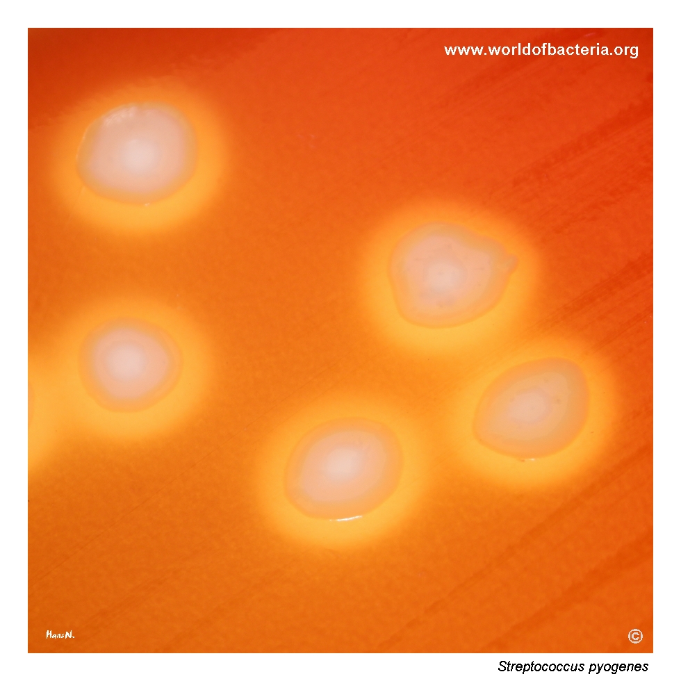
Close-up view of Streptococcus pyogenes colonies isolated from pus obtained by paracentesis from a 21-year-old patient with the diagnosis H65.0, acute serous otitis media. Culture conditions: 48 hours at 36 °C in 5% CO2. The colonies are large, measuring about 5–7 mm in diameter. Sequencing of the emm gene encoding M protein assigned this isolate to the emm3 group, a lineage often associated with marked virulence.
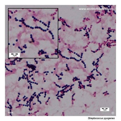
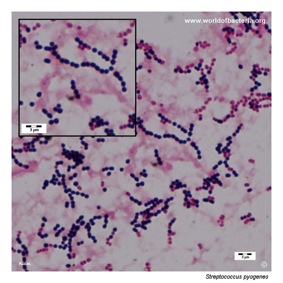
Gram-stained blood culture showing numerous short chains of Gram-positive cocci. Cells with an insufficiently matured cell wall may stain Gram-negative, producing partial Gram variability in the smear. This finding represents a serious, potentially life-threatening condition, because the presence of Streptococcus pyogenes in blood culture is consistent with invasive infection requiring urgent clinical attention.
Diagnostic and Clinical Notes
This page set captures several complementary faces of Streptococcus pyogenes: near-pure growth from the female genital tract, the textbook beta-haemolytic phenotype from a throat swab, a markedly large-colony emm3 isolate from purulent material, and invasive disease documented directly in blood culture.
The throat-swab image is the classical teaching picture of GAS on blood agar: medium-sized colonies with clear beta-haemolysis after overnight incubation. This remains one of the most useful visual patterns in routine streptococcal plate reading.
The genital-specimen image is historically important as well as microbiologically interesting. Although such material may seem less typical from a modern routine-laboratory perspective, infections caused by S. pyogenes in the female genital tract were historically among the major causes of fatal puerperal sepsis, and Semmelweis' work on hand hygiene remains one of the defining milestones in understanding their transmission.
The large emm3 isolate is didactically valuable because it shows how striking some GAS colonies can become after longer incubation and because emm3 lineages are often associated with increased virulence. Even without molecular typing, unusually robust beta-haemolytic growth from purulent material deserves attention.
The blood-culture smear is the most clinically urgent image in the set. Invasive GAS disease is a severe condition, and once S. pyogenes is detected in blood culture, the result must be communicated and interpreted as a major clinical finding rather than as incidental flora.
Laboratory Identification
Colony Morphology
On blood agar, Streptococcus pyogenes typically forms smooth greyish to translucent colonies surrounded by a clear zone of beta-haemolysis. Colony size can vary with strain and incubation time, as shown here by both a more standard 2–3 mm throat-swab isolate and a much larger emm3 isolate from purulent material.
Microscopy
Gram staining usually shows Gram-positive cocci in pairs and especially in chains. In blood-culture smears, some cells may stain weakly or even appear partly Gram negative when the cell wall is not fully matured, but the overall pattern still strongly supports a streptococcal invasive isolate.
Key Identification Clues
Classic beta-haemolytic colonies on blood agar
Catalase-negative streptococcal morphology
Association with pharyngeal, purulent, or invasive clinical material
Group A streptococcal identity in the appropriate laboratory context
Need to distinguish from other beta-haemolytic streptococci when morphology alone is considered
Modern Identification Methods
Modern identification is straightforward with MALDI-TOF MS or other validated laboratory methods, but the real diagnostic challenge is often not species recognition itself but correct clinical interpretation of the isolate. This is especially true for unusual specimen types and for invasive disease, where rapid recognition and communication matter most.
Antibiotic Characteristics
The photographed colonies should not be used on their own to infer therapy. For Streptococcus pyogenes, treatment depends on the clinical syndrome, specimen type, and severity of disease, especially when invasive infection is suspected.
In routine diagnostics, the main value of the plate phenotype is rapid presumptive recognition of a clinically important beta-haemolytic streptococcus rather than prediction of resistance from colony appearance alone.
Note: In this page set, one plate also shows material removed for susceptibility testing, reminding the reader that even when the morphology looks classical, laboratory confirmation remains part of good clinical microbiology practice.