Shigella sonnei is a non-motile Gram-negative enteric pathogen belonging to serogroup D and a major cause of shigellosis, particularly in regions with well-developed sanitation systems.
Basic Characteristics
Taxonomy
Domain: Bacteria
Phylum: Pseudomonadota (Proteobacteria)
Class: Gammaproteobacteria
Order: Enterobacterales
Family: Enterobacteriaceae
Genus:Shigella
Species:Shigella sonnei
Serogroup: D
Microscopy & Gram Stain
Small, short Gram-negative rods
Cells usually occur singly
Microscopy does not distinguish Shigella from Escherichia coli
Oxygen Relationship
Facultatively anaerobic
Rapid Identification Tests
Oxidase: negative
Catalase: usually positive
Motility: negative
Lactose fermentation: absent during routine early incubation; delayed fermentation may occur
ONPG: positive
Ornithine decarboxylase: positive
Hydrogen sulphide: not produced
Urease: negative
Ecology and Clinical Relevance
Natural Habitat
Human intestinal tract during infection or temporary carriage
Humans are the principal reservoir
Contaminated hands, food, water, and surfaces involved in faecal–oral transmission
Common Clinical Specimens
Stool
Rectal swabs
Stool enrichment cultures
Public-health isolates submitted for serotyping and molecular surveillance
Clinical Significance
Important cause of shigellosis and bacillary dysentery
May produce watery or bloody diarrhoea, abdominal pain, fever, and tenesmus
Highly transmissible because a small inoculum may be sufficient for infection
Person-to-person spread and outbreaks are epidemiologically important
Bloodstream infection and other extraintestinal complications are uncommon but may occur in vulnerable patients
Differential Considerations
Escherichia coli, especially enteroinvasive E. coli (EIEC)
Shigella flexneri and other Shigella species
Salmonella enterica and other lactose-non-fermenting Enterobacterales
Routine MALDI-TOF MS may identify Shigella isolates as Escherichia coli or provide an ambiguous result
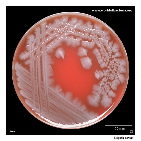
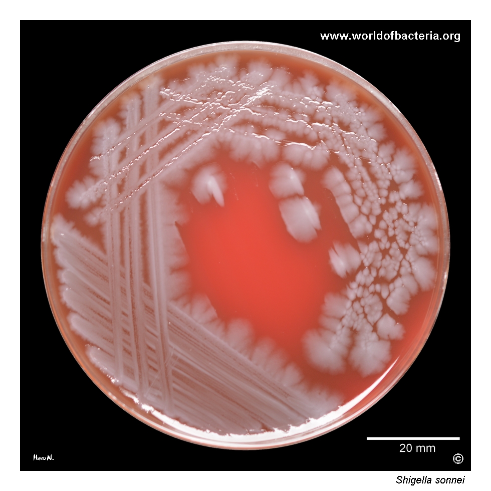
Shigella sonnei on blood agar after 24 hours of incubation at 36 °C in ambient air. In this culture, the organism forms large, irregular colonies spreading across the surface of the medium. This colony type may occur in S. sonnei, but it is not species-specific and can also be observed in other Shigella species and in some strains of Escherichia coli. Colony morphology alone is therefore insufficient for reliable identification.
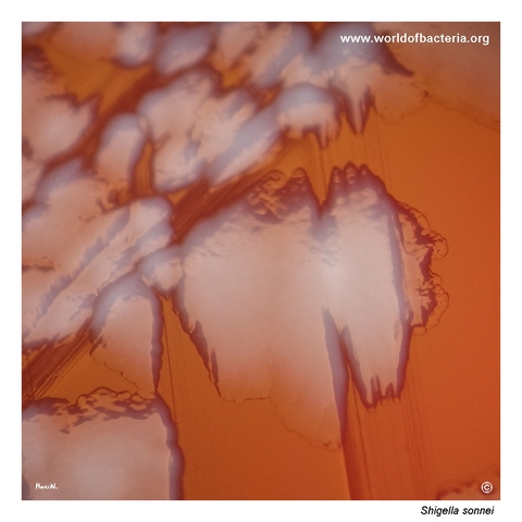
Colonies of Shigella sonnei on blood agar after 24 hours at 36 °C in ambient air. The colonies are large, flat, grey-white, and irregular, with uneven margins. Similar isolates identified as Escherichia coli by routine MALDI-TOF MS should be examined further, because this method may not reliably distinguish Escherichia from Shigella. Additional biochemical testing and serological agglutination are therefore required for confirmation.
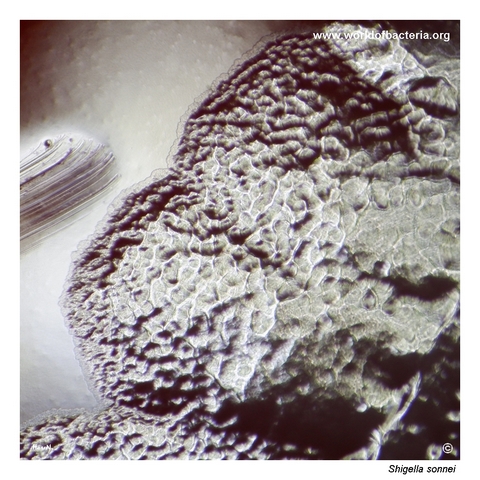
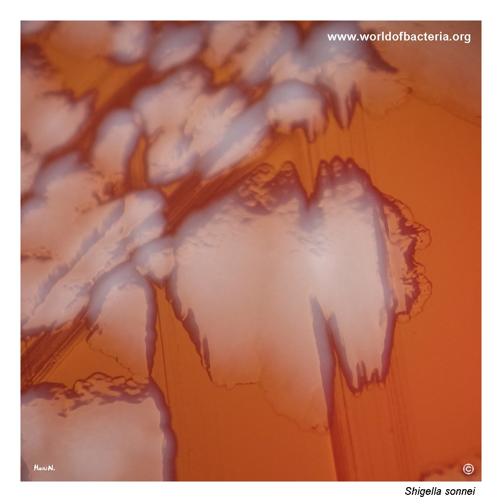
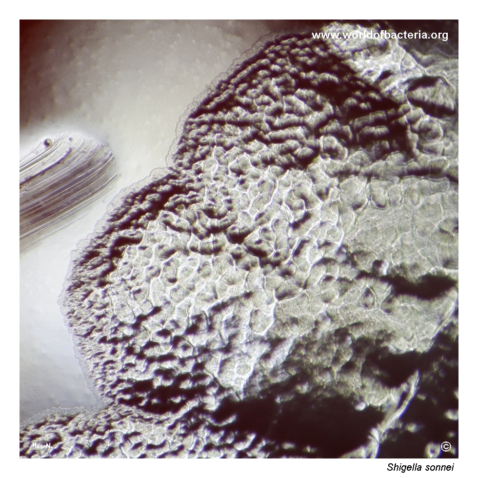
Highly magnified view of a Shigella sonnei colony on blood agar, photographed in reflected light. The colony surface shows a coarse texture resembling a sheet of beaten metal. Irregular projections extend from the colony margin and rise above the surrounding surface, contributing to the partially blurred appearance seen in the lower-magnification image. Culture conditions: 24 hours at 36 °C in ambient air.
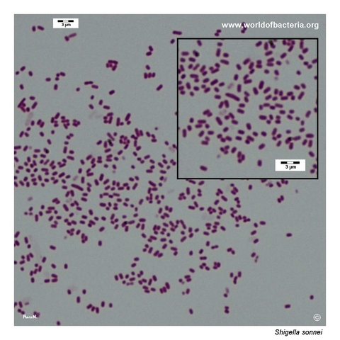
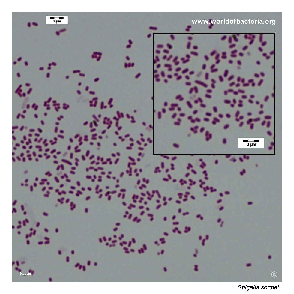
Gram-stained smear prepared from a colony of Shigella sonnei grown on blood agar. The organism appears as small, short Gram-negative rods. This microscopic morphology is typical of enteric Gram-negative bacteria but does not distinguish Shigella from Escherichia coli or other closely related members of the Enterobacterales. Reliable identification therefore depends on additional biochemical, serological, or molecular testing.
Diagnostic and Clinical Notes
Shigella sonnei is one of the four medically recognized Shigella species and belongs to serogroup D. It is a major cause of shigellosis and is especially prominent in countries with relatively well-developed water and sanitation systems.
The organism is highly adapted to humans and spreads primarily by the faecal–oral route. Its low infectious dose facilitates direct person-to-person transmission, household spread, and outbreaks in childcare, institutional, travel-related, and other close-contact settings.
Clinical illness ranges from mild watery diarrhoea to inflammatory diarrhoea with blood, abdominal cramps, fever, and tenesmus. Most infections are self-limited, but prolonged disease, seizures in young children, and bloodstream infection in immunocompromised or otherwise vulnerable patients may occur.
The illustrated isolate forms unusually large, flat, irregular colonies with a coarse surface. This appearance is visually instructive but is not sufficiently specific to distinguish S. sonnei from other Shigella species or morphologically variable strains of Escherichia coli.
The close genetic and proteomic relationship between Shigella and Escherichia coli is a major diagnostic limitation. Routine MALDI-TOF MS cannot be relied upon as the sole method for separating these organisms, so apparently inactive or unusual E. coli isolates should undergo appropriate biochemical, serological, and molecular confirmation.
Laboratory Identification
Colony Morphology
On routine blood agar, Shigella sonnei may form grey-white, non-haemolytic colonies whose size and shape vary between isolates. The strain illustrated here forms unusually large, flat, spreading colonies with irregular margins and a coarse beaten-metal surface. On lactose-differential media, early colonies are generally pale because lactose fermentation is absent or delayed.
Microscopy
Gram staining reveals small, short Gram-negative rods occurring mainly singly. This appearance is indistinguishable from Escherichia coli and many other Enterobacterales and therefore provides only broad morphological classification.
Key Identification Clues
Small, short Gram-negative rods
Facultatively anaerobic growth
Oxidase negative
Non-motile
Hydrogen sulphide and urease negative
ONPG and ornithine decarboxylase positive
Delayed lactose fermentation may occur
Serogroup D agglutination
Modern Identification Methods
Presumptive identification combines selective and differential culture with biochemical reactions, particularly non-motility, ONPG activity, ornithine decarboxylase activity, and the absence of hydrogen sulphide production. Serological agglutination confirms the group D antigenic profile. Because routine MALDI-TOF MS may not reliably separate Shigella from Escherichia coli, molecular assays, reference-laboratory confirmation, or whole-genome sequencing may be needed for definitive identification, surveillance, and differentiation from enteroinvasive E. coli.
Antibiotic Characteristics
Many uncomplicated cases of shigellosis resolve without antimicrobial therapy, and supportive management with attention to hydration is sufficient.
Antibiotic treatment may be appropriate for severe disease, vulnerable patients, prolonged illness, or epidemiological situations in which reducing the duration of symptoms and bacterial shedding is important.
Resistance to commonly used agents, including azithromycin, fluoroquinolones, trimethoprim–sulfamethoxazole, ampicillin, and third-generation cephalosporins, is an increasing public-health concern in Shigella.
Note: When treatment is planned, antimicrobial selection should be guided by susceptibility testing of the individual isolate and current local or outbreak-specific recommendations. Antimotility drugs should be avoided because they may worsen shigellosis.