Shigella flexneri is a non-motile, lactose-non-fermenting Gram-negative enteric pathogen and an important cause of shigellosis, including inflammatory diarrhoea and bacillary dysentery.
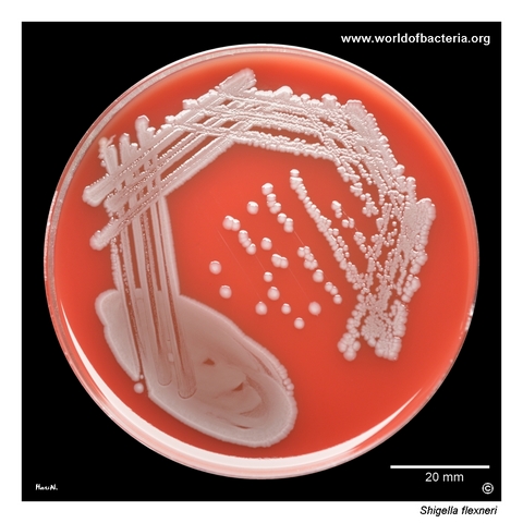
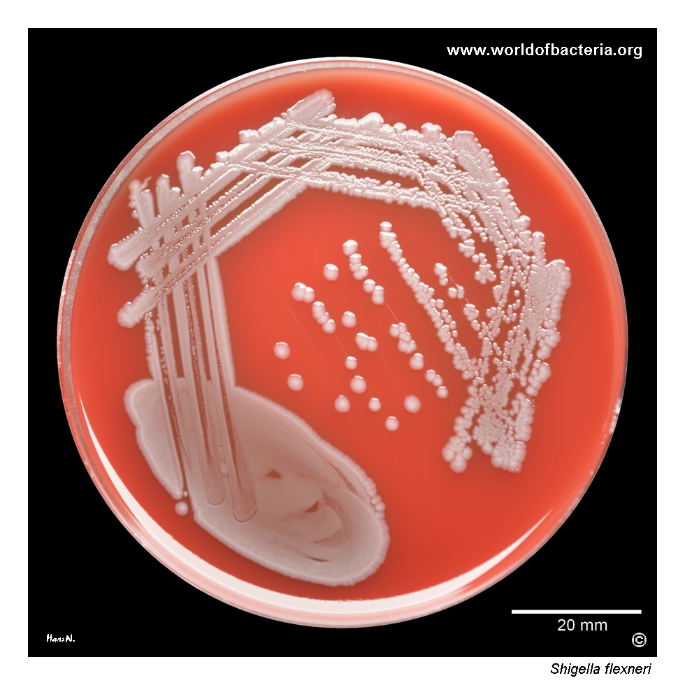
Shigella flexneri on blood agar after 24 hours of incubation at 36 °C in ambient air. Strains of S. flexneri are non-haemolytic and may be visually indistinguishable from non-haemolytic strains of Escherichia coli. The image shows collection strain CCM 4422, antigenic structure IV:3,4, serovar 4a. Colony appearance on blood agar therefore provides no reliable basis for differentiating these closely related organisms.
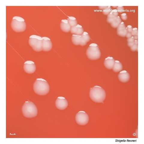
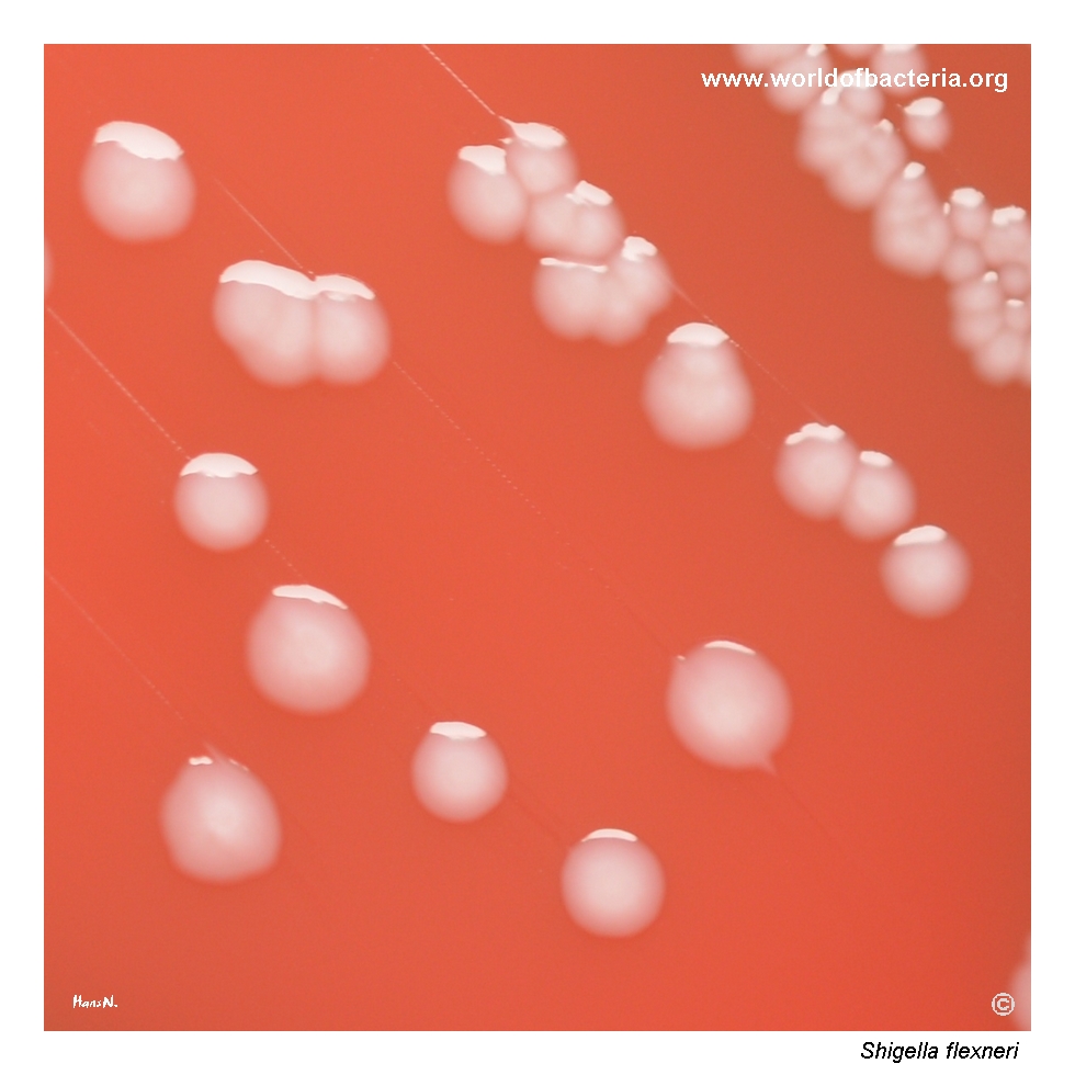
Colonies of Shigella flexneri on blood agar after 24 hours at 36 °C in ambient air. The colonies are inconspicuous, smooth, convex, grey-white, and round with entire margins. Their appearance closely resembles that of ordinary Escherichia coli strains forming relatively small non-haemolytic colonies. Reliable identification consequently requires evaluation beyond morphology on routine blood agar.
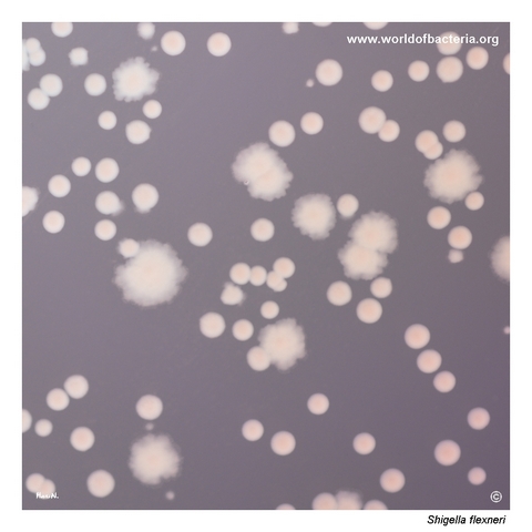
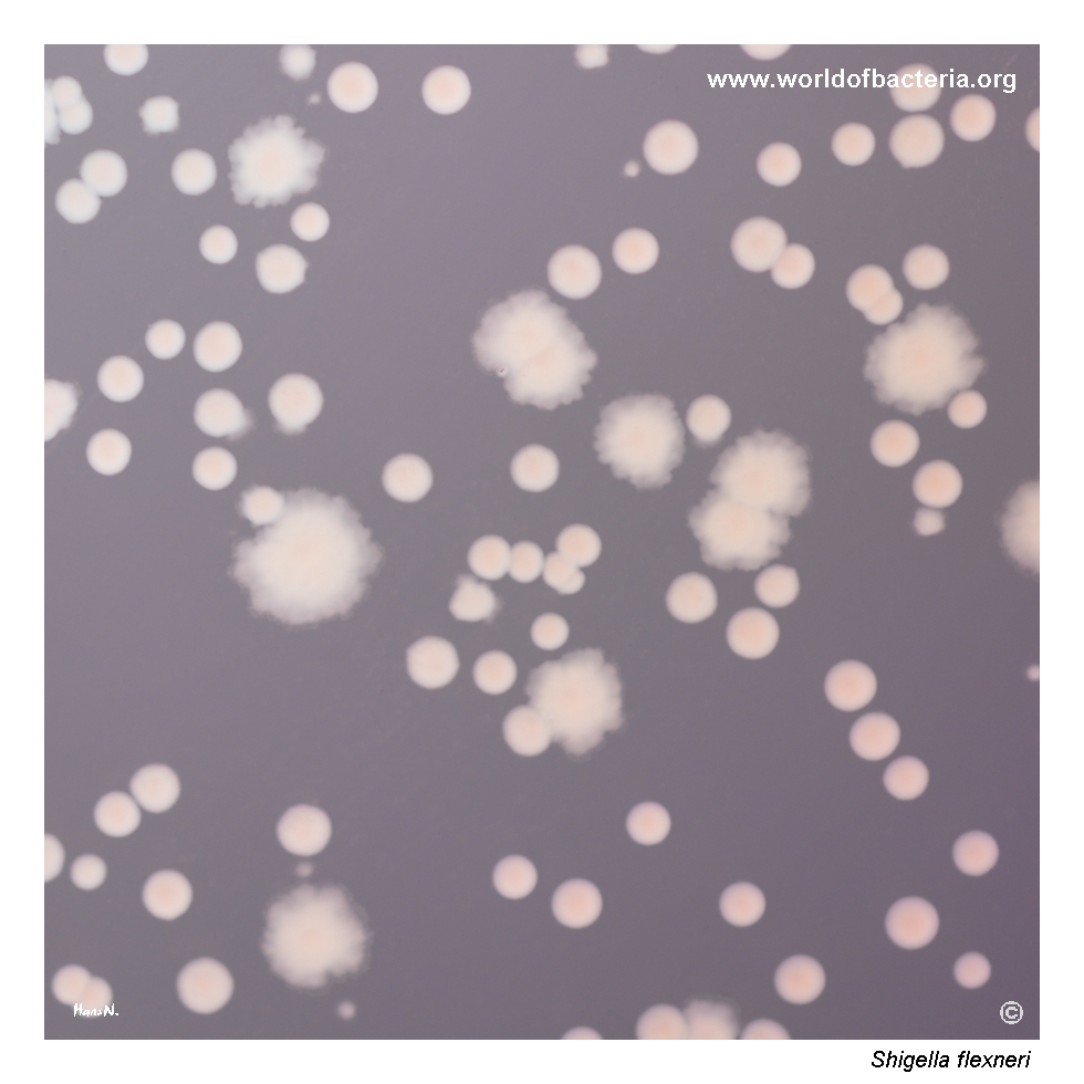
Close-up view of lactose-non-fermenting Shigella flexneri colonies on MacConkey agar after 24 hours at 36 °C in ambient air. Two colony forms are visible: smaller, smooth, convex colonies with entire margins and larger, flat colonies with irregular margins. Both variants are produced by the same collection strain, S. flexneri CCM 4422, illustrating morphological variation within a single bacterial culture.
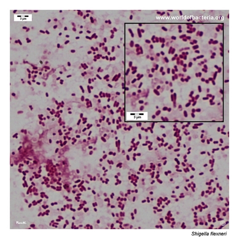
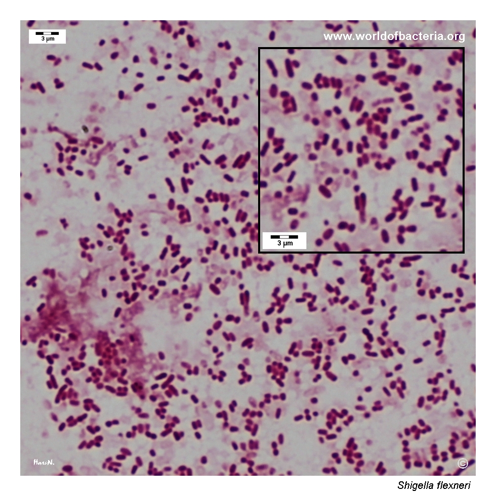
Gram-stained smear prepared from a colony of Shigella flexneri grown on blood agar. The organism appears as short Gram-negative rods. This microscopic morphology is typical of enteric Gram-negative bacteria but does not distinguish Shigella from Escherichia coli or related taxa. Species identification therefore depends on culture characteristics and additional biochemical, antigenic, or molecular methods.
Diagnostic and Clinical Notes
Shigella flexneri is one of the four medically recognized Shigella species and belongs to serogroup B. It is a major cause of shigellosis in many regions and is particularly associated with inflammatory diarrhoea and bacillary dysentery.
The organism is highly adapted to humans and spreads predominantly by the faecal–oral route. A relatively small infectious dose facilitates direct person-to-person transmission, household spread, and outbreaks in settings where hygiene is difficult to maintain.
Clinical disease may include watery or bloody diarrhoea, abdominal cramps, fever, and tenesmus. Most infections are self-limited, but severe illness, prolonged symptoms, seizures in young children, bloodstream infection in vulnerable patients, and post-infectious arthritis may occur.
The close genetic and phenotypic relationship between Shigella and Escherichia coli creates a major identification challenge. Ordinary colony morphology, Gram staining, and routine MALDI-TOF MS may report ambiguous results, especially when trying to distinguish Shigella from enteroinvasive E. coli.
The strain illustrated here, CCM 4422, is a collection strain used for media testing. It belongs to serovar 4a and demonstrates that even a single strain may produce more than one colony form on MacConkey agar.
Laboratory Identification
Colony Morphology
On blood agar after 18–24 hours at 35–37 °C, Shigella flexneri usually forms small to medium-sized, smooth, convex, grey-white, non-haemolytic colonies. On MacConkey agar, colonies are typically pale or colourless because lactose is not fermented during routine incubation. Colony size, elevation, and margins may vary even within a single culture.
Microscopy
Gram staining reveals short Gram-negative rods occurring mainly singly. This appearance is indistinguishable from Escherichia coli and many other Enterobacterales, so microscopy provides only a broad morphological classification.
Key Identification Clues
Short Gram-negative rods
Facultatively anaerobic growth
Oxidase negative
Non-motile
Lactose non-fermenting during routine incubation
Hydrogen sulphide and urease negative
Serogroup B; the illustrated strain is serovar 4a
Modern Identification Methods
Presumptive identification combines selective and differential culture, biochemical testing, and serological agglutination. Because Shigella and Escherichia coli are exceptionally closely related, molecular assays, reference-laboratory confirmation, and whole-genome sequencing may be required for reliable discrimination, outbreak investigation, and differentiation from enteroinvasive E. coli. Positive culture-independent tests should be followed by culture whenever possible to obtain an isolate for susceptibility testing and public-health characterization.
Antibiotic Characteristics
Many cases of shigellosis resolve without antimicrobial therapy, and supportive management with attention to hydration is sufficient for uncomplicated disease.
Antibiotic treatment may be indicated for severe disease, vulnerable patients, prolonged illness, or situations in which shortening the duration of symptoms and bacterial shedding is clinically or epidemiologically important.
Resistance to commonly used agents, including azithromycin, fluoroquinolones, trimethoprim–sulfamethoxazole, ampicillin, and third-generation cephalosporins, has become an important public-health problem in Shigella.
Note: When treatment is planned, antimicrobial selection should be guided by susceptibility testing of the individual isolate and current local or outbreak-specific recommendations. Antimotility drugs should be avoided in shigellosis because they may worsen illness.