Proteus mirabilis is a highly motile, urease-producing Gram-negative rod best known for swarming growth and for its important role in complicated and catheter-associated urinary tract infections, urinary stone formation, and occasional invasive disease.
Basic Characteristics
Taxonomy
Domain: Bacteria
Phylum: Pseudomonadota (Proteobacteria)
Class: Gammaproteobacteria
Order: Enterobacterales
Family: Morganellaceae
Genus:Proteus
Species:Proteus mirabilis
Microscopy & Gram Stain
Gram-negative rods
Vegetative cells usually occur singly
Elongated, highly flagellated swarmer cells may develop on solid surfaces
Oxygen Relationship
Facultatively anaerobic
Rapid Identification Tests
Oxidase: negative
Catalase: positive
Urease: strongly positive
Motility: strongly positive; swarming is characteristic
Lactose fermentation: negative
Hydrogen sulphide: usually positive
Indole: usually negative
Phenylalanine deaminase: positive
Ecology and Clinical Relevance
Natural Habitat
Human and animal intestinal tracts
Soil, water, sewage, and environments contaminated with organic material
May colonize the urinary tract, particularly in patients with long-term catheters
Common Clinical Specimens
Urine and catheter urine
Urinary stones and catheter material
Blood cultures
Wound and soft-tissue specimens
Rectal or stool specimens
Clinical Significance
Important cause of complicated and catheter-associated urinary tract infection
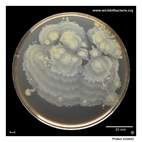
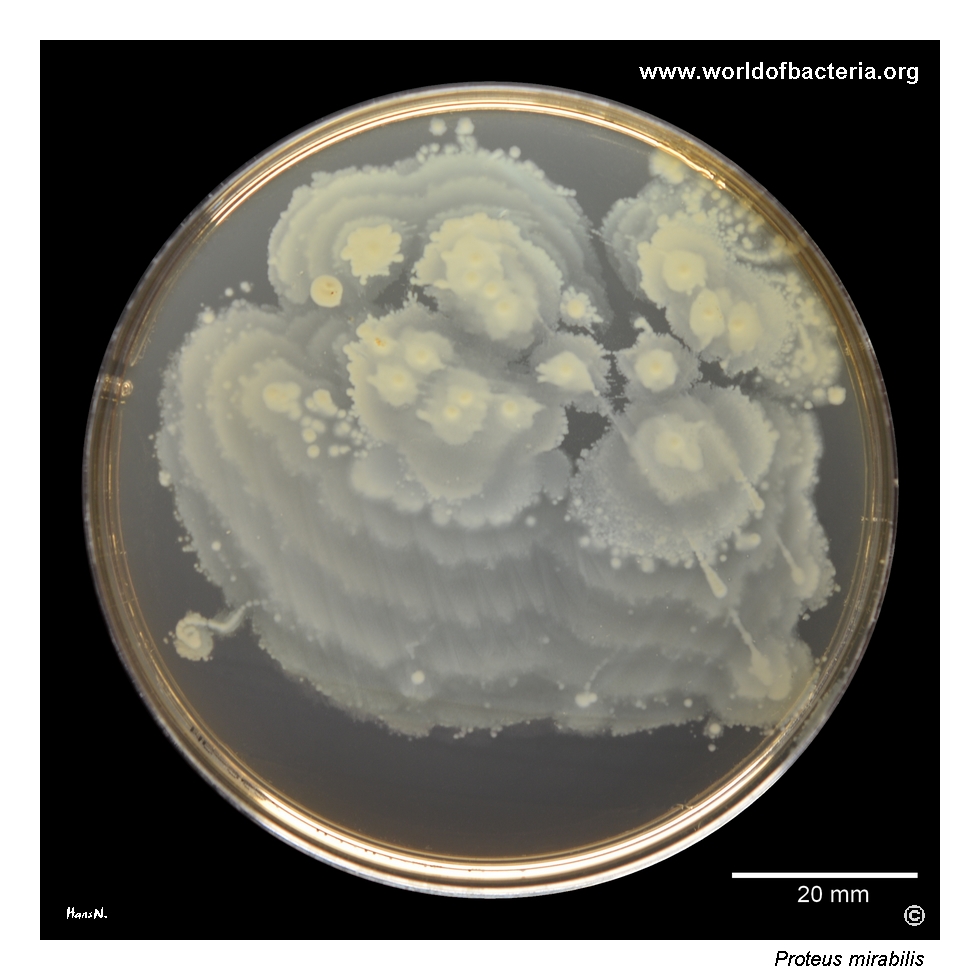
Proteus mirabilis on MacConkey agar after 24 hours of incubation at 36 °C in ambient air. The transparent medium clearly demonstrates the characteristic swarming growth, also known as the Rauss phenomenon, produced by most strains of P. mirabilis and P. vulgaris. The bacteria spread progressively across the agar surface rather than remaining confined to individual colonies. P. mirabilis does not ferment lactose and therefore forms pale colonies on MacConkey agar.
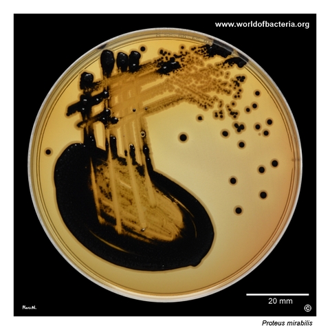
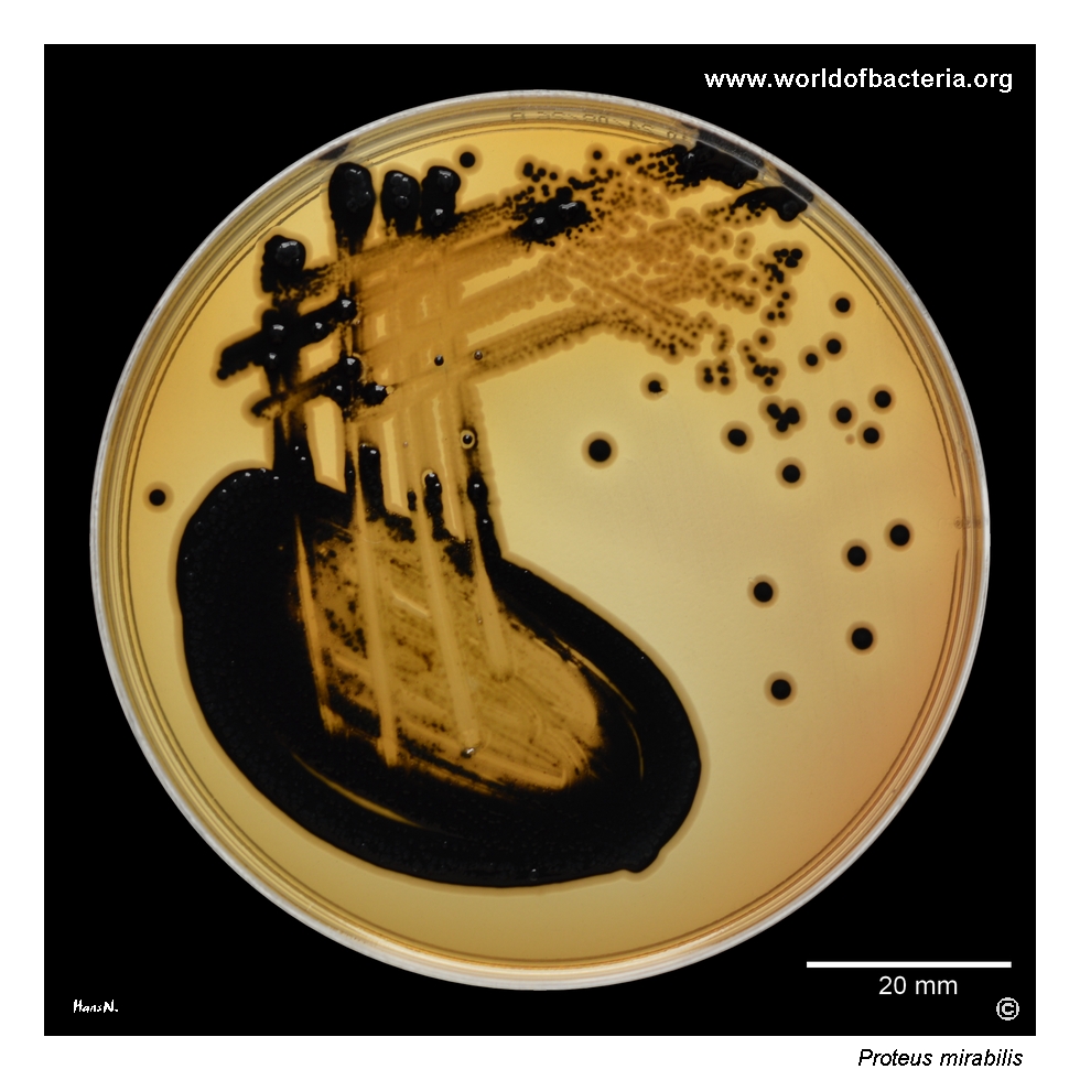
Proteus mirabilis on deoxycholate citrate agar after 24 hours at 36 °C in ambient air. The isolate forms hydrogen sulphide-positive colonies with black centres, closely resembling the appearance of many salmonellae on this medium. The strain was recovered from a rectal swab. Black-centred colonies provide a useful preliminary clue, but this feature is not species-specific and must be followed by additional identification tests.
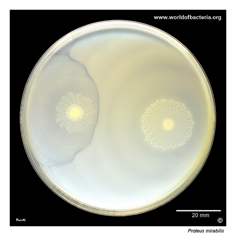
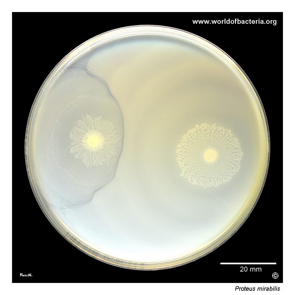
Two different strains of Proteus mirabilis inoculated at separate points on Mueller–Hinton agar. Both cultures spread across the surface and grow towards each other. Where the advancing populations meet, a visible demarcation zone forms between the incompatible strains, producing the Dienes phenomenon. The image also shows that the two isolates differ in their rate of swarming across the agar. Culture conditions: 24 hours at 36 °C in ambient air.
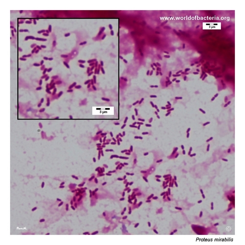
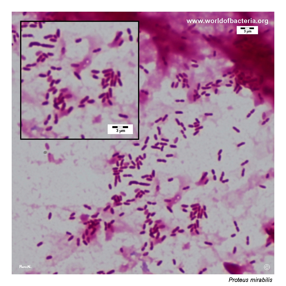
Proteus mirabilis in a positive blood culture from an 85-year-old man admitted to the emergency department with the diagnosis R50.9, fever, unspecified. The Gram-stained preparation shows Gram-negative rods. Recovery of P. mirabilis from blood indicates bacteraemia and may be associated with a urinary or another invasive focus of infection, requiring prompt clinical assessment and antimicrobial susceptibility testing.
Diagnostic and Clinical Notes
Proteus mirabilis is best recognized in routine culture by its ability to differentiate into elongated swarmer cells and spread rapidly across suitable agar surfaces. Repeated cycles of migration and consolidation may produce broad waves or concentric rings, historically described as the Rauss phenomenon.
Swarming is visually striking but is not unique to P. mirabilis. It is also common in P. vulgaris, and its extent depends on the strain, agar composition, moisture, incubation conditions, and inhibitory components of the medium.
The illustrated black-centred colonies on deoxycholate citrate agar reflect hydrogen sulphide production. This may mimic salmonellae and some Citrobacter isolates, so the finding is presumptive rather than diagnostic.
The Dienes phenomenon is a visible boundary that may form when swarming populations of two non-identical Proteus strains meet. It is a useful demonstration of strain-level self-recognition but should not be treated as a species-identification test.
The major clinical association is complicated urinary tract infection, especially in patients with long-term urinary catheters. Urease hydrolyses urea and raises urinary pH, encouraging crystalline biofilm formation, catheter blockage, and struvite or carbonate-apatite stones. A positive blood culture is always clinically significant and frequently prompts investigation of a urinary source.
Laboratory Identification
Colony Morphology
On suitable non-selective agar, Proteus mirabilis commonly produces rapidly spreading swarming growth rather than discrete colonies. On lactose-differential media it forms pale, lactose-non-fermenting colonies. Hydrogen sulphide production may create black-centred colonies on media containing an appropriate iron indicator.
Microscopy
Gram staining usually shows Gram-negative rods occurring singly. Cells in liquid culture or clinical specimens are generally short to medium-sized, whereas growth on solid surfaces may differentiate into markedly elongated, hyperflagellated swarmer cells.
Key Identification Clues
Gram-negative rod and facultative anaerobe
Oxidase negative and catalase positive
Strongly urease positive
Highly motile with characteristic swarming growth
Lactose non-fermenting
Hydrogen sulphide usually produced
Phenylalanine deaminase positive
Usually indole negative, helping distinguish it from P. vulgaris
Modern Identification Methods
Routine species identification is usually achieved by MALDI-TOF mass spectrometry or automated biochemical systems. Conventional testing remains useful for demonstrating urease, indole, hydrogen sulphide, phenylalanine deaminase, and motility. Molecular methods and whole-genome sequencing may be used for outbreak investigation, strain comparison, resistance characterization, and detailed analysis of virulence determinants.
Antibiotic Characteristics
Proteus mirabilis has an expected resistant phenotype to nitrofurantoin, making this agent unsuitable for treatment even when the organism is recovered from urine.
Clinical isolates may acquire extended-spectrum beta-lactamases, plasmid-mediated AmpC enzymes, fluoroquinolone resistance, aminoglycoside resistance, and carbapenemases. Resistance patterns vary substantially between community and healthcare-associated isolates.
The species is also naturally poorly susceptible or resistant to polymyxins, and susceptibility results should be interpreted using current laboratory standards and expected-phenotype guidance.
Note: Treatment should be based on the infection site, source control, patient factors, and antimicrobial susceptibility testing of the individual isolate. Catheter removal or replacement and management of obstructing stones may be essential in complicated urinary infection.