Citrobacter koseri is a facultatively anaerobic Gram-negative rod of the family Enterobacteriaceae that may cause urinary and bloodstream infections and has a distinctive association with neonatal meningitis complicated by cerebral abscess formation.
Basic Characteristics
Taxonomy
Domain: Bacteria
Phylum: Pseudomonadota (Proteobacteria)
Class: Gammaproteobacteria
Order: Enterobacterales
Family: Enterobacteriaceae
Genus:Citrobacter
Species:Citrobacter koseri
Microscopy & Gram Stain
Gram-negative rods, sometimes very short or coccobacillary
Cells occur singly, in pairs, and occasionally in short groupings
Capsular or extracellular polysaccharide material may contribute to a mucoid phenotype in some isolates
Oxygen Relationship
Facultatively anaerobic
Rapid Identification Tests
Oxidase: negative
Catalase: positive
Motility: usually positive
Lactose fermentation: variable or delayed
Indole: positive
Citrate: usually positive
Urease: variable
Hydrogen sulphide: usually negative
Ecology and Clinical Relevance
Natural Habitat
Human and animal gastrointestinal tracts
Water, soil, sewage, food, and other environmental reservoirs
May colonize hospitalized patients and moist healthcare environments
Common Clinical Specimens
Urine and catheter urine
Blood cultures
Cerebrospinal fluid
Respiratory specimens
Wound, abscess, and other purulent material
Other normally sterile specimens
Clinical Significance
Opportunistic cause of urinary tract infection, especially in older, hospitalized, or instrumented patients
May cause bacteraemia, sepsis, respiratory infection, and wound or intra-abdominal infection
A particularly important cause of neonatal meningitis with a strong tendency toward cerebral abscess formation
Clinical importance depends on specimen quality, quantity of growth, host factors, and recovery from normally sterile sites
Differential Considerations
Citrobacter freundii and other members of the genus Citrobacter
Klebsiella pneumoniae and other encapsulated or strongly mucoid Enterobacterales
Escherichia coli, Enterobacter, and other lactose-variable enteric rods
Important clues include indole positivity, usual motility, and absence of hydrogen sulphide production in typical isolates
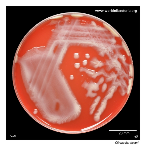
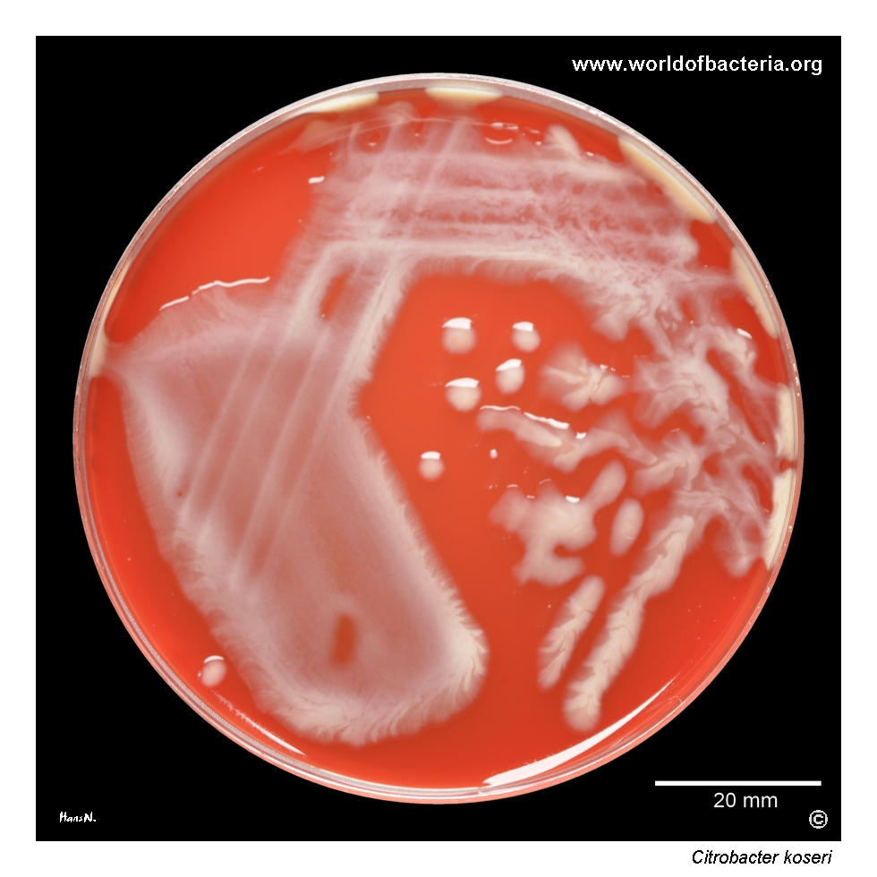
Citrobacter koseri on blood agar. Ten microlitres of urine were inoculated and streaked across the plate. Markedly mucoid, encapsulated variants may occur among many members of the Enterobacterales and are often encountered among urinary isolates. Culture conditions: 24 hours at 36 °C in ambient air. The abundant extracellular polysaccharide gives the colonies a glistening appearance and may substantially alter their usual morphology.
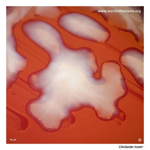
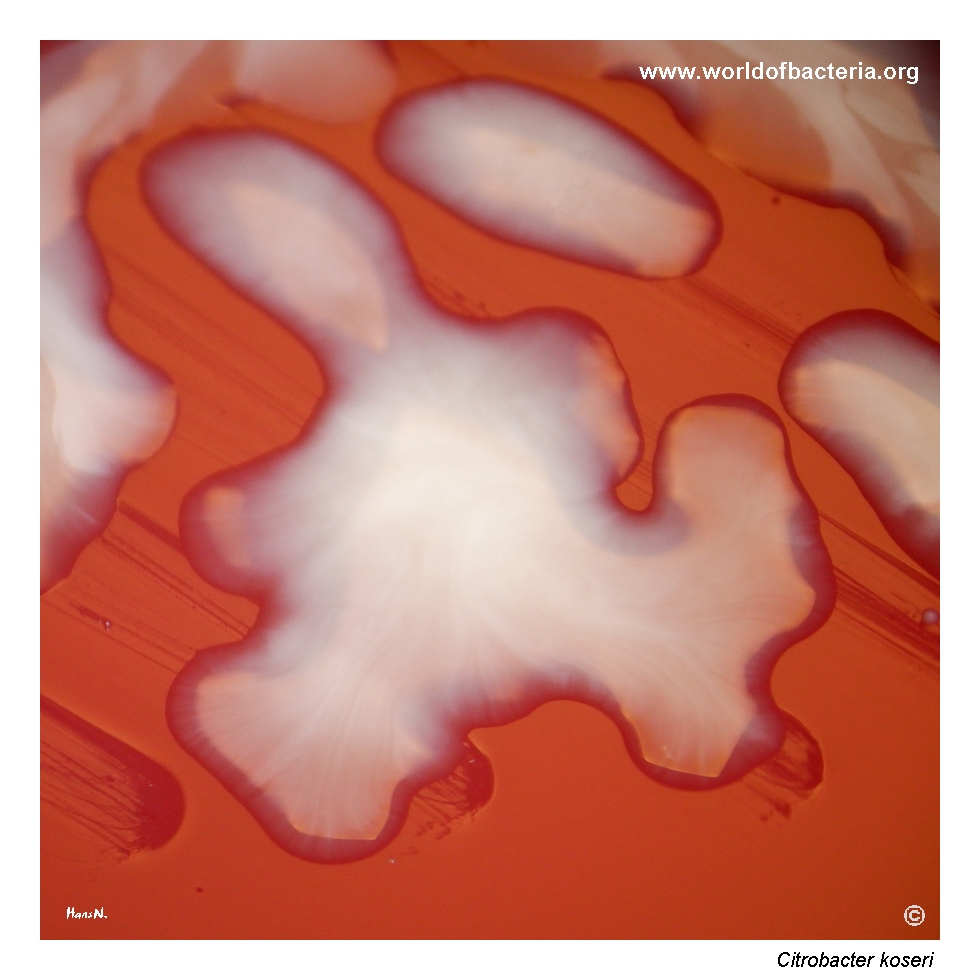
Strongly mucoid and highly glistening colonies of Citrobacter koseri on blood agar after 24 hours of incubation at 36 °C in ambient air. The colonies show a pronounced tendency to spread across the agar surface because of their soft, viscous consistency. This appearance reflects abundant production of capsular or extracellular polysaccharide material and may make individual colonies difficult to separate during routine manipulation.
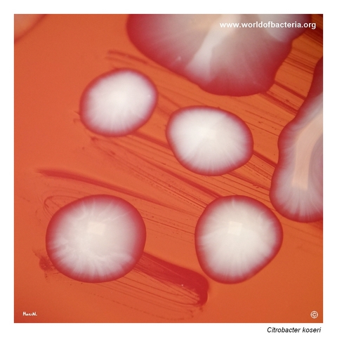
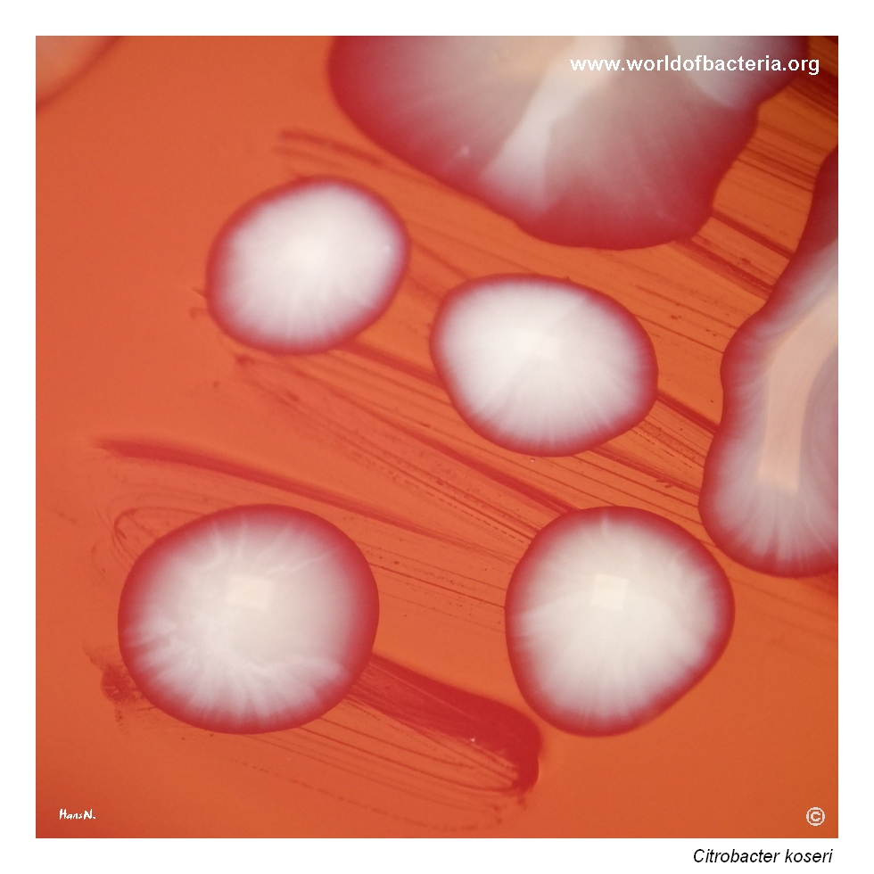
Close-up view of Citrobacter koseri colonies on blood agar after 24 hours at 36 °C in ambient air. The colonies measure approximately 5 mm in diameter and are strongly mucoid, smooth, convex, and round with entire margins. This striking phenotype is not present in every isolate and should not be regarded as species-specific, but it provides a useful demonstration of the considerable morphological variability found among urinary Enterobacterales.
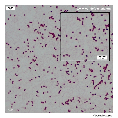
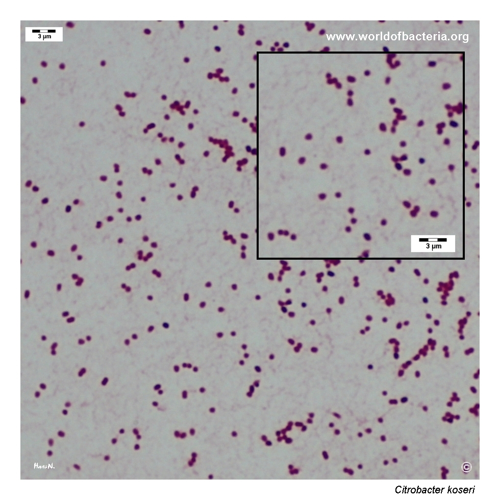
Gram-stained preparation made by directly spreading a mucoid colony of Citrobacter koseri onto a microscope slide. The organism appears as very short Gram-negative rods, with some cells showing an almost coccobacillary form. The compact cellular morphology may initially suggest cocci at low magnification, but closer examination reveals the short rod-shaped appearance characteristic of this isolate.
Diagnostic and Clinical Notes
Citrobacter koseri is an opportunistic member of the Enterobacterales that may colonize the intestinal tract but can also cause clinically significant urinary, bloodstream, respiratory, wound, and intra-abdominal infections. Urinary isolates are particularly common among adult clinical recoveries.
Its most characteristic disease association is neonatal meningitis, which has a marked tendency to progress to ventriculitis and one or more cerebral abscesses. This association is disproportionately strong compared with most other Enterobacterales and may result in substantial mortality or long-term neurological sequelae.
The strongly mucoid isolate illustrated here demonstrates phenotype rather than taxonomy. Abundant capsular or extracellular polysaccharide may produce large, glistening, viscous colonies, but similarly mucoid forms occur in Klebsiella, Escherichia, Enterobacter, and other enteric bacteria.
In urine culture, the significance of C. koseri depends on quantitative growth, symptoms, pyuria, specimen quality, catheter status, and accompanying organisms. Marked mucoidy should not be used to infer virulence, invasiveness, or antimicrobial resistance.
The historical name Citrobacter diversus may appear in older medical literature and is now treated as a later heterotypic synonym of Citrobacter koseri.
Laboratory Identification
Colony Morphology
On blood agar after 18–24 hours at 35–37 °C, Citrobacter koseri commonly forms smooth, grey-white, moist colonies that may be convex and round with entire margins. Colony size and consistency vary, and some urinary isolates produce abundant extracellular polysaccharide resulting in a strikingly glistening, spreading, or strongly mucoid appearance.
Microscopy
Gram staining shows Gram-negative rods. Depending on growth conditions and preparation technique, cells may be very short and can appear coccobacillary, as in the illustrated isolate. Microscopic morphology alone does not distinguish the species from other Enterobacterales.
Key Identification Clues
Gram-negative facultatively anaerobic rod
Oxidase negative and catalase positive
Usually motile
Indole positive
Usually citrate positive
Lactose fermentation variable or delayed
Hydrogen sulphide usually not produced
Colony morphology may range from ordinary enteric growth to markedly mucoid variants
Modern Identification Methods
Routine species identification is commonly achieved using MALDI-TOF mass spectrometry or automated biochemical systems. Difficult or epidemiologically important isolates may be confirmed with targeted molecular methods or whole-genome sequencing. In neonatal invasive disease, rapid species identification and assessment for central nervous system complications are clinically important.
Antibiotic Characteristics
Citrobacter koseri may show resistance to ampicillin and other beta-lactams through chromosomal or acquired beta-lactamases, while susceptibility to cephalosporins, fluoroquinolones, aminoglycosides, trimethoprim-sulfamethoxazole, and other agents varies between isolates.
Acquired extended-spectrum beta-lactamases, plasmid-mediated quinolone resistance determinants, and carbapenemases have been reported in clinical isolates, including urinary strains.
Resistance cannot be inferred from colony morphology, degree of mucoidy, source, or species identification alone. This is especially important in healthcare-associated infection and in isolates recovered after previous antimicrobial exposure.
Note: Antimicrobial treatment should be guided by the infection site, patient age and condition, and susceptibility testing of the individual isolate. Neonatal meningitis or cerebral abscess requires specialist management with agents that achieve appropriate central nervous system penetration.