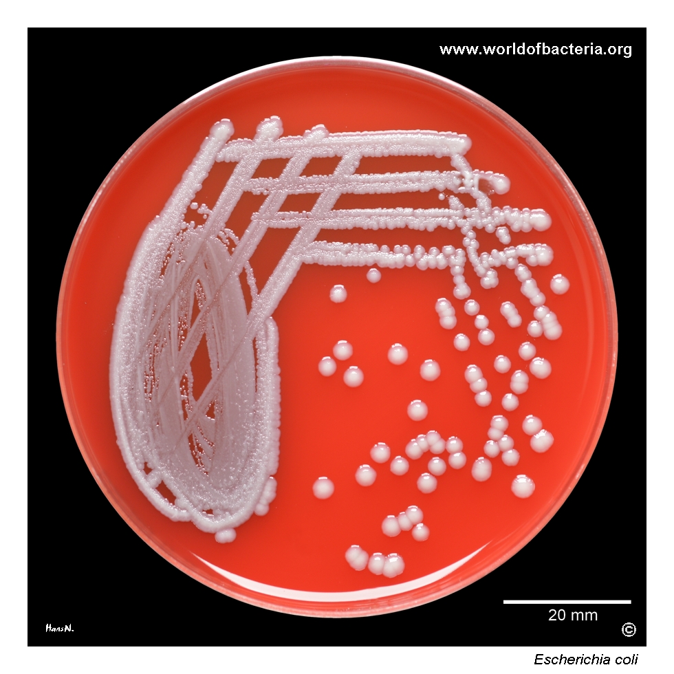
A non-haemolytic urinary isolate of Escherichia coli, the most frequently encountered bacterial cause of urinary tract infection and a highly diverse species whose strains range from harmless intestinal commensals to important extraintestinal and intestinal pathogens.
Basic Characteristics
Taxonomy
Domain: Bacteria
Phylum: Pseudomonadota (Proteobacteria)
Class: Gammaproteobacteria
Order: Enterobacterales
Family: Enterobacteriaceae
Genus:Escherichia
Species:Escherichia coli
Microscopy & Gram Stain
Gram-negative rods
Cells occur mainly singly, with pairs and short arrangements also possible
Oxygen Relationship
Facultatively anaerobic
Rapid Identification Tests
Oxidase: negative
Catalase: positive
Indole: usually positive
Lactose fermentation: usually positive
Motility: usually positive
Haemolysis on blood agar: absent in this isolate
Ecology and Clinical Relevance
Natural Habitat
Human and animal intestinal tract
Common member of the normal colonic microbiota
May transiently contaminate the perineal and periurethral region
Widely present in environments contaminated by faecal material
Common Clinical Specimens
Urine
Blood cultures
Wound and tissue specimens
Intra-abdominal material
Stool when intestinal pathogenic strains are suspected
Cerebrospinal fluid in neonatal infection
Clinical Significance
The most frequent bacterial cause of community-acquired urinary tract infection
Important cause of catheter-associated and healthcare-associated urinary infection
May cause pyelonephritis, urosepsis, bloodstream infection, and intra-abdominal infection
Certain specialized pathotypes cause diarrhoeal disease or neonatal meningitis
Differential Considerations
Klebsiella pneumoniae, Citrobacter, Enterobacter, and other lactose-fermenting Enterobacterales
Proteus mirabilis and other common urinary Gram-negative rods
Shigella species, which are genetically very closely related but usually non-motile and non-lactose-fermenting
Colony appearance on blood agar is variable and must be combined with biochemical or instrumental identification
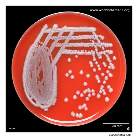
Ten microlitres of midstream urine were inoculated and streaked onto blood agar. The specimen was obtained from a 50-year-old woman with the diagnosis N39.0, urinary tract infection, site not specified. A pure culture of Escherichia coli grew at an estimated concentration exceeding 105 CFU/ml after 24 hours at 36 °C in ambient air. This level of growth is compatible with significant bacteriuria when interpreted together with the clinical findings.
Colonies of Escherichia coli on blood agar after 24 hours of incubation at 36 °C in ambient air. In this isolate, the colonies are convex and non-haemolytic. Colony morphology among urinary strains of E. coli is highly variable: isolates may differ in size, surface texture, elevation, margins, and haemolytic activity. Visual appearance may therefore provide an initial clue but is not sufficiently specific for reliable identification.
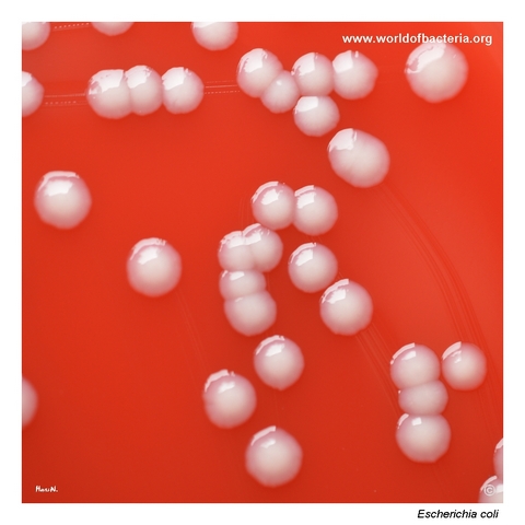
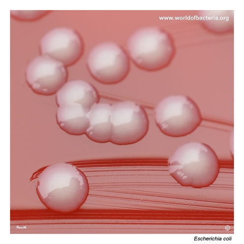
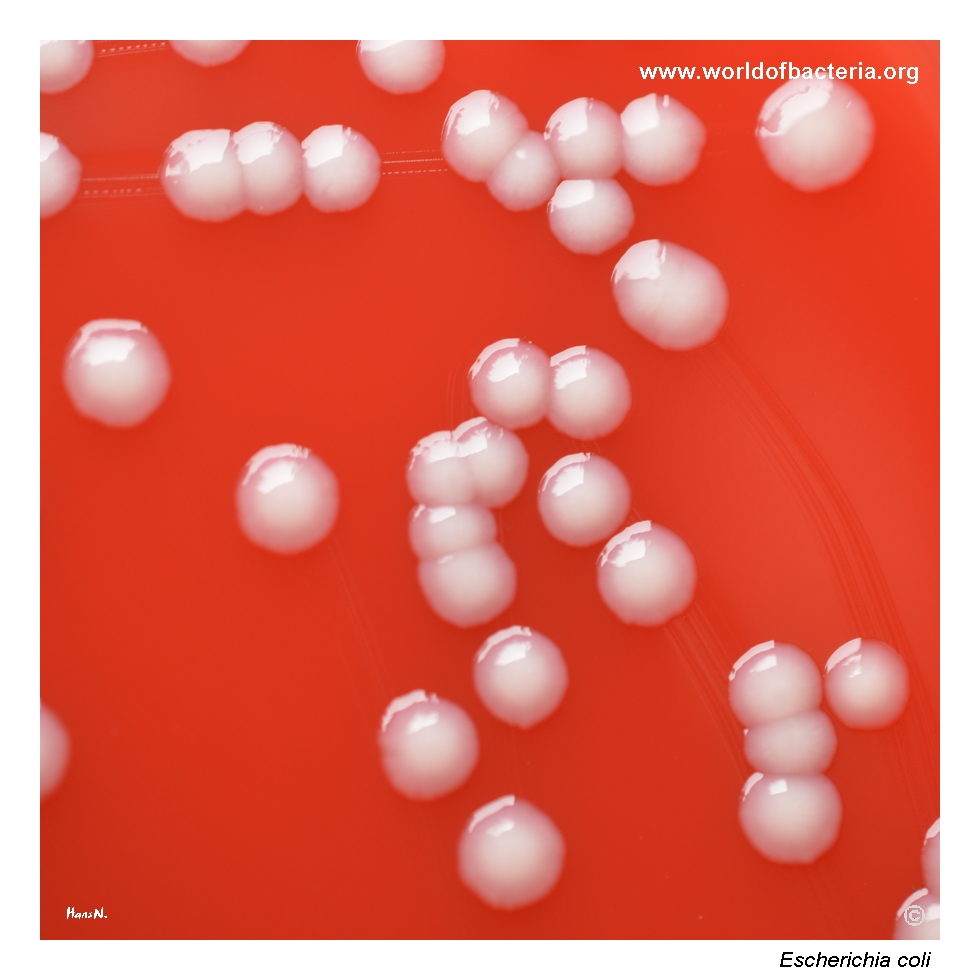
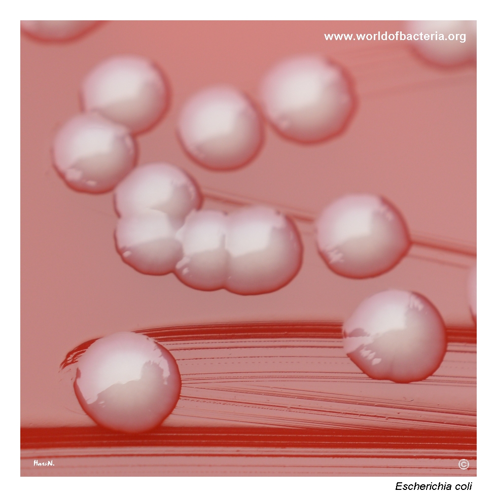
Close-up view of Escherichia coli colonies on blood agar after 24 hours at 36 °C in ambient air. The colonies are smooth, convex, grey-white, and round with entire margins. In some diagnostic laboratories, this represents one of the most frequently encountered colony types among urinary E. coli isolates. Nevertheless, different strains of the same species may show markedly different appearances on primary culture media.
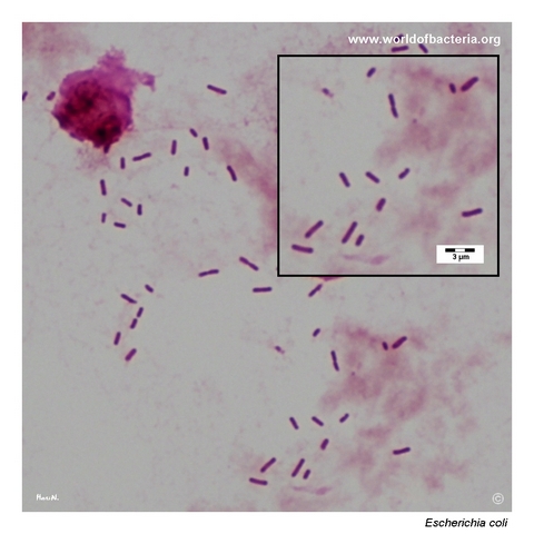
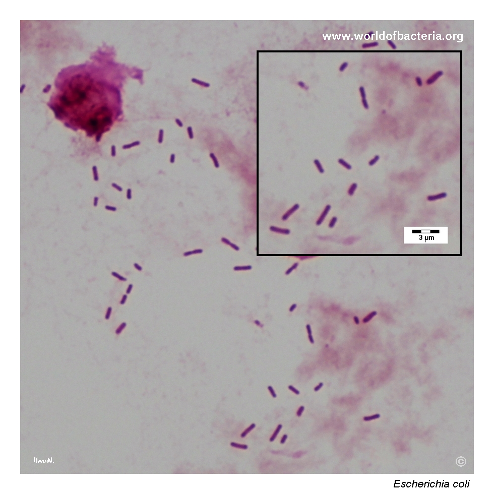
Gram-stained smear of the original urine specimen, from which a pure culture of Escherichia coli was subsequently isolated. Numerous Gram-negative rods are visible throughout the preparation. Marked leukocyturia is not present in this sample. The combination of abundant bacteria with only limited inflammatory cells illustrates why microscopy should be interpreted together with quantitative culture and the patient’s clinical presentation.
Diagnostic and Clinical Notes
Escherichia coli is a highly diverse species. Most strains colonize the intestinal tract without causing disease, whereas specialized intestinal and extraintestinal lineages possess virulence factors that allow them to cause distinct clinical syndromes.
Uropathogenic E. coli is the leading bacterial cause of urinary tract infection. Infection usually develops by an ascending route after organisms from the intestinal microbiota colonize the periurethral region and enter the urinary tract.
A colony count exceeding 105 CFU/ml in a pure midstream urine culture has traditionally been associated with significant bacteriuria. Its meaning must nevertheless be assessed together with symptoms, specimen quality, patient factors, and the presence or absence of inflammatory cells.
Absence of haemolysis does not reduce the possible clinical significance of a urinary isolate. Many clinically important uropathogenic strains are non-haemolytic, while other strains may produce alpha- or beta-haemolysis.
In this specimen, abundant Gram-negative rods were present without marked leukocyturia. This combination may occur for several reasons and should not be used alone either to confirm or exclude urinary tract infection.
Laboratory Identification
Colony Morphology
On blood agar after 18–24 hours at 35–37 °C, Escherichia coli commonly forms medium-sized grey-white colonies that may be smooth, convex, and round with entire margins. Considerable strain-to-strain variation occurs, including differences in colony size, surface, mucoidity, margin, and haemolysis. On lactose-containing differential media, most isolates ferment lactose and produce coloured colonies.
Microscopy
Gram staining shows Gram-negative rods, usually occurring singly. Direct urine microscopy may demonstrate numerous organisms, but the number of leukocytes can vary and microscopic findings must be correlated with quantitative culture and clinical information.
Key Identification Clues
Gram-negative rods
Facultatively anaerobic growth
Oxidase negative
Usually lactose fermenting
Usually indole positive
Usually motile
Colony morphology and haemolysis are variable and not species-specific
Modern Identification Methods
Routine species identification is commonly achieved using MALDI-TOF mass spectrometry or automated biochemical systems. Additional testing may be required to characterize intestinal pathotypes, Shiga toxin production, serotype, virulence genes, resistance mechanisms, or epidemiologically important clones.
Antibiotic Characteristics
Antimicrobial susceptibility in Escherichia coli varies substantially between patients, geographical regions, clinical settings, and individual lineages.
Urinary isolates may be resistant to commonly used agents, including aminopenicillins, trimethoprim-sulfamethoxazole, and fluoroquinolones. Local resistance data are therefore important when empirical treatment is considered.
Extended-spectrum beta-lactamase production is an important resistance mechanism in both community and healthcare-associated isolates. Some strains also acquire carbapenemases or other mechanisms that further restrict treatment options.
Note: The non-haemolytic appearance of this isolate provides no information about its antimicrobial susceptibility. Treatment decisions should be guided by susceptibility testing, the site and severity of infection, and the clinical context.