Klebsiella pneumoniae is an encapsulated, non-motile, lactose-fermenting Gram-negative rod that colonizes the human gastrointestinal tract and is an important cause of urinary tract infection, pneumonia, bloodstream infection, and healthcare-associated disease.
Basic Characteristics
Taxonomy
Domain: Bacteria
Phylum: Pseudomonadota (Proteobacteria)
Class: Gammaproteobacteria
Order: Enterobacterales
Family: Enterobacteriaceae
Genus:Klebsiella
Species:Klebsiella pneumoniae
Microscopy & Gram Stain
Gram-negative, relatively short and plump rods
Cells occur singly, in pairs, and occasionally in short chains
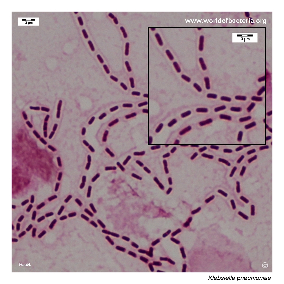
Capsular material may appear as a pale, unstained halo around the cells
Oxygen Relationship
Facultatively anaerobic
Rapid Identification Tests
Oxidase: negative
Catalase: positive
Motility: negative
Lactose fermentation: positive
Urease: usually positive
Indole: usually negative for typical K. pneumoniae isolates
Citrate: positive
Hydrogen sulphide: negative
Ecology and Clinical Relevance
Natural Habitat
Human gastrointestinal tract
May colonize the oropharynx and other mucosal sites
Soil, water, plants, sewage, and other environmental reservoirs
Healthcare environments and colonized patients may serve as sources of transmission
Common Clinical Specimens
Urine and catheter urine
Blood cultures
Sputum, tracheal aspirates, and other respiratory specimens
Wound and surgical-site specimens
Abscess material, including liver abscesses
Cerebrospinal fluid and other normally sterile specimens
Clinical Significance
Common cause of urinary tract infection, particularly in hospitalized or catheterized patients
Important cause of healthcare-associated pneumonia and bloodstream infection
May cause wound infection, intra-abdominal infection, neonatal disease, and meningitis
Hypervirulent lineages may cause community-onset invasive disease, including liver abscess with metastatic spread
Carbapenem-resistant and other multidrug-resistant lineages are major infection-control and public-health concerns
Differential Considerations
Other members of the Klebsiella pneumoniae species complex, especially K. variicola and K. quasipneumoniae
Klebsiella oxytoca, which is usually indole positive
Escherichia coli, which is motile and usually less prominently mucoid
Enterobacter, Raoultella, and other lactose-fermenting Enterobacterales
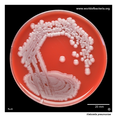
Klebsiella pneumoniae in a urine specimen. Ten microlitres of urine were inoculated and streaked onto blood agar. After 24 hours of incubation at 36 °C, the organism typically forms medium-sized to large grey-white colonies with a conspicuously smooth and glistening surface. This appearance reflects the production of abundant extracellular polysaccharide material and is a useful preliminary clue when examining urinary cultures.
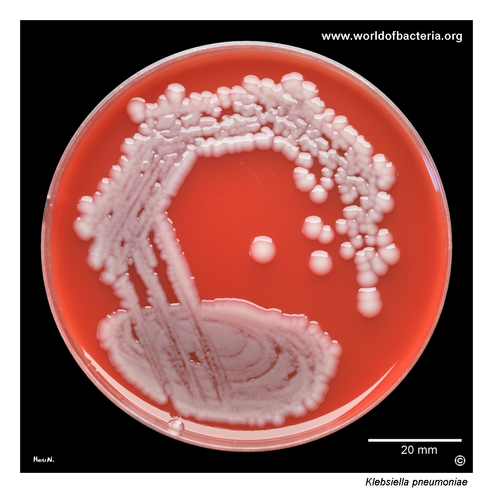
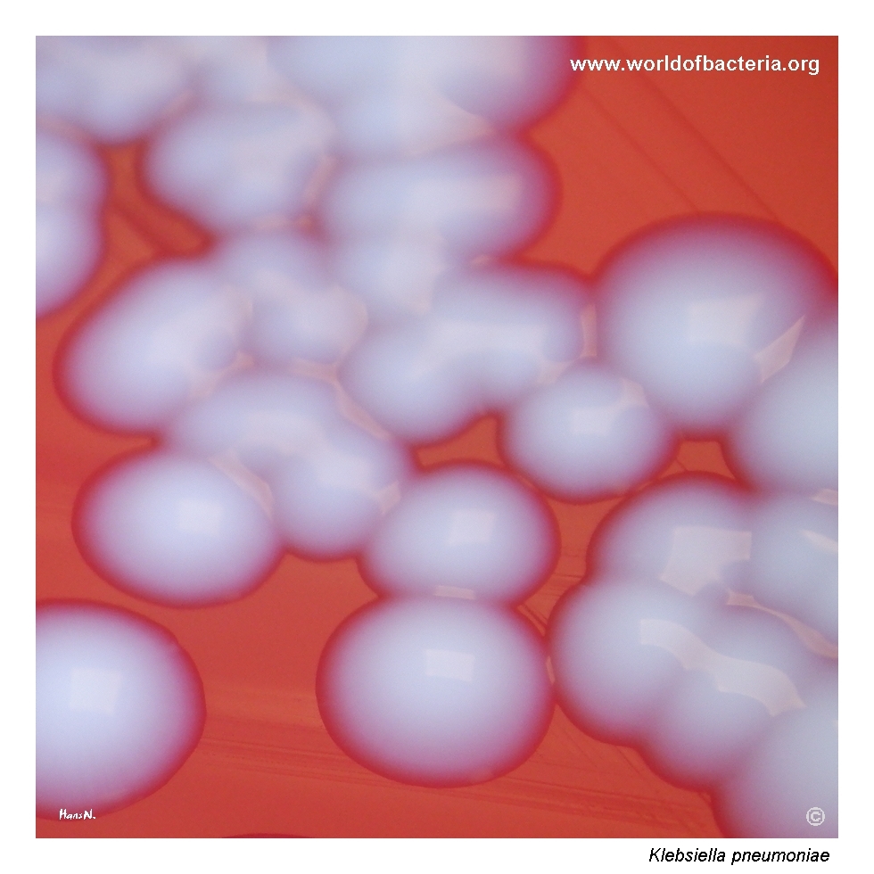
Colonies of Klebsiella pneumoniae on blood agar after 24 hours at 36 °C in ambient air. Under the same culture conditions, they are usually larger and more glistening than colonies of most Escherichia coli strains. The shiny, sometimes mucoid appearance results from the production of capsular and extracellular polysaccharide material surrounding the bacterial cells. This phenotype may vary in intensity between individual isolates.
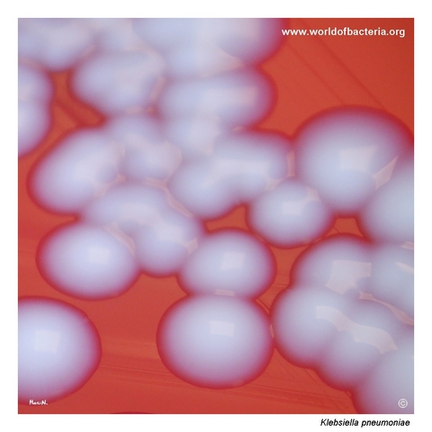
Close-up view of Klebsiella pneumoniae colonies on blood agar after 24 hours of incubation at 36 °C in ambient air. The colonies are medium-sized to large, smooth, convex, and round with entire margins and a markedly glistening surface. Some strains may produce even more pronounced mucoid growth, but colony texture alone does not provide a reliable indication of virulence or antimicrobial resistance.
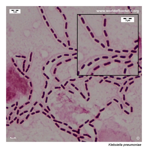
Gram-stained urine specimen containing Klebsiella pneumoniae. The organism appears as Gram-negative rods, in this preparation sometimes arranged in short chains. Pale halos are visible around some cells, consistent with the presence of capsular material that is not directly stained by the Gram method. The polysaccharide capsule protects the bacteria from environmental stresses and contributes to their ability to evade host immune defences.
Diagnostic and Clinical Notes
Klebsiella pneumoniae is a common intestinal colonizer and an important opportunistic pathogen in both community and healthcare settings. It is particularly prominent in urinary tract infections, pneumonia, bloodstream infections, and infections involving critically ill or instrumented patients.
Abundant capsular and extracellular polysaccharide production gives many isolates their characteristic smooth, glistening, or mucoid appearance. The degree of mucoidy varies substantially and may be affected by strain background and culture conditions.
A strongly mucoid colony or a positive string test should not be treated as proof of hypervirulence. Hypervirulent and classical lineages overlap phenotypically, and reliable characterization requires clinical correlation and detection of appropriate molecular markers.
The species belongs to a closely related complex that includes K. variicola and K. quasipneumoniae. Routine systems identify most clinical isolates correctly, but difficult or epidemiologically important isolates may require updated MALDI-TOF databases, targeted molecular tests, or whole-genome sequencing.
In urine culture, colony quantity, specimen quality, pyuria, symptoms, catheter status, and accompanying organisms remain essential for distinguishing infection from colonization or contamination. Colony texture does not predict susceptibility: ordinary-looking isolates may carry extended-spectrum beta-lactamases or carbapenemases.
Laboratory Identification
Colony Morphology
On blood agar after 18–24 hours at 35–37 °C, Klebsiella pneumoniae usually forms medium-sized to large, grey-white, smooth, convex, glistening colonies with entire margins. Capsule and extracellular polysaccharide production may create a distinctly mucoid consistency, although the intensity of this phenotype varies between isolates.
Microscopy
Gram staining shows relatively short, plump Gram-negative rods occurring singly, in pairs, or occasionally in short chains. Capsular material may be seen indirectly as a pale halo, but ordinary Gram staining is not a definitive capsule demonstration method.
Key Identification Clues
Gram-negative, encapsulated rod
Facultatively anaerobic
Oxidase negative and catalase positive
Non-motile
Lactose fermenting
Usually urease and citrate positive
Usually indole negative for typical K. pneumoniae
Smooth, glistening, and sometimes markedly mucoid colonies
Modern Identification Methods
Routine identification is commonly performed by MALDI-TOF mass spectrometry or automated biochemical systems. Updated reference databases improve recognition within the K. pneumoniae species complex. Molecular assays are used to detect important resistance genes and selected hypervirulence-associated markers, while whole-genome sequencing provides the highest resolution for species-complex assignment, outbreak investigation, resistance analysis, and lineage characterization.
Antibiotic Characteristics
Klebsiella pneumoniae is intrinsically resistant to ampicillin and related narrow-spectrum aminopenicillins because of a chromosomally encoded beta-lactamase.
Clinical isolates may acquire extended-spectrum beta-lactamases, plasmid-mediated AmpC enzymes, aminoglycoside and fluoroquinolone resistance, and carbapenemases such as KPC, NDM, OXA-48-like, VIM, or IMP enzymes.
Carbapenem-resistant K. pneumoniae belongs to the most important multidrug-resistant Gram-negative threats in healthcare. Convergence of carbapenem resistance with hypervirulent lineages is of particular concern because it combines invasive potential with limited treatment options.
Resistance to last-line agents may also occur through additional acquired genes or chromosomal alterations, and the expected activity of an antimicrobial agent cannot be inferred from colony morphology or source alone.
Note: Treatment and infection-control decisions should be guided by the infection site, patient factors, susceptibility testing, and, where relevant, laboratory detection of extended-spectrum beta-lactamases or carbapenemases.