Staphylococcus intermedius is a Gram-positive, coagulase-positive coccus that is recognized primarily as an animal pathogen, but may occasionally cause zoonotic or opportunistic infections in humans.
Basic Characteristics
Taxonomy
Domain: Bacteria
Phylum: Bacillota (Firmicutes)
Class: Bacilli
Order: Bacillales
Family: Staphylococcaceae
Genus:Staphylococcus
Species:Staphylococcus intermedius
Microscopy & Gram Stain
Gram-positive cocci
Cells occur singly, in pairs, and in irregular clusters
Oxygen Relationship
Facultatively anaerobic
Rapid Identification Tests
Catalase: positive
Coagulase: positive
Haemolysis on blood agar: usually present and often pronounced
Colony type: large-colony staphylococcus
Ecology and Clinical Relevance
Natural Habitat
Primarily associated with animals rather than humans
Commonly discussed as part of animal-associated staphylococci
May be encountered in zoonotic settings or mixed infections linked to animal flora
Common Clinical Specimens
Abscess material
Wound swabs
Skin and soft tissue specimens
Animal-associated bite or contact-related material
Occasionally blood cultures or deeper invasive samples
Clinical Significance
Primarily an animal pathogen
Only occasional human pathogen, usually in zoonotic or opportunistic contexts
May cause skin, soft tissue, wound, or abscess infections
Can occur in mixed infections together with anaerobes or other animal-associated bacteria
Differential Considerations
Staphylococcus aureus, because both species are coagulase positive and often beta-haemolytic
Other members of the Staphylococcus intermedius group
Large-colony, haemolytic coagulase-positive staphylococci in routine culture
Important clues include animal association, large colony size, and the need for reliable species-level identification beyond morphology alone
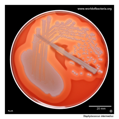
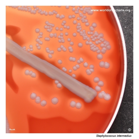
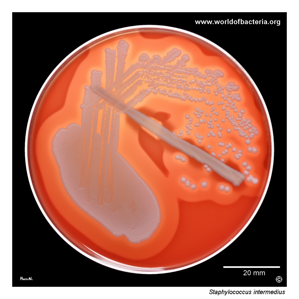
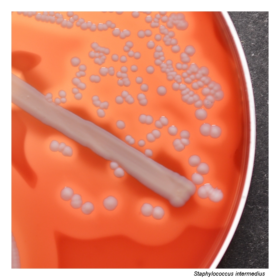
A skin abscess from the lower limb of a 60-year-old woman was enriched overnight in nutrient broth at 36 °C and subcultured onto blood agar. After 24 hours at 36 °C in 5% CO2, Staphylococcus intermedius formed grey-white colonies approximately 3–4 mm in diameter, surrounded by a broad zone of beta-haemolysis. Anaerobic culture also revealed significant growth of Bacteroides pyogenes and Fusobacterium canifelinum, indicating a mixed infection.
This isolate of Staphylococcus intermedius shows intensified haemolysis immediately adjacent to the colonies, forming a narrow zone of complete haemolysis, while a wider outer zone of incomplete beta-haemolysis extends farther into the agar. The appearance of haemolysis depends on which haemolysin, or combination of haemolysins, the strain produces and in what proportion. Such variation can markedly influence the visual phenotype on blood agar.
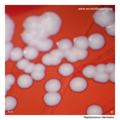
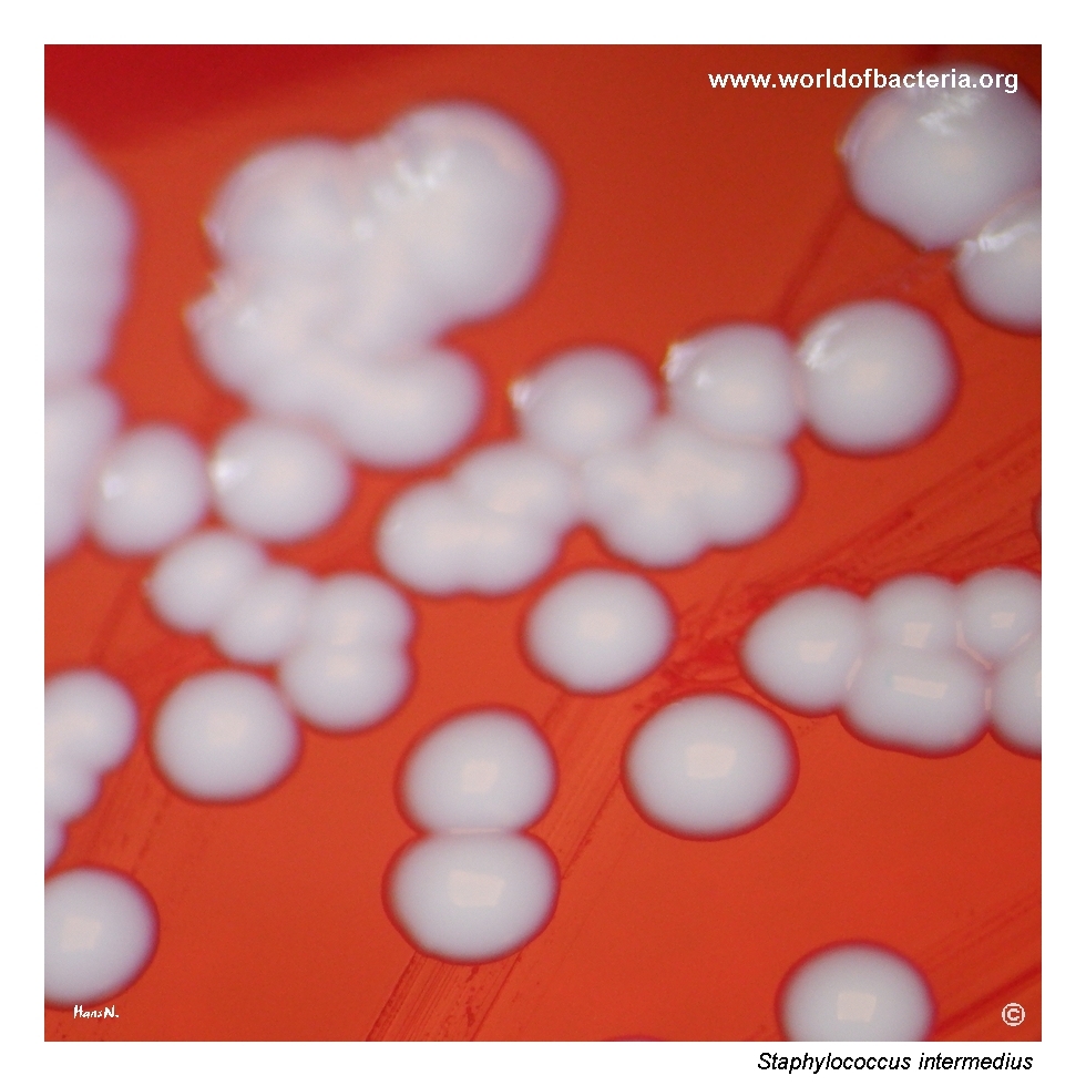
Staphylococcus intermedius forms smooth, convex, non-pigmented colonies that usually reach 3–4 mm in diameter after 24 hours of incubation at 36 °C. It belongs to the group of large-colony staphylococci, and with prolonged incubation of about five days, colony diameter may reach or exceed 6 mm. Colony size, together with haemolysis and pigmentation, can provide useful preliminary clues during routine culture examination.
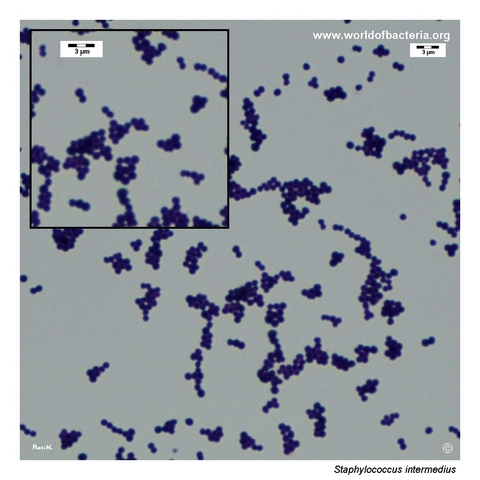
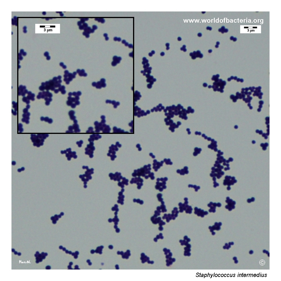
Gram-stained smear prepared from a colony of Staphylococcus intermedius grown on blood agar. The organism appears as Gram-positive cocci occurring singly, in pairs, and in clusters, the typical microscopic arrangement of staphylococci. As in other members of the genus, Gram stain morphology is not sufficient for species identification and must be correlated with colony features, haemolysis, and additional laboratory tests.
Diagnostic and Clinical Notes
Staphylococcus intermedius is important to note as a species associated primarily with animals, and human infection is considerably less common than infection caused by major human-adapted staphylococci. This animal association should be explicitly considered whenever the organism is recovered from human material.
In human microbiology, isolates reported as S. intermedius are often linked to zoonotic exposure or to infections arising in settings where contact with companion animals is plausible. Soft tissue infections, abscesses, and wound infections are among the best-recognized presentations.
A practical diagnostic challenge is that S. intermedius may resemble Staphylococcus aureus on routine culture because it is coagulase positive, frequently beta-haemolytic, and may form relatively large colonies. For this reason, colony appearance alone is not sufficient for reliable species identification.
The species may also appear in mixed infections together with anaerobic bacteria or other components of animal-associated flora, particularly in abscesses and contaminated wounds. Careful interpretation of specimen type and polymicrobial growth is therefore important.
Although this page uses the traditional name S. intermedius, laboratories should be aware that species-level differentiation within the Staphylococcus intermedius group may require modern identification methods.
Laboratory Identification
Colony Morphology
On blood agar incubated at 35–37 °C, colonies of Staphylococcus intermedius are typically smooth, convex, grey-white, and non-pigmented, usually reaching about 3–4 mm after 24 hours. The species belongs among large-colony staphylococci, and prolonged incubation may lead to markedly larger colonies. Beta-haemolysis is usually evident and may vary in intensity and pattern.
Microscopy
Gram staining reveals Gram-positive cocci occurring singly, in pairs, and in irregular clusters. As with other staphylococci, the Gram stain appearance is typical for the genus but not diagnostic at species level.
Key Identification Clues
Gram-positive cocci in clusters
Strongly catalase positive
Coagulase positive
Usually beta-haemolytic on blood agar
Large-colony staphylococcus that can mimic S. aureus
Animal association is an important epidemiological clue
Modern Identification Methods
Species-level identification in clinical microbiology laboratories is commonly achieved using MALDI-TOF mass spectrometry and may be further refined by molecular methods when needed. This is particularly relevant because phenotypic resemblance to S. aureus and overlap within the Staphylococcus intermedius group can complicate conventional identification.
Antibiotic Characteristics
Staphylococcus intermedius may show variable antimicrobial susceptibility depending on strain origin and clinical context. As with other staphylococci, susceptibility testing is necessary for clinically significant isolates.
Because human infections are relatively uncommon and some isolates belong to animal-associated lineages, empirical expectations based only on common human staphylococcal patterns may be misleading.
In polymicrobial soft tissue infections, therapy must also take into account accompanying anaerobes and other copathogens identified in the specimen.
Note: Antimicrobial therapy should always be guided by laboratory susceptibility testing, specimen context, and overall clinical evaluation.