Pseudomonas aeruginosa is an oxidase-positive, non-fermenting Gram-negative rod widely distributed in moist environments and notable for green pigment production, biofilm formation, opportunistic infections, and extensive intrinsic and acquired antimicrobial resistance.
Basic Characteristics
Taxonomy
Domain: Bacteria
Phylum: Pseudomonadota (Proteobacteria)
Class: Gammaproteobacteria
Order: Pseudomonadales
Family: Pseudomonadaceae
Genus:Pseudomonas
Species:Pseudomonas aeruginosa
Microscopy & Gram Stain
Slender Gram-negative rods
Cells usually occur singly or in pairs
Motile by means of polar flagella; microscopic morphology is not species-specific
Oxygen Relationship
Primarily aerobic and non-fermenting
Can grow anaerobically when suitable alternative electron acceptors, such as nitrate, are available
Rapid Identification Tests
Oxidase: positive
Catalase: positive
Glucose metabolism: oxidative, non-fermentative
Motility: positive
Growth at 42 °C: usually positive
Pigments: pyocyanin and pyoverdine may produce blue-green to yellow-green coloration
Ecology and Clinical Relevance
Natural Habitat
Soil, freshwater, wastewater, and other moist environmental habitats
Hospital water systems, sinks, drains, and wet equipment
Plant, animal, and human-associated moist surfaces
May transiently colonize the skin, respiratory tract, gastrointestinal tract, or other body sites
Common Clinical Specimens
Respiratory specimens, including material from ventilated patients and people with cystic fibrosis
Wound, burn, and soft-tissue specimens
Urine and catheter-associated urinary specimens
Blood cultures
Ear, eye, and other localized specimens
Clinical Significance
Important opportunistic and healthcare-associated pathogen
Associated with pneumonia, bloodstream infection, urinary tract infection, surgical-site infection, and device-related infection
A major cause of chronic respiratory colonization and infection in cystic fibrosis and other structural lung diseases
Biofilm formation promotes persistence on tissues, medical devices, and moist environmental surfaces
Differential Considerations
Stenotrophomonas maltophilia, Achromobacter xylosoxidans, and Acinetobacter baumannii
Other fluorescent and non-fluorescent Pseudomonas species
Aeromonas and Vibrio species, which are oxidase-positive but ferment glucose
Helpful preliminary clues include oxidase positivity, non-fermentative metabolism, growth at 42 °C, characteristic pigments, and growth on cetrimide agar
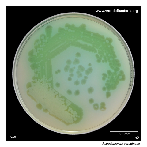
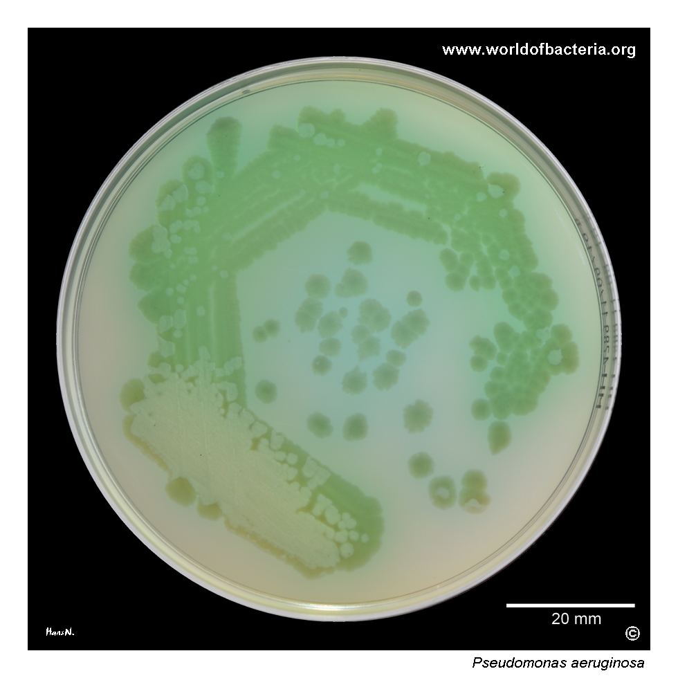
Pseudomonas aeruginosa on Mueller–Hinton agar after 24 hours of incubation at 36 °C in ambient air. The culture produces a conspicuous green pigment that diffuses into the surrounding medium. The coloration may result from a combination of blue-green pyocyanin and yellow-green pyoverdine. The species name aeruginosa refers to a blue-green or verdigris-like colour, reflecting this characteristic pigment production.
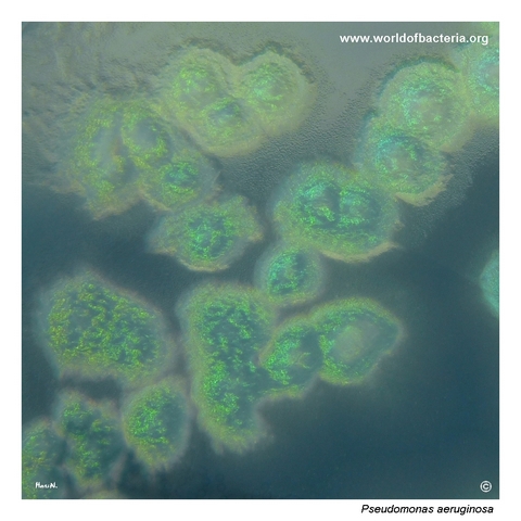
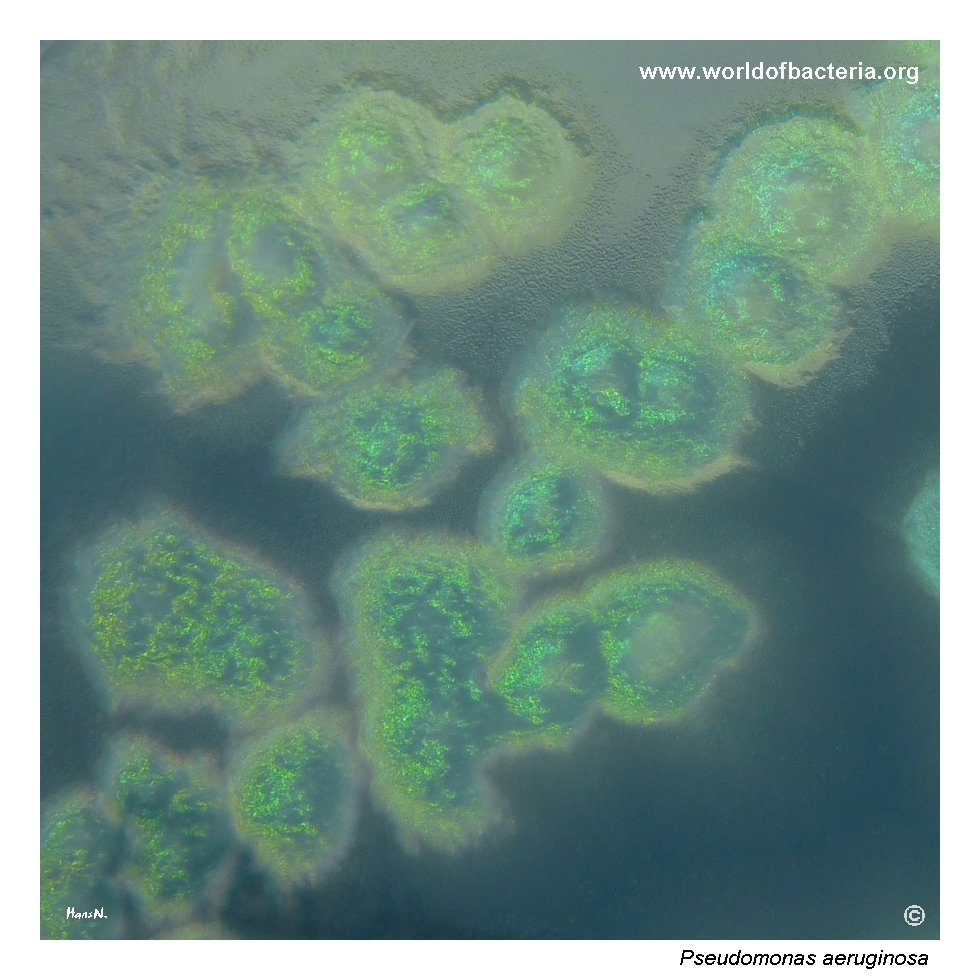
Colonies of Pseudomonas aeruginosa on cetrimide agar. Cetrimide is a quaternary ammonium compound that inhibits many accompanying bacteria, whereas P. aeruginosa shows comparatively high tolerance and can grow on the medium. Cetrimide agar also promotes the production of characteristic pigments, making the green coloration of the culture more conspicuous and supporting presumptive recognition of the species.
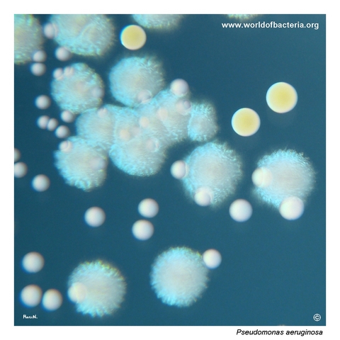
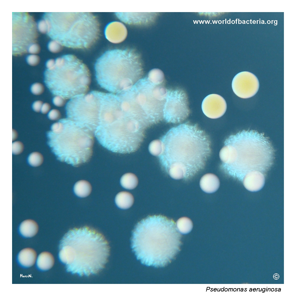
Close-up view of Pseudomonas aeruginosa, Staphylococcus aureus, and Enterococcus faecalis on tryptic soy agar. P. aeruginosa forms large bluish colonies, while the colonies of S. aureus are yellow and those of E. faecalis are white. All three isolates also showed strong biofilm formation. Culture conditions: 24 hours at 36 °C in ambient air. The colour contrast allows the three organisms to be distinguished readily in this mixed culture.
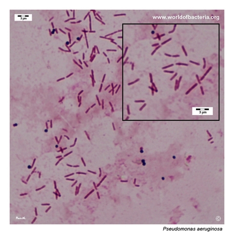
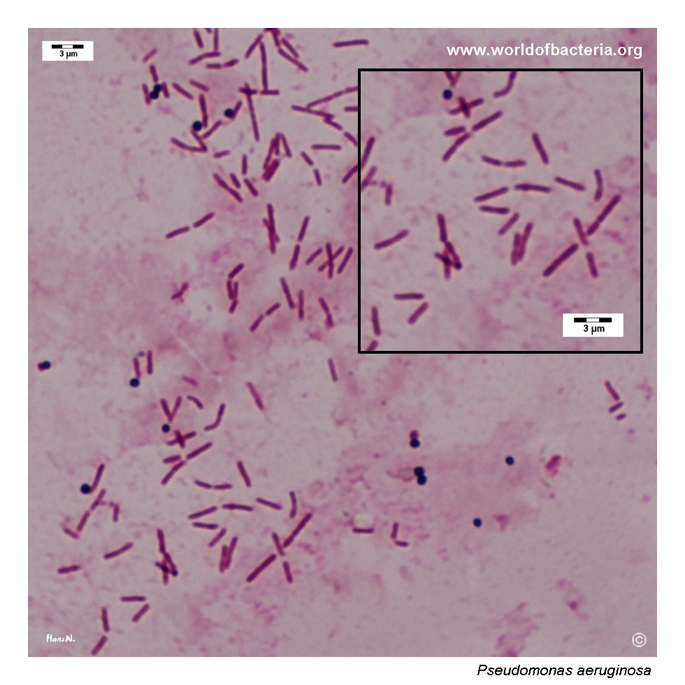
Gram-stained smear of a urine specimen containing numerous slender Gram-negative rods subsequently identified as Pseudomonas aeruginosa. Occasional Gram-positive cocci belonging to Staphylococcus epidermidis are also visible. The thin rod-shaped morphology is compatible with P. aeruginosa but is not species-specific, so microscopic findings must be interpreted together with culture characteristics, pigment production, and identification testing.
Diagnostic and Clinical Notes
Pseudomonas aeruginosa is widely distributed in water, soil, and other moist habitats and is especially well adapted to persist in healthcare water systems, sinks, drains, respiratory equipment, and other wet environmental reservoirs.
Pigment production is one of the most recognizable laboratory features. Blue-green pyocyanin is strongly associated with P. aeruginosa, while fluorescent yellow-green pyoverdine is produced by this and several other fluorescent pseudomonads. Pigmentation varies between isolates, and some clinically important strains may appear weakly pigmented or non-pigmented.
Cetrimide agar is useful as a selective and pigment-enhancing medium. Growth with characteristic coloration provides a valuable presumptive clue, but definitive identification should integrate oxidase activity, non-fermentative metabolism, temperature tolerance, and an instrumental or molecular identification method.
Biofilm formation contributes to persistent colonization of medical devices, chronic wounds, and the respiratory tract. In cystic fibrosis and other chronic lung diseases, long-term adaptation may produce mucoid variants with abundant alginate and altered colony morphology.
Clinical interpretation depends on the specimen and host. Recovery may represent colonization, particularly from non-sterile respiratory or device-associated material, but isolation from blood or compatible invasive specimens is clinically significant and requires prompt susceptibility testing.
Laboratory Identification
Colony Morphology
On routine agar after approximately 18–24 hours at 35–37 °C, Pseudomonas aeruginosa commonly forms medium-sized to large, flat or slightly raised colonies that may show irregular margins, metallic sheen, beta-haemolysis, and blue-green, yellow-green, or occasionally brown pigmentation. Colony morphology and pigment intensity vary considerably between strains.
Microscopy
Gram staining shows slender Gram-negative rods occurring mainly singly or in pairs. The morphology is compatible with a non-fermenting Gram-negative bacillus but does not permit species identification.
Key Identification Clues
Slender Gram-negative, motile rod
Oxidase and catalase positive
Non-fermentative oxidative metabolism
Usually capable of growth at 42 °C
Pyocyanin and pyoverdine may produce blue-green to fluorescent yellow-green coloration
Growth on cetrimide agar with enhanced pigment production
Strong biofilm-forming capacity; mucoid variants may occur in chronic respiratory infection
Modern Identification Methods
Species identification is commonly achieved by MALDI-TOF mass spectrometry or validated automated biochemical systems. Molecular or genome-based methods may be used for unusual isolates, outbreak investigation, strain typing, and characterization of resistance determinants, including carbapenemase genes.
Antibiotic Characteristics
Pseudomonas aeruginosa has substantial intrinsic resistance because of low outer-membrane permeability, active multidrug efflux pumps, chromosomal AmpC beta-lactamase, and the protective effects of biofilm growth.
Additional resistance may arise through porin loss or modification, target mutations, increased efflux or AmpC expression, aminoglycoside resistance mechanisms, and acquisition of carbapenemases such as VIM, IMP, or NDM enzymes.
Multidrug-resistant and carbapenem-resistant isolates are important healthcare-associated threats and may leave very limited therapeutic options. Resistance patterns vary considerably between institutions, patient populations, and individual strains.
Note: Clinically significant isolates should undergo antimicrobial susceptibility testing using a validated method. Antimicrobial selection must be based on the individual result, infection site, severity, and clinical context rather than on colony colour or another phenotypic feature.
For broader information about Pseudomonas aeruginosa, environmental persistence, human infection, laboratory identification, antimicrobial resistance, and current taxonomy, see: