Aeromonas veronii is an oxidase-positive, facultatively anaerobic Gram-negative rod associated with aquatic environments. It is one of the Aeromonas species most frequently recovered from human clinical material and may be involved in gastrointestinal, wound, bloodstream, hepatobiliary, and occasionally urinary tract infections.
Basic Characteristics
Taxonomy
Domain: Bacteria
Phylum: Pseudomonadota (Proteobacteria)
Class: Gammaproteobacteria
Order: Aeromonadales
Family: Aeromonadaceae
Genus:Aeromonas
Species:Aeromonas veronii
Microscopy & Gram Stain
Gram-negative rods
Cells usually occur singly or in pairs
Motile by means of polar flagella; microscopic morphology is not species-specific
Oxygen Relationship
Facultatively anaerobic
Grows readily under routine aerobic culture conditions
Rapid Identification Tests
Oxidase: positive
Catalase: positive
Motility: usually positive
Indole: usually positive
Glucose metabolism: fermentative
Haemolysis on blood agar: frequently beta-haemolytic, but variable between isolates
Ecology and Clinical Relevance
Natural Habitat
Freshwater, brackish water, and aquatic sediments
Fish and other aquatic animals
Sewage, soil, and water-associated environmental niches
Occasionally present in the gastrointestinal tract of humans and animals
Common Clinical Specimens
Stool and other gastrointestinal specimens
Wound and soft-tissue specimens after water exposure
Blood cultures and hepatobiliary specimens
Urine and catheter-associated urinary specimens, although urinary infection is uncommon
Other localized specimens, including respiratory and ear material
Clinical Significance
Associated with gastroenteritis, although recovery from stool must be interpreted with the clinical findings and exclusion of other enteric pathogens
May cause wound and soft-tissue infection following exposure to freshwater or contaminated environments
Can cause bacteraemia, sepsis, hepatobiliary infection, and other invasive disease, particularly in patients with underlying illness or impaired host defences
Urinary tract infection is uncommon but has been documented, especially in patients with predisposing factors or urinary devices
Differential Considerations
Aeromonas hydrophila, A. caviae, and A. dhakensis
Vibrio species and Plesiomonas shigelloides
Pseudomonas aeruginosa and other oxidase-positive Gram-negative rods
Enterobacterales, which are usually oxidase-negative
Helpful preliminary clues include oxidase positivity, glucose fermentation, motility, aquatic association, and frequently visible beta-haemolysis
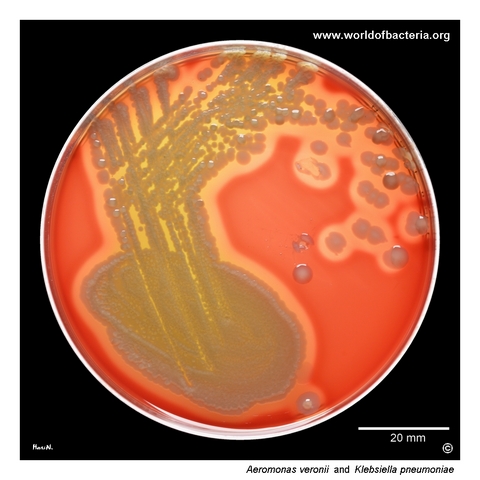
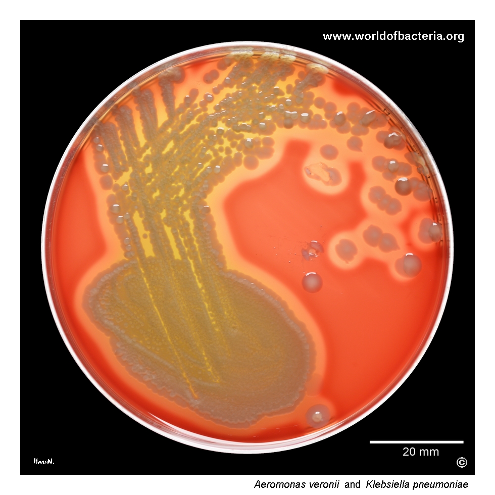
Aeromonas veronii in a urine specimen. Ten microlitres of midstream urine were inoculated and streaked onto blood agar. Beta-haemolytic colonies of A. veronii are present in mixed culture with non-haemolytic colonies of Klebsiella pneumoniae. Culture conditions: 24 hours at 36 °C in ambient air. The contrasting haemolytic patterns make the two organisms readily distinguishable during primary plate examination.
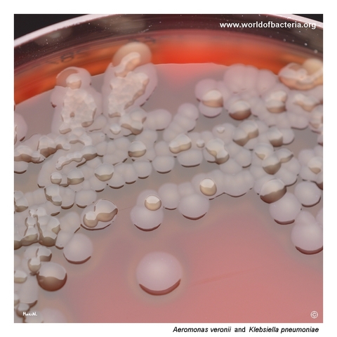
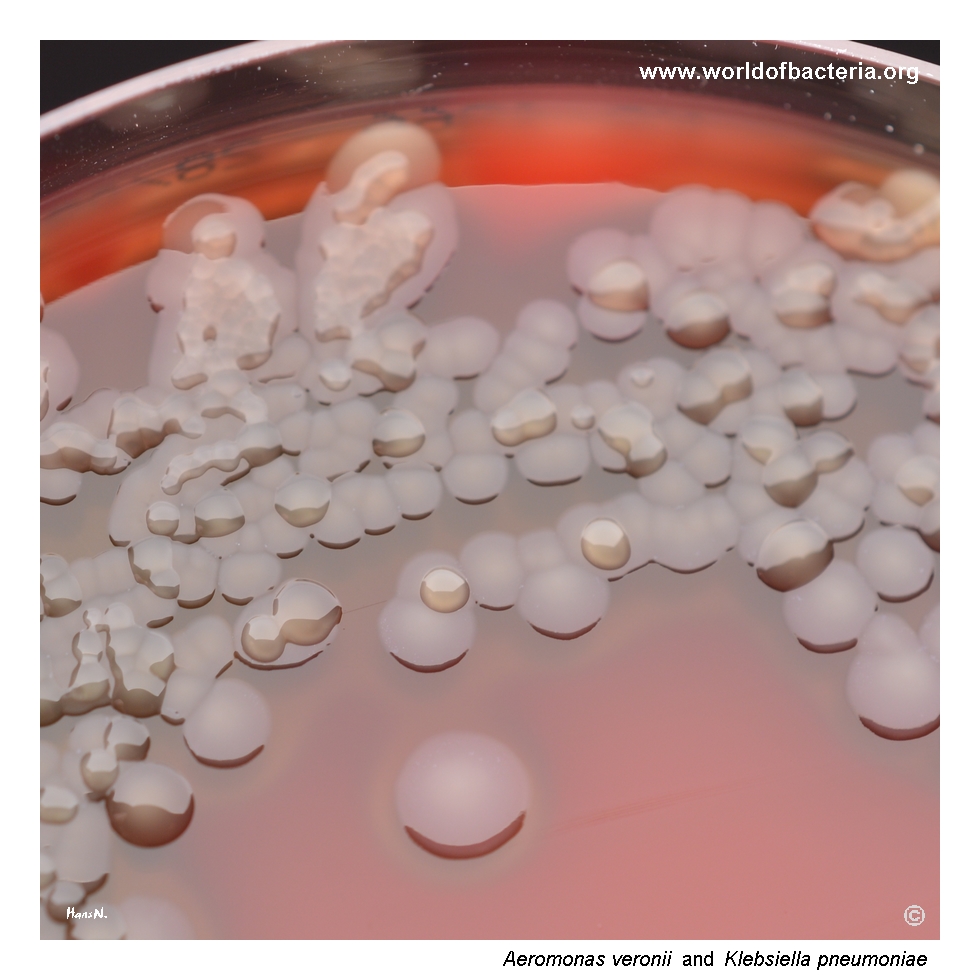
Colonies of Aeromonas veronii and Klebsiella pneumoniae on blood agar after 24 hours of incubation at 36 °C in ambient air. The K. pneumoniae isolate shows a strongly hypermucoid phenotype, producing large, glistening colonies that contrast clearly with the less mucoid colonies of A. veronii. This mixed culture demonstrates how colony texture and haemolysis can assist preliminary differentiation on routine medium.
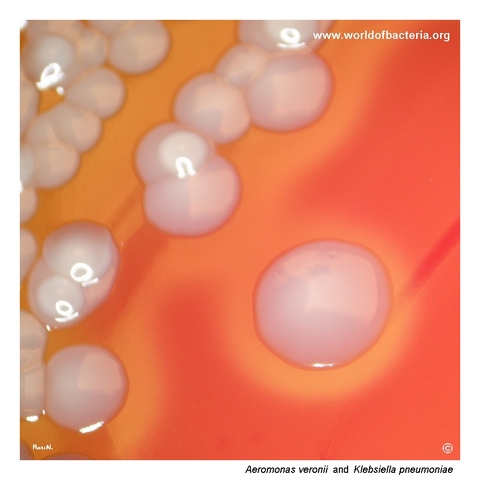
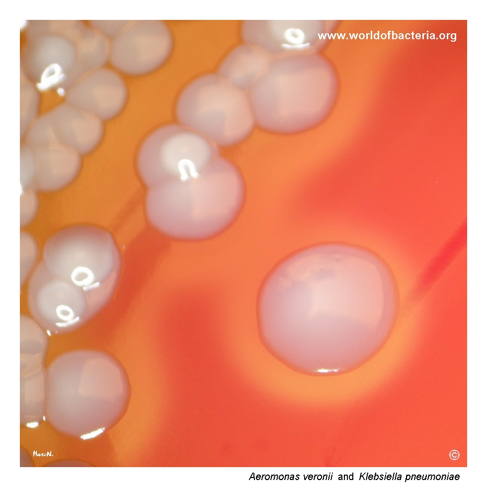
Close-up view of Aeromonas veronii colonies on blood agar. The colonies are smooth, convex, and round with entire margins, surrounded by a distinct zone of beta-haemolysis. The accompanying Klebsiella pneumoniae colonies are readily distinguished by their strongly mucoid appearance and absence of haemolysis. Culture conditions: 24 hours at 36 °C in ambient air.
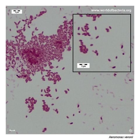
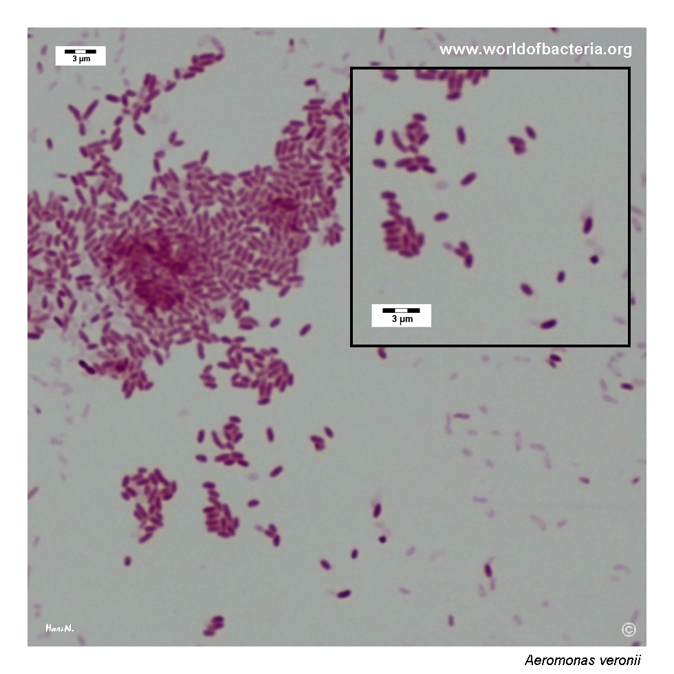
Gram-stained smear prepared from a colony of Aeromonas veronii grown on blood agar. The organism appears as Gram-negative rods. This microscopic morphology is not specific and may resemble many other Gram-negative bacteria. Reliable identification therefore requires correlation with colony morphology, haemolytic activity, oxidase testing, and additional biochemical or instrumental methods.
Diagnostic and Clinical Notes
Aeromonas veronii is an aquatic Gram-negative organism and one of the Aeromonas species commonly recovered from human clinical material. Human exposure may occur through contaminated water, food, aquatic animals, or direct inoculation of damaged tissue.
The illustrated isolate was recovered from a urine specimen in mixed culture with Klebsiella pneumoniae. Urinary tract infection due to Aeromonas is uncommon, so clinical interpretation should consider symptoms, quantitative growth, specimen quality, accompanying organisms, and predisposing factors such as urinary instrumentation or underlying disease.
Prominent beta-haemolysis makes the A. veronii colonies visually distinct from the non-haemolytic, hypermucoid K. pneumoniae colonies in this culture. Haemolysis and colony texture are useful descriptive clues but do not provide definitive species identification or establish pathogenic significance.
Historical literature frequently used the designations A. veronii biovar sobria and A. veronii biovar veronii. Modern identification increasingly treats A. veronii at the species level, while older biovar terminology may still appear in clinical and epidemiological publications.
Species-level identification within the genus can be difficult. MALDI-TOF MS and automated biochemical methods are useful when their reference databases are current, but sequencing of discriminatory housekeeping genes or genome-based analysis may be required for unusual, invasive, or epidemiologically important isolates.
Laboratory Identification
Colony Morphology
On blood agar after 18–24 hours at approximately 35–37 °C, Aeromonas veronii commonly forms smooth, convex, grey-white colonies that may be surrounded by clear beta-haemolysis. Colony size, pigmentation, and haemolytic intensity vary between isolates and are not sufficient for species identification.
Microscopy
Gram staining shows Gram-negative rods occurring mainly singly or in pairs. The microscopic appearance overlaps with many other Gram-negative bacteria and cannot distinguish A. veronii from related Aeromonas species.
Key Identification Clues
Gram-negative, facultatively anaerobic rod
Oxidase positive, unlike Enterobacterales
Usually motile, catalase positive, and indole positive
Ferments glucose
Frequently beta-haemolytic on blood agar
Aquatic association and recovery from gastrointestinal, wound, bloodstream, hepatobiliary, or occasional urinary specimens
Modern Identification Methods
Routine identification may be achieved by MALDI-TOF mass spectrometry with an up-to-date database or validated automated biochemical systems. Difficult isolates may require sequencing of genes such as rpoB, gyrB, or rpoD, or whole-genome analysis, because 16S rRNA sequencing and older phenotypic systems may not reliably separate closely related Aeromonas species.
Antibiotic Characteristics
Aeromonas veronii commonly possesses chromosomal beta-lactamases and is usually resistant to ampicillin and several older beta-lactam agents.
Susceptibility to extended-spectrum cephalosporins, fluoroquinolones, trimethoprim-sulfamethoxazole, aminoglycosides, carbapenems, and other potentially active drugs varies between isolates.
Some strains may carry inducible cephalosporinases, metallo-beta-lactamases, or acquired resistance determinants, and resistance may emerge or become apparent during therapy with selected beta-lactam agents.
Note: Clinically significant urinary, wound, bloodstream, hepatobiliary, and other invasive isolates should undergo antimicrobial susceptibility testing using an appropriate validated method. Treatment should be selected according to the individual result and clinical context.