Stenotrophomonas maltophilia is an aerobic, non-fermenting Gram-negative rod found in water, soil, plants, and moist healthcare environments and recognized as an opportunistic pathogen, particularly in patients with chronic lung disease, intensive-care exposure, indwelling devices, malignancy, or impaired immunity.
Basic Characteristics
Taxonomy
Domain: Bacteria
Phylum: Pseudomonadota (Proteobacteria)
Class: Gammaproteobacteria
Order: Xanthomonadales
Family: Xanthomonadaceae
Genus:Stenotrophomonas
Species:Stenotrophomonas maltophilia
Microscopy & Gram Stain
Gram-negative rods
Cells usually occur singly or in pairs
Microscopic appearance is non-specific and resembles other non-fermenting Gram-negative bacilli
Oxygen Relationship
Aerobic
Non-fermenting; carbohydrates are used oxidatively rather than by fermentation
Rapid Identification Tests
Oxidase: usually negative
Catalase: positive
Motility: positive
Glucose metabolism: oxidative, non-fermentative
Haemolysis on blood agar: usually absent
Pigment: yellow pigmentation may develop and intensify with prolonged incubation
MacConkey agar: growth as a non-lactose-fermenting organism
Ecology and Clinical Relevance
Natural Habitat
Freshwater, soil, plant-associated habitats, and the rhizosphere
Domestic and healthcare water systems and other persistently moist environments
Respiratory tract of some patients with cystic fibrosis, bronchiectasis, or other chronic lung disease
May colonize medical devices, sinks, tubing, and contaminated fluids
Common Clinical Specimens
Sputum, tracheal aspirates, and bronchoalveolar lavage
Blood cultures and intravascular catheter specimens
Wound, soft-tissue, and burn specimens
Urine and other device-associated specimens
Pleural fluid and other normally sterile materials
Clinical Significance
Opportunistic pathogen most relevant in patients with chronic lung disease, intensive-care exposure, malignancy, immunocompromise, or indwelling devices
May represent either respiratory colonization or true lower respiratory tract infection
Associated with pneumonia, bloodstream infection, catheter-related infection, urinary infection, wound infection, and other invasive disease
Clinical interpretation depends strongly on specimen quality, quantity of growth, repeated isolation, accompanying organisms, and the condition of the patient
Differential Considerations
Achromobacter xylosoxidans, Pseudomonas aeruginosa, and other non-fermenting Gram-negative rods
Burkholderia cepacia complex in respiratory specimens from patients with cystic fibrosis
Acinetobacter baumannii and related oxidase-negative non-fermenters
Helpful clues include oxidase negativity, motility, non-fermentative metabolism, yellow pigmentation, and usually non-haemolytic colonies
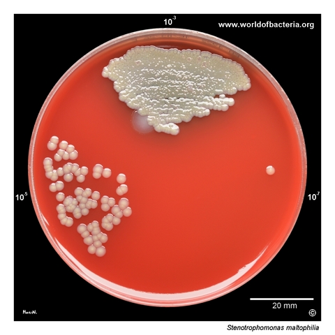
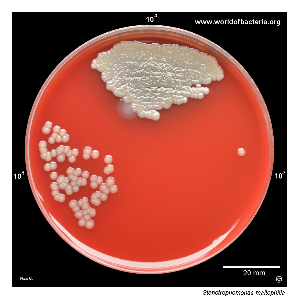
Stenotrophomonas maltophilia recovered from the sputum of a patient hospitalized in the intensive care unit. The specimen was diluted in buffered saline, and 10−3, 10−5, and 10−7 dilutions were inoculated with a calibrated 10 µl loop onto separate sectors of the agar plate. A larger colony of Pseudomonas aeruginosa is also visible in the 10−3 dilution. Culture conditions: 48 hours at 36 °C in ambient air.
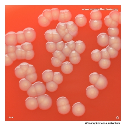
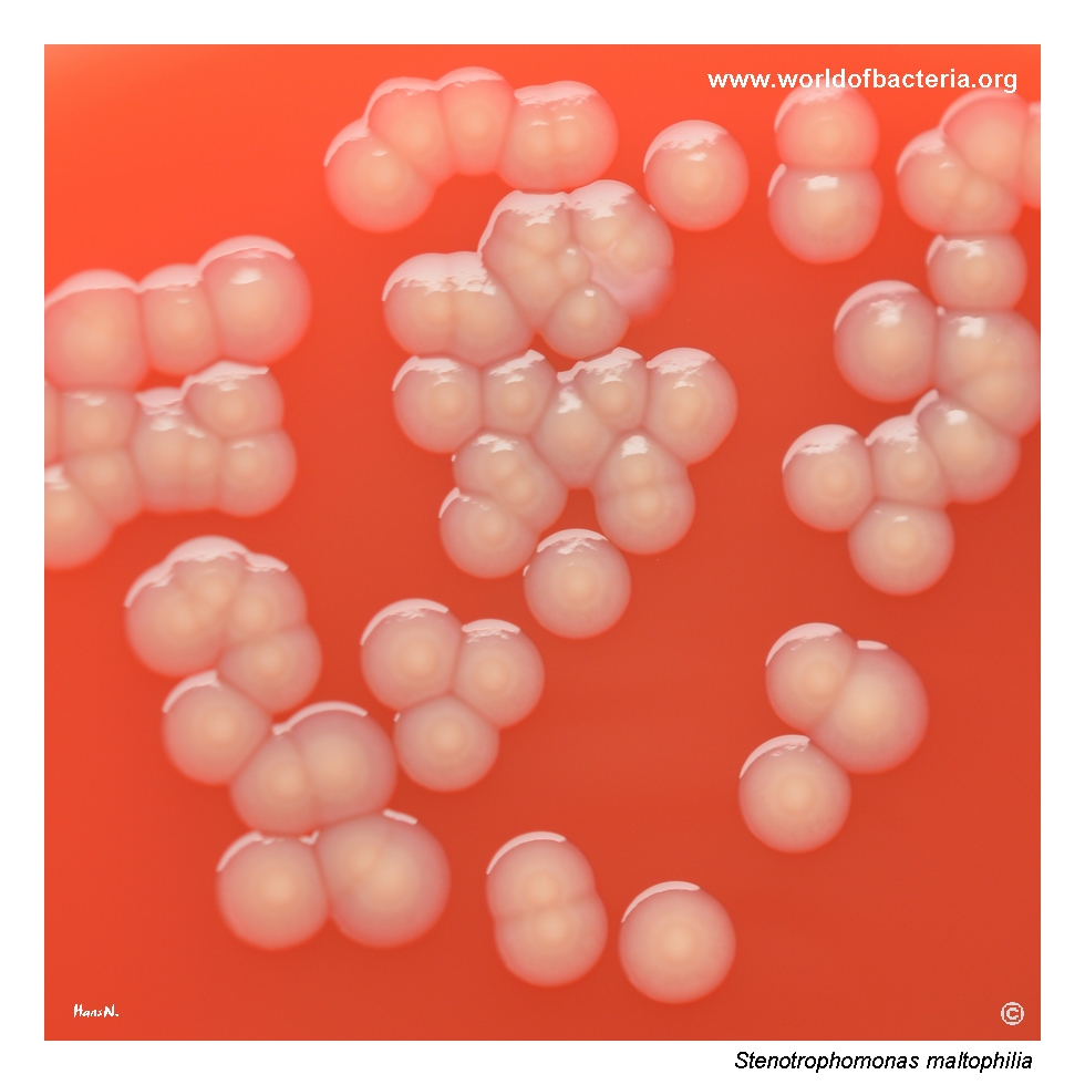
Colonies of Stenotrophomonas maltophilia on blood agar after 48 hours at 36 °C in ambient air. The colonies have entire margins and commonly develop yellow pigmentation, which becomes more pronounced during prolonged incubation. In this isolate, the surface is slightly uneven. The species shows high intrinsic resistance to many antibiotics, particularly β-lactams, through chromosomal L1 and L2 β-lactamases, efflux pumps, and other resistance mechanisms.
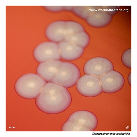
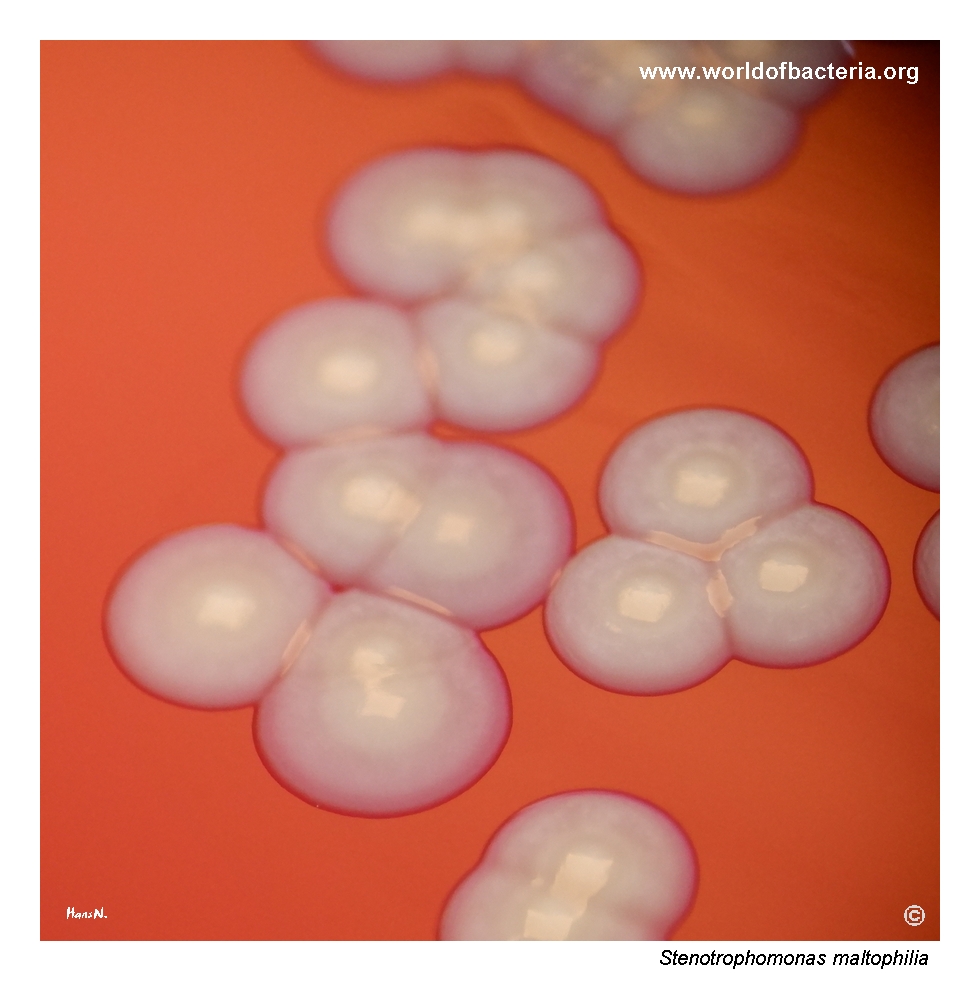
Close-up view of Stenotrophomonas maltophilia colonies on blood agar after 24 hours of incubation at 36 °C in ambient air. The colonies are non-haemolytic and show a raised central area, described as umbonate. Yellow pigment production is most pronounced in the elevated centre of the colonies. Pigmentation and surface morphology may vary between isolates and become more conspicuous after prolonged incubation.
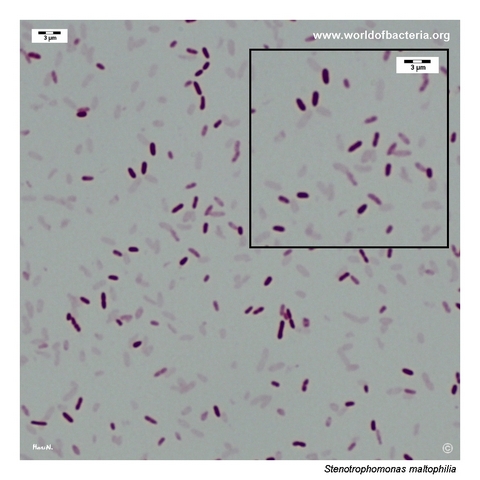
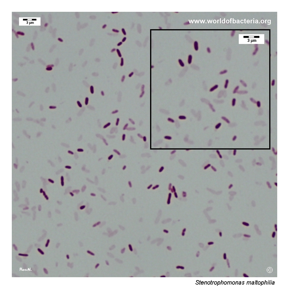
Gram-stained smear prepared from a colony of Stenotrophomonas maltophilia grown on blood agar. The organism appears as Gram-negative rods. This microscopic morphology is non-specific and may resemble that of other non-fermenting Gram-negative bacteria. Reliable identification therefore requires correlation with colony characteristics and additional biochemical or instrumental methods.
Diagnostic and Clinical Notes
Stenotrophomonas maltophilia is an environmental, aerobic, non-fermenting Gram-negative rod that can persist in water systems and other moist habitats. In healthcare settings, it may colonize sinks, tubing, devices, and aqueous solutions and can be selected by broad-spectrum antimicrobial exposure.
The sputum culture shown here was examined quantitatively after serial dilution and also contained a colony of Pseudomonas aeruginosa. In respiratory specimens, recovery of S. maltophilia may represent colonization, chronic airway persistence, or true infection; interpretation therefore requires specimen quality, abundance, repeated recovery, radiological findings, inflammatory response, and the overall clinical picture.
Yellow pigmentation, non-haemolytic growth, and a subtly uneven or umbonate colony surface may provide useful descriptive clues. These features are variable, however, and cannot replace species-level identification.
The organism has major diagnostic and therapeutic importance because it is intrinsically resistant to many antimicrobial classes. Chromosomal L1 metallo-beta-lactamase and L2 serine beta-lactamase activity, multidrug efflux systems, reduced permeability, biofilm formation, and additional mechanisms contribute to its broad resistance profile.
MALDI-TOF mass spectrometry commonly supports routine identification, but the result should be correlated with the oxidase-negative non-fermenting phenotype and the clinical context. Molecular or genomic methods may be useful in unusual isolates, outbreak investigations, and detailed resistance analysis.
Laboratory Identification
Colony Morphology
After 24–48 hours on blood agar at 35–37 °C, colonies of Stenotrophomonas maltophilia are usually small to medium-sized, grey-white to yellow, smooth or slightly uneven, and round with entire margins. Raised or umbonate centres may occur, pigmentation often intensifies with prolonged incubation, and visible haemolysis is usually absent.
Microscopy
Gram staining shows Gram-negative rods occurring mainly singly or in pairs. The microscopic appearance is non-specific and overlaps with Achromobacter, Pseudomonas, Burkholderia, and other aerobic non-fermenting Gram-negative bacilli.
Key Identification Clues
Gram-negative rods
Aerobic, non-fermenting metabolism
Oxidase usually negative and catalase positive
Motile
Yellow pigmentation may develop on blood agar
Usually non-haemolytic
Growth on MacConkey agar as a non-lactose-fermenting organism
Broad intrinsic resistance, including resistance to many beta-lactams and carbapenems
Modern Identification Methods
MALDI-TOF mass spectrometry and contemporary automated identification systems are commonly used for routine species identification. Updated reference databases are important because closely related environmental Stenotrophomonas taxa may be difficult to resolve phenotypically. Targeted sequencing or whole-genome methods can provide additional resolution for epidemiological, taxonomic, and resistance investigations.
Antibiotic Characteristics
Stenotrophomonas maltophilia has extensive intrinsic resistance. The chromosomal L1 metallo-beta-lactamase and L2 serine beta-lactamase reduce the activity of many beta-lactams, including carbapenems, while efflux systems, reduced permeability, aminoglycoside resistance mechanisms, biofilm growth, and acquired determinants further complicate treatment.
Trimethoprim–sulfamethoxazole has historically been one of the most frequently used active agents, but susceptibility is not universal and adverse effects or clinical circumstances may limit its use. Minocycline, levofloxacin, cefiderocol, and selected combination regimens may be considered when supported by susceptibility results and current clinical guidance.
Respiratory recovery does not automatically require treatment because colonization is common, particularly in patients with chronic lung disease or prolonged hospitalization. Antimicrobial therapy should be reserved for a clinically supported infection rather than a culture result alone.
Note: Clinically significant isolates require antimicrobial susceptibility testing with validated methods and appropriate interpretive criteria. Treatment selection should consider the complete susceptibility profile, infection site, disease severity, source control, previous antimicrobial exposure, and current specialist guidance.