Achromobacter xylosoxidans is an aerobic, oxidase-positive, non-fermenting Gram-negative rod found in water and moist environments and recognized as an opportunistic pathogen, particularly in patients with cystic fibrosis, indwelling devices, repeated healthcare exposure, or impaired immunity.
Basic Characteristics
Taxonomy
Domain: Bacteria
Phylum: Pseudomonadota (Proteobacteria)
Class: Betaproteobacteria
Order: Burkholderiales
Family: Alcaligenaceae
Genus:Achromobacter
Species:Achromobacter xylosoxidans
Microscopy & Gram Stain
Relatively small Gram-negative rods
Cells usually occur singly or in pairs
Microscopic appearance is non-specific and resembles other non-fermenting Gram-negative bacilli
Oxygen Relationship
Aerobic
Non-fermenting; carbohydrates are used oxidatively rather than by fermentation
Rapid Identification Tests
Oxidase: positive
Catalase: positive
Motility: positive
Glucose metabolism: oxidative, non-fermentative
Xylose oxidation: usually positive
Haemolysis on blood agar: usually absent
MacConkey agar: growth as a non-lactose-fermenting organism
Ecology and Clinical Relevance
Natural Habitat
Freshwater, soil, and other moist environmental habitats
Domestic and healthcare water systems, humidifiers, solutions, and wet equipment
Respiratory tract of some patients with cystic fibrosis or other chronic lung disease
May transiently colonize human mucosal surfaces and medical devices
Common Clinical Specimens
Respiratory specimens, especially from patients with cystic fibrosis
Blood cultures and intravascular catheter specimens
Wound, soft-tissue, and ear specimens
Urine, peritoneal fluid, and other normally sterile materials
Ocular specimens and contaminated medical solutions or devices
Clinical Significance
Opportunistic pathogen most relevant in patients with chronic lung disease, immunocompromise, indwelling devices, or repeated healthcare exposure
May cause persistent airway colonization or infection in cystic fibrosis
Associated with bloodstream infection, pneumonia, device-related infection, urinary infection, wound infection, otitis, keratitis, and peritonitis
Clinical significance depends strongly on specimen type, quantity of growth, repeated recovery, and the condition of the patient
Differential Considerations
Stenotrophomonas maltophilia, Pseudomonas aeruginosa, and other non-fermenting Gram-negative rods
Burkholderia cepacia complex and related organisms in cystic fibrosis respiratory specimens
Alcaligenes faecalis and other members of the family Alcaligenaceae
Helpful clues include oxidase positivity, motility, non-fermentative metabolism, xylose oxidation, and usually non-haemolytic grey-white colonies
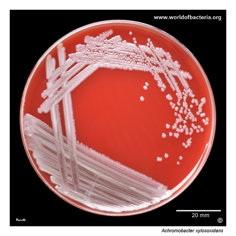
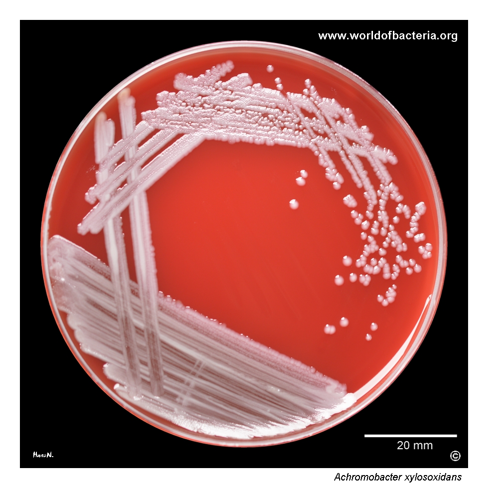
Achromobacter xylosoxidans on blood agar after 24 hours of incubation at 36 °C in ambient air. The isolate originated from an ear canal swab from a 30-year-old patient with the diagnosis H60.9, otitis externa, unspecified, and was present in significant quantity. Although this species is only occasionally recovered from ear specimens, abundant growth may support its possible involvement in the infectious process.
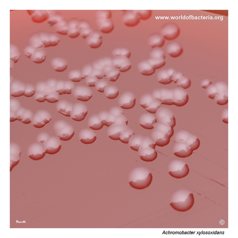
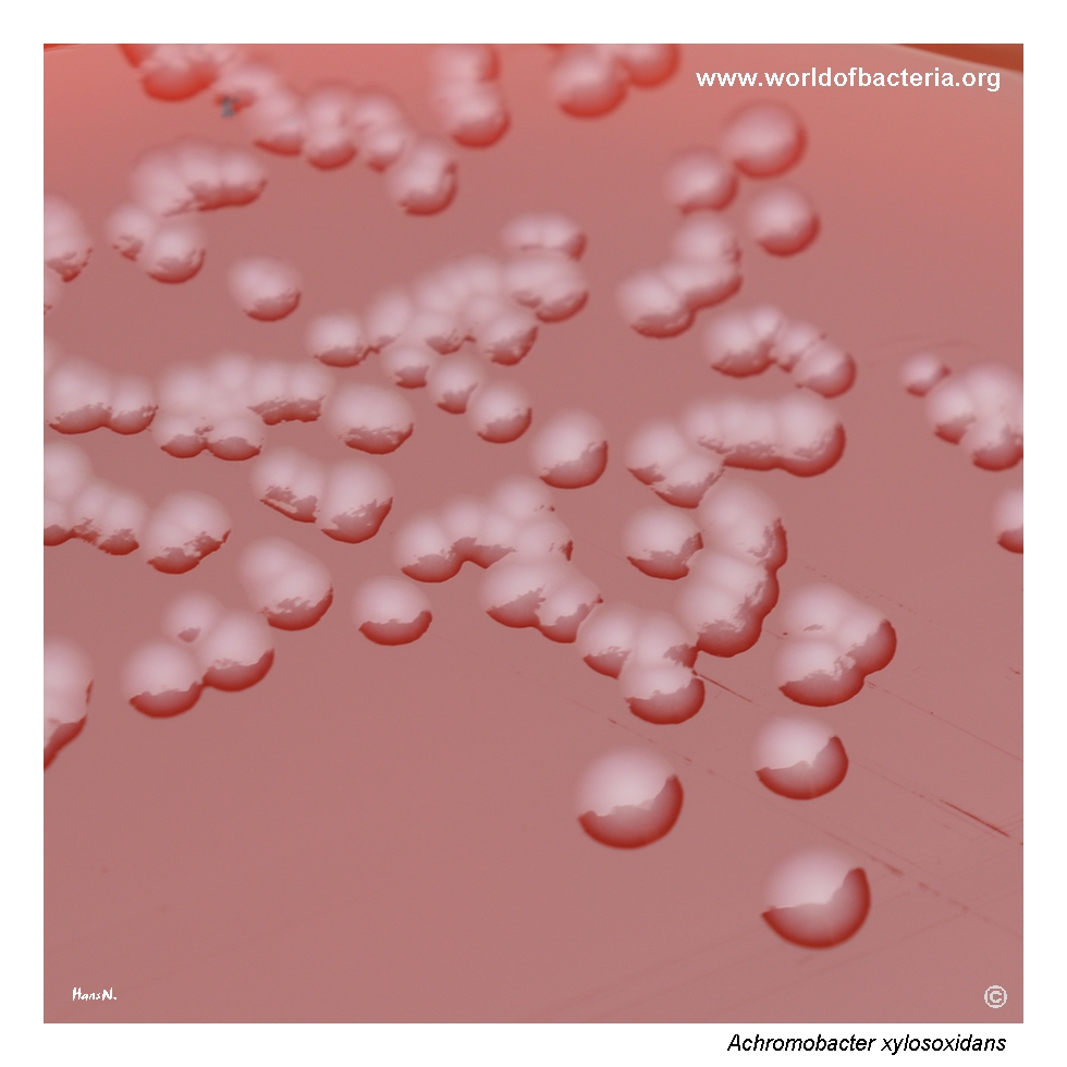
Colonies of Achromobacter xylosoxidans on blood agar after 24 hours at 36 °C in ambient air. At this stage, the colonies remain relatively small, reflecting slower growth than that of many Enterobacterales, as is common among non-fermenting Gram-negative rods. At higher magnification, the colony surface is not completely smooth and may resemble the subtly uneven texture seen in some isolates of Stenotrophomonas maltophilia.
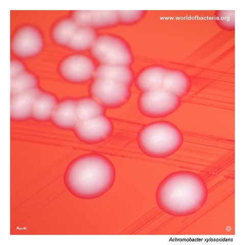
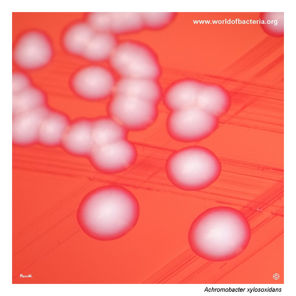
Close-up view of Achromobacter xylosoxidans colonies on blood agar after 24 hours of incubation at 36 °C in ambient air. The colonies are grey-white, convex, and round with entire margins and show no visible haemolysis. This relatively inconspicuous morphology provides few species-specific clues and may resemble that of several other non-fermenting Gram-negative bacteria encountered in clinical specimens.
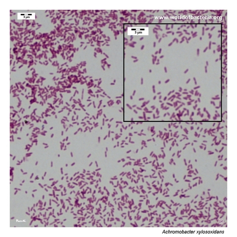
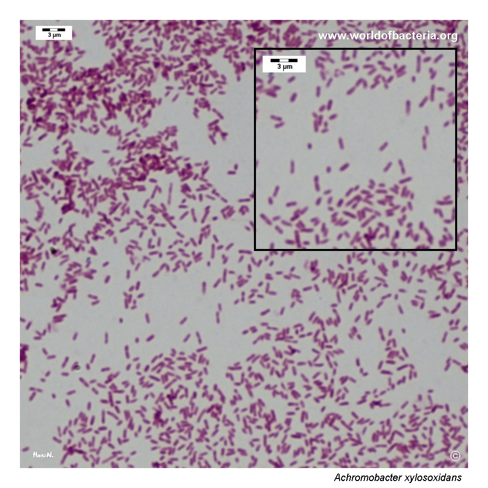
Gram-stained smear prepared from a colony of Achromobacter xylosoxidans grown on blood agar. The organism appears as relatively small Gram-negative rods. This microscopic morphology is non-specific and does not distinguish the species from other non-fermenting bacteria. Reliable identification therefore depends on culture characteristics together with biochemical or instrumental identification methods.
Diagnostic and Clinical Notes
Achromobacter xylosoxidans is the type species of the genus Achromobacter and was originally described from human ear discharge. It is an environmental, aerobic, non-fermenting Gram-negative rod that can persist in water systems, moist hospital environments, and contaminated solutions or devices.
In the ear specimen shown here, the isolate was present in significant quantity. Because A. xylosoxidans is only occasionally recovered from otic material, abundant or repeated growth together with compatible clinical findings is more informative than the unusual source alone.
The species has particular clinical importance in cystic fibrosis, where it may establish chronic respiratory colonization or infection and may acquire increasing antimicrobial resistance during prolonged persistence and repeated treatment.
Outside cystic fibrosis, clinically significant infection occurs most often in patients with immunocompromise, indwelling vascular devices, recurrent healthcare exposure, or other major underlying disease. Pneumonia and bloodstream infection are among the better documented presentations, but urinary, ocular, wound, ear, central nervous system, and peritoneal infections have also been reported.
Species-level identification within Achromobacter remains difficult. Older biochemical systems and some MALDI-TOF databases may report non-xylosoxidans species as A. xylosoxidans; updated databases, targeted gene sequencing, or whole-genome methods may therefore be needed for definitive classification.
Laboratory Identification
Colony Morphology
After approximately 24 hours on blood agar at 35–37 °C, colonies of Achromobacter xylosoxidans are usually small, grey-white, convex, and round with entire margins. The surface may be smooth or subtly uneven, and visible haemolysis is usually absent. Colony appearance alone does not distinguish the species from other non-fermenting Gram-negative rods.
Microscopy
Gram staining shows relatively small Gram-negative rods occurring mainly singly or in pairs. This morphology is non-specific and overlaps with Stenotrophomonas, Pseudomonas, Burkholderia, and other aerobic non-fermenters.
Key Identification Clues
Small Gram-negative rods
Aerobic, non-fermenting metabolism
Oxidase and catalase positive
Motile
Usually oxidizes xylose
Usually non-haemolytic on blood agar
Growth on MacConkey agar as a non-lactose-fermenting organism
Modern Identification Methods
MALDI-TOF mass spectrometry commonly provides reliable genus-level identification, but species-level resolution depends strongly on the reference library and may remain unreliable among closely related Achromobacter species. Sequencing of discriminatory targets such as nrdA, multilocus approaches, or whole-genome sequencing can provide more definitive identification and are also useful for epidemiological and resistance investigations.
Antibiotic Characteristics
Achromobacter xylosoxidans has substantial intrinsic resistance mediated by multidrug efflux systems, beta-lactamases, reduced permeability, and other mechanisms. Resistance is commonly observed to aminoglycosides, aztreonam, and many cephalosporins, while susceptibility to fluoroquinolones and other agents is variable.
Piperacillin–tazobactam, meropenem or imipenem, trimethoprim–sulfamethoxazole, and sometimes ceftazidime may retain activity, but no agent should be assumed to be active without testing. Respiratory isolates from chronically treated patients, particularly those with cystic fibrosis, may show broader resistance than isolates from other sources.
Resistance can increase during chronic infection and antimicrobial exposure. Species misidentification and differences between interpretive standards may also complicate comparison of susceptibility results between laboratories.
Note: Clinically significant isolates require antimicrobial susceptibility testing with validated methods and appropriate interpretive criteria. Treatment should be selected according to the complete susceptibility profile, infection site, disease severity, source control, and current clinical guidance.