Vibrio cholerae is an oxidase-positive, motile, curved Gram-negative rod found in aquatic environments. Toxigenic strains of serogroups O1 and O139 cause epidemic cholera, whereas non-O1/non-O139 strains may cause sporadic diarrhoeal, wound, or invasive infections.
Basic Characteristics
Taxonomy
Domain: Bacteria
Phylum: Pseudomonadota (Proteobacteria)
Class: Gammaproteobacteria
Order: Vibrionales
Family: Vibrionaceae
Genus:Vibrio
Species:Vibrio cholerae
Microscopy & Gram Stain
Gram-negative rods, often slightly curved or comma-shaped
Cells usually occur singly, although short chains may occasionally be seen
Curvature may be subtle and some cells may appear almost straight in colony smears
Oxygen Relationship
Facultatively anaerobic
Grows readily under routine aerobic conditions
Rapid Identification Tests
Oxidase: positive
Motility: positive, usually with a single polar flagellum
Urease: negative
Indole: usually positive
Sucrose fermentation: usually positive, producing yellow colonies on TCBS agar
Haemolysis: variable; many non-O1/non-O139 and El Tor-associated isolates may be haemolytic
Ecology and Clinical Relevance
Natural Habitat
Freshwater, brackish water, and coastal aquatic environments
Association with plankton, chitinous surfaces, shellfish, and other aquatic organisms
Environmental persistence is influenced by temperature, salinity, nutrients, and plankton abundance
Common Clinical Specimens
Stool and rectal swabs from patients with diarrhoeal disease
Blood cultures in invasive infection
Wound and soft-tissue specimens after water exposure
Food, seafood, and environmental water samples during surveillance or outbreak investigations
Clinical Significance
Toxigenic serogroups O1 and O139 are responsible for epidemic cholera
Severe cholera causes profuse watery diarrhoea, rapid dehydration, electrolyte loss, shock, and potentially death without prompt rehydration
Non-O1/non-O139 strains may cause sporadic gastroenteritis, cholera-like diarrhoea, wound infection, or bacteraemia
Species identification alone does not establish epidemic potential; serogroup and toxin status are essential
Differential Considerations
Vibrio mimicus, which is closely related but usually does not ferment sucrose on TCBS agar
Vibrio parahaemolyticus and other halophilic vibrios
Aeromonas species, which are also oxidase-positive aquatic Gram-negative rods
Helpful clues include curved motile rods, oxidase positivity, sucrose fermentation, and appropriate serological or molecular confirmation
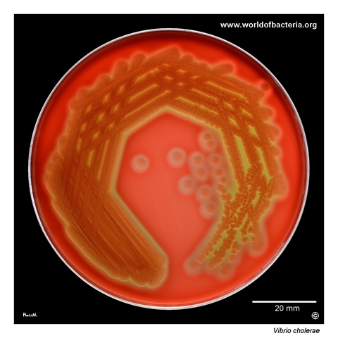
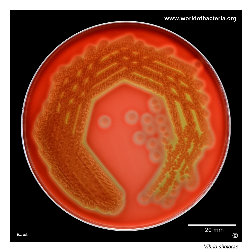
A non-O1 strain of Vibrio cholerae with an El Tor-like haemolytic phenotype on sheep blood agar. The culture has a slightly yellowish appearance and is surrounded by prominent beta-haemolysis. Culture conditions: 24 hours at 36 °C in ambient air. This striking phenotype is useful for demonstrating the variability of V. cholerae, although haemolysis on blood agar alone does not determine the serogroup or toxigenic potential of an isolate.
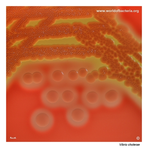
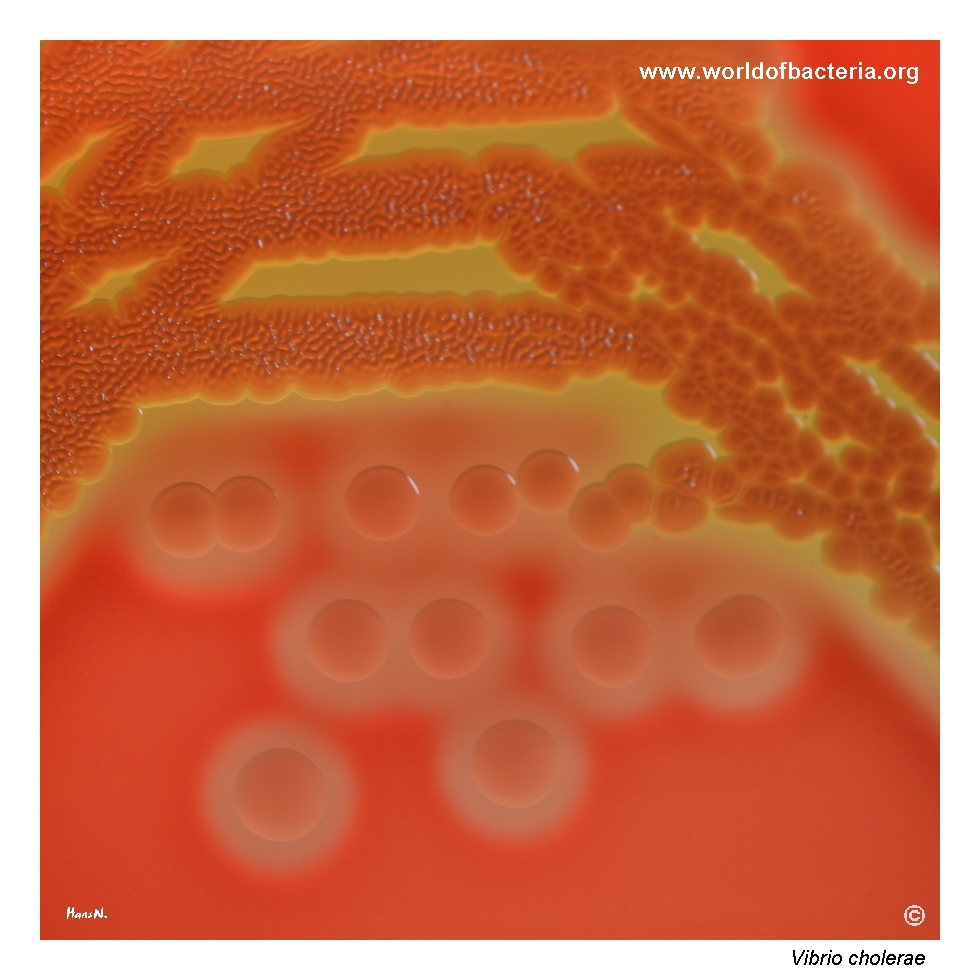
Colonies of Vibrio cholerae on blood agar after 24 hours of incubation at 36 °C in ambient air. The colonies are smooth and convex, with marked haemolysis in the surrounding agar. In the area of confluent growth, the culture surface shows a conspicuous wrinkled texture. Such surface morphology may become more apparent where individual colonies merge into a continuous bacterial layer.
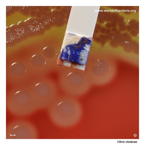
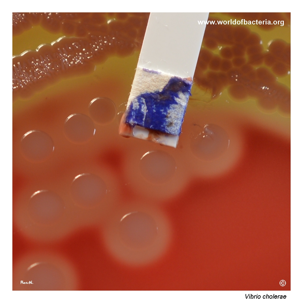
Positive oxidase test performed with a culture of Vibrio cholerae. Bacterial growth was transferred onto a diagnostic strip containing an oxidase reagent, producing rapid blue coloration and confirming the presence of cytochrome c oxidase. Most clinically relevant members of the genus Vibrio are oxidase-positive, making this reaction a useful preliminary clue, although it is not sufficient for identification at the species level.
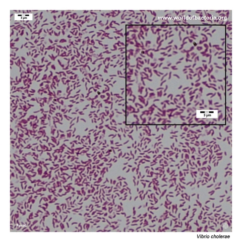
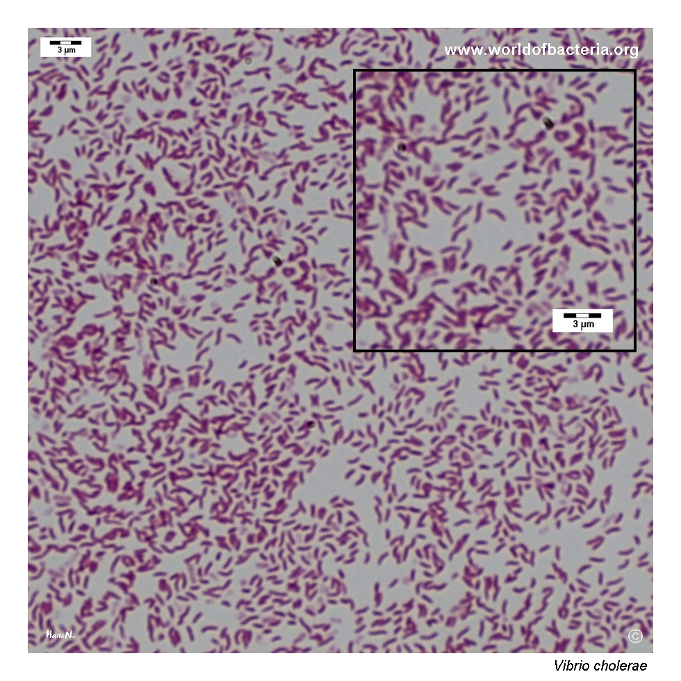
Gram-stained smear prepared from a colony of Vibrio cholerae grown on blood agar. The organism appears as Gram-negative rods, many of which show the slightly curved or comma-like shape traditionally associated with vibrios. The degree of curvature varies between individual cells and may be subtle, so some organisms may appear almost straight in preparations made from cultures grown on solid media.
Diagnostic and Clinical Notes
The illustrated organism is a non-O1 strain with a conspicuous El Tor-like haemolytic phenotype. This is visually striking but must not be interpreted as evidence of serogroup O1, El Tor biotype, or cholera-toxin production without specific confirmation.
Vibrio cholerae comprises many serogroups. Toxigenic strains belonging to O1 and O139 are responsible for widespread epidemic cholera, while other serogroups may cause sporadic diarrhoeal disease, wound infection, or invasive infection and may be either toxigenic or non-toxigenic.
On TCBS agar, typical V. cholerae colonies are yellow because most isolates ferment sucrose. Selective culture is especially useful for stool specimens, but presumptive colonies still require species confirmation followed by O1/O139 serogrouping and toxin or toxin-gene testing when cholera is suspected.
The positive oxidase reaction provides a rapid orientation away from oxidase-negative Enterobacterales. It does not distinguish V. cholerae from other vibrios, Aeromonas, or several additional aquatic Gram-negative organisms.
Curved or comma-shaped cells are a classic teaching feature, but the curvature is variable and may be subtle. Reliable interpretation therefore depends on the combined culture phenotype, biochemical or instrumental identification, serological findings, and molecular detection of relevant virulence genes.
Laboratory Identification
Colony Morphology
On blood agar, Vibrio cholerae usually forms smooth, moist, convex colonies that may appear grey-white to slightly yellowish. Haemolysis is variable and can be prominent in non-O1/non-O139 or El Tor-associated isolates. On TCBS agar, sucrose-fermenting isolates typically produce yellow colonies, but this remains a presumptive rather than definitive feature.
Microscopy
Gram staining shows slender Gram-negative rods, often with a slight curve or comma-like appearance. Some cells may appear nearly straight, especially in preparations from solid media. The organism is motile, classically by means of a single polar flagellum.
Key Identification Clues
Oxidase-positive, motile Gram-negative rod
Often curved or comma-shaped, although curvature may be subtle
Facultatively anaerobic
Usually sucrose positive, producing yellow colonies on TCBS agar
Urease negative and usually indole positive
Can grow without an added requirement for high sodium chloride concentrations
Serogrouping and toxin testing are required to determine cholera-associated potential
Modern Identification Methods
Routine species identification can be achieved by MALDI-TOF mass spectrometry, validated biochemical systems, or molecular assays. When cholera is suspected, the laboratory investigation must continue beyond species identification to O1 and O139 serogrouping and detection of cholera toxin or the corresponding ctx genes. Molecular typing and whole-genome sequencing may be used for outbreak investigation, resistance surveillance, and epidemiological comparison.
Antibiotic Characteristics
For cholera, rapid replacement of water and electrolytes is the central lifesaving intervention. Mild illness may require only oral rehydration, whereas severe dehydration requires urgent and carefully monitored fluid therapy.
Antibiotics may shorten the duration and volume of diarrhoea in severe cholera, but the choice of agent depends on current local resistance patterns, patient factors, and public-health guidance.
Non-O1/non-O139 isolates causing wound infection, bacteraemia, or other extraintestinal disease should be managed according to the infection site and antimicrobial susceptibility of the individual isolate.
Note: Antimicrobial resistance varies geographically and over time. Colony haemolysis, serogroup, and visible morphology do not predict susceptibility, so clinically significant isolates should be tested using a validated method and current interpretive standards.