Legionella pneumophila is a fastidious, strictly aerobic Gram-negative rod associated with natural and engineered water systems and is the principal cause of Legionnaires’ disease, a potentially severe form of pneumonia.
Basic Characteristics
Taxonomy
Domain: Bacteria
Phylum: Pseudomonadota (Proteobacteria)
Class: Gammaproteobacteria
Order: Legionellales
Family: Legionellaceae
Genus:Legionella
Species:Legionella pneumophila
Microscopy & Gram Stain
Slender, pleomorphic Gram-negative rods that may stain weakly by the Gram method
Cells usually occur singly; elongated and filamentous forms may appear in older cultures
Many strains are motile by means of polar or subpolar flagella
Oxygen Relationship
Strictly aerobic
Facultative intracellular organism that multiplies in free-living amoebae and human macrophages
Rapid Identification Tests
Growth requirement: L-cysteine and iron-enriched media
Growth on routine blood agar: absent
Catalase: positive
Oxidase: usually positive or weakly positive
Hippurate hydrolysis: positive
Urease: negative
Ecology and Clinical Relevance
Natural Habitat
Freshwater environments and aquatic biofilms
Hot-water systems, showers, cooling towers, spas, and other engineered water systems
Intracellular multiplication within free-living amoebae
Persistence in complex water-system biofilms
Common Clinical Specimens
Sputum and other lower respiratory tract specimens
Bronchoalveolar lavage and tracheal aspirates
Lung tissue
Urine for Legionella antigen testing
Environmental water samples during surveillance and outbreak investigations
Clinical Significance
Principal cause of Legionnaires’ disease, a potentially severe atypical pneumonia
May also cause Pontiac fever, a self-limited non-pneumonic illness
Risk is increased in older adults, smokers, people with chronic lung disease, and immunocompromised patients
Usually acquired by inhalation or aspiration of contaminated water aerosols rather than by person-to-person spread
Differential Considerations
Other Legionella species and non-pneumophila serogroups
Other fastidious Gram-negative rods that fail to grow on routine media
Other causes of severe community-acquired or healthcare-associated atypical pneumonia
A negative urinary antigen test does not exclude non-serogroup 1 L. pneumophila or other Legionella species
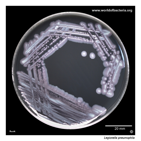
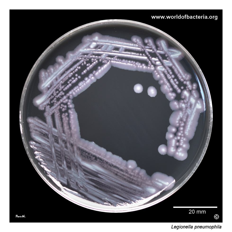
Legionella pneumophila on Legionella GVPC selective medium after 5 days of incubation at 36 °C in 5% CO2. The strain was isolated during screening of a water sample for the presence of legionellae. Unlike most clinically important bacteria, Legionella species do not grow on routine blood agar and require specially formulated culture media. Their recovery therefore depends strongly on selecting the appropriate medium and allowing sufficient incubation time.
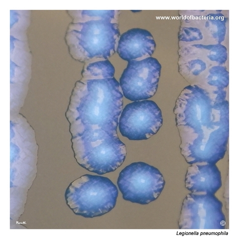
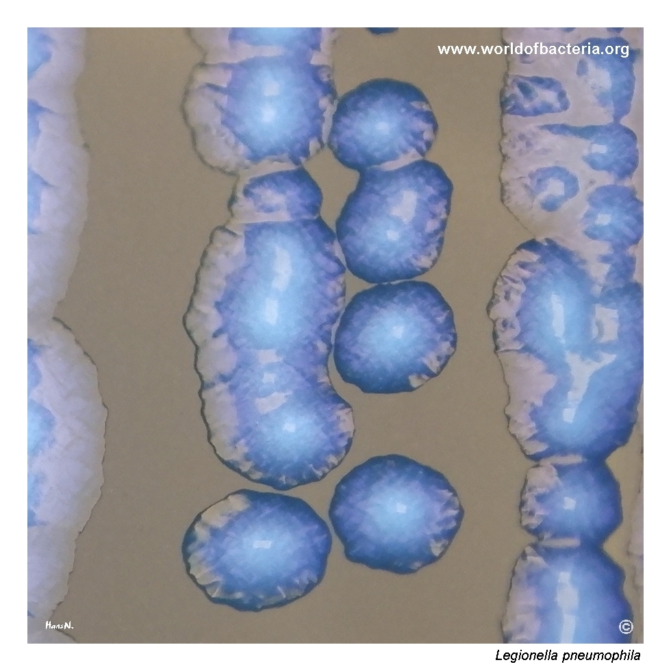
Colonies of Legionella pneumophila on Legionella GVPC selective medium. The organism grows relatively slowly, with visible colonies generally appearing after about 2 days of incubation. Mature colonies may show a raised centre and an uneven surface with a characteristic ground-glass appearance. Culture conditions: 5 days at 36 °C in 5% CO2. These distinctive features become easier to appreciate as the colonies develop.
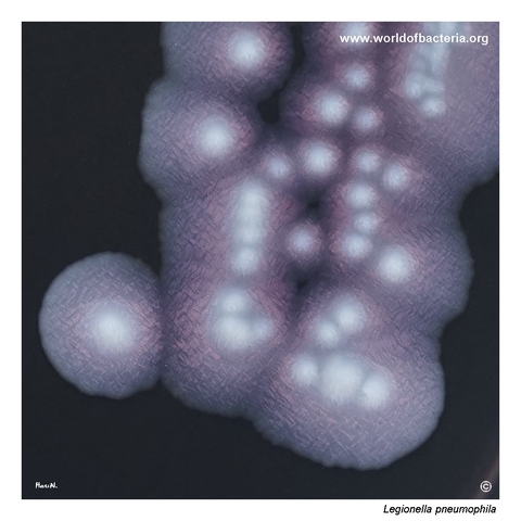
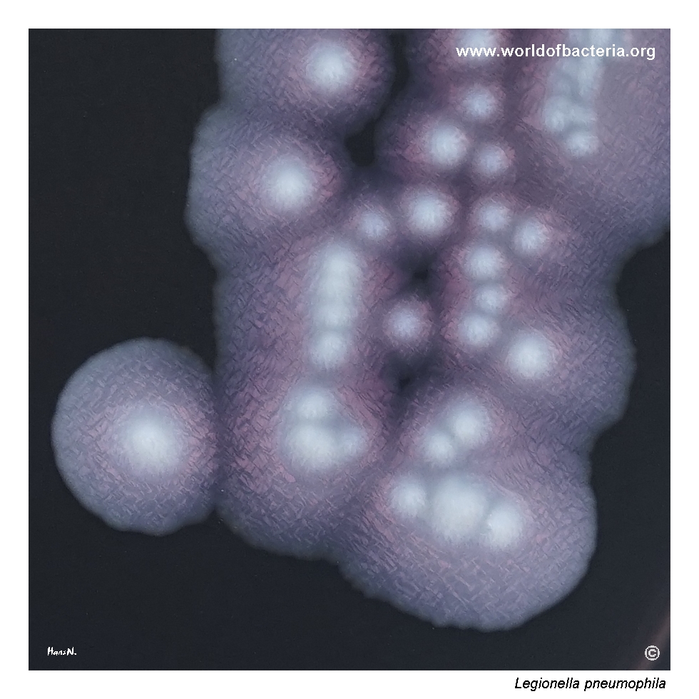
Close-up view of Legionella pneumophila colonies. Some strains form convex, iridescent colonies that appear opalescent when examined with a dissecting microscope. This characteristic optical effect and the fine surface structure are often most evident between the third and fourth days of incubation. The appearance can provide a useful preliminary clue, although definitive identification requires additional laboratory testing.
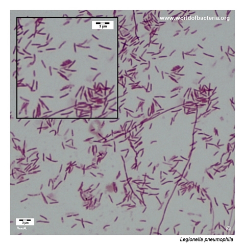
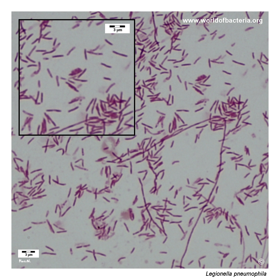
Gram-stained smear prepared from a colony grown on Legionella GVPC selective medium. Although Legionella pneumophila may stain poorly in direct clinical material, a preparation made from colonies after 5 days of incubation clearly shows long, slender Gram-negative rods. Filamentous forms are also visible in some areas. The microscopic appearance may vary with culture age and growth conditions.
Diagnostic and Clinical Notes
Legionella pneumophila is an aquatic bacterium that persists in natural and engineered water systems, often within biofilms and free-living amoebae. Multiplication inside protozoa helps the organism survive environmental stress and prepares it for intracellular replication in human macrophages.
Culture requires buffered charcoal yeast extract agar or a related medium supplemented with L-cysteine and iron. GVPC medium adds selective agents that suppress competing environmental flora. Failure to inoculate appropriate media is a major reason why legionellae may remain undetected in routine cultures.
Colonies usually become visible after several days and may show a ground-glass, finely textured, opalescent, or iridescent appearance. These features are useful preliminary clues but remain presumptive and should be confirmed by antigenic, mass-spectrometric, or molecular methods.
For suspected Legionnaires’ disease, urinary antigen testing should be paired with testing of a lower respiratory specimen by culture or a molecular method. The commonly used urinary antigen test primarily detects L. pneumophila serogroup 1, so a negative result does not exclude infection caused by other serogroups or species.
Environmental recovery documents contamination of a water system but does not by itself prove the source of a human infection. Outbreak investigation may require comparison of clinical and environmental isolates by molecular typing or whole-genome sequencing.
Laboratory Identification
Colony Morphology
On BCYE or selective GVPC medium incubated at approximately 35–37 °C, Legionella pneumophila usually produces visible colonies after 2–5 days. Mature colonies may be small to medium-sized, convex or centrally raised, finely granular, ground-glass, opalescent, or iridescent. The organism does not grow on routine blood agar because it requires L-cysteine and iron.
Microscopy
Gram staining shows slender, pleomorphic Gram-negative rods that may stain faintly, particularly in direct clinical material. Preparations from mature colonies may contain elongated or filamentous forms. Microscopy supports recognition of the organism but is not sufficiently sensitive or specific for definitive diagnosis.
Key Identification Clues
Fastidious, strictly aerobic Gram-negative rod
Requires L-cysteine and iron-enriched BCYE-based media
No growth on routine blood agar or on BCYE lacking L-cysteine
Catalase positive and usually oxidase positive or weakly positive
Hippurate hydrolysis positive
Slow-growing ground-glass, opalescent, or iridescent colonies
Urinary antigen testing mainly detects L. pneumophila serogroup 1
Modern Identification Methods
Diagnosis may combine urinary antigen testing with culture or nucleic acid amplification from a lower respiratory specimen. Cultured isolates can be identified by MALDI-TOF mass spectrometry when the reference database is suitable, serological methods, or molecular assays. Whole-genome sequencing and other high-resolution typing methods are particularly useful for linking clinical and environmental isolates during outbreak investigations.
Antibiotic Characteristics
Legionella pneumophila replicates intracellularly, so effective therapy requires antimicrobial agents that penetrate host cells and remain active within the intracellular compartment.
Macrolides, particularly azithromycin, and respiratory fluoroquinolones such as levofloxacin are commonly used for Legionnaires’ disease. Prompt treatment is especially important in severe pneumonia and in high-risk patients.
Many beta-lactam agents are unsuitable because of limited intracellular activity and bacterial beta-lactamase production. In vitro susceptibility results do not always predict clinical efficacy for this intracellular organism.
Note: Routine susceptibility testing is not commonly performed for individual Legionella isolates. Treatment should follow current clinical guidance and take account of disease severity, host factors, contraindications, and local recommendations.