Streptococcus anginosus is a member of the anginosus group often linked with purulent infections and abscess formation, yet on blood agar its colonies may be surprisingly variable, ranging from small non-haemolytic growth to more conspicuous beta-haemolytic isolates.
Basic Characteristics
Taxonomy
Domain: Bacteria
Phylum: Bacillota
Class: Bacilli
Order: Lactobacillales
Family: Streptococcaceae
Genus:Streptococcus
Species:Streptococcus anginosus
Microscopy & Gram Stain
Gram-positive oval cocci that may occur singly, in pairs, and in short chains, although anginosus-group isolates can show a somewhat variable microscopic arrangement
In this page set, the colony smear also shows small irregular groupings, which can make the slide look less classically streptococcal at first glance
The overall morphology is fully compatible with the genus Streptococcus, but microscopy alone cannot securely distinguish S. anginosus from other small Gram-positive cocci
Correlation with the colony phenotype, the clinical specimen, and modern identification methods remains essential
Oxygen Relationship
Facultatively anaerobic streptococcus that grows well on routine blood agar under capnophilic incubation
In this page set, the organism produced clearly visible colonies after 24 hours at 36 °C in 5% CO2
Routine culture conditions therefore readily support growth, even though the visual phenotype may still be subtle and easy to underestimate
Rapid Identification Tests
Catalase: negative
Haemolysis: variable; non-haemolytic, alpha-haemolytic, or beta-haemolytic isolates may all occur
Group context: belongs to the Streptococcus anginosus group (SAG)
Key caution: colony appearance alone does not reliably separate this species from other small streptococci or from other SAG members
Practical implication: definitive species identification is best based on reliable modern laboratory methods
Ecology and Clinical Relevance
Natural Habitat
Streptococcus anginosus belongs to the anginosus group and forms part of the normal human microbiota, especially in the oral cavity, gastrointestinal tract, and genitourinary tract
This broad ecological distribution helps explain why the species may appear in specimens from several anatomical sites while still differing greatly in clinical relevance
In this page set, one isolate came from gallbladder pus and another from the female urogenital tract
Common Clinical Specimens
Purulent material and abscess-associated specimens
Intra-abdominal samples, including biliary or gallbladder material
Urogenital specimens
Other mixed or invasive clinical materials in which small streptococci must be interpreted carefully
Clinical Significance
Streptococcus anginosus is clinically important because members of the anginosus group are well known for their association with suppurative infections and abscess formation
At the same time, the organism is not tied to one single plate phenotype: some isolates remain small and non-haemolytic, whereas others show much more conspicuous haemolysis
This combination of clinical relevance and phenotypic variability makes the species particularly instructive for routine microbiology
Differential Considerations
Other members of the Streptococcus anginosus group, especially S. constellatus and S. intermedius
Other small streptococci with non-haemolytic or alpha-haemolytic colonies on blood agar
Beta-haemolytic small streptococci when the isolate produces a more striking haemolytic phenotype
Mixed purulent specimens in which the clinical source may be more informative than colony appearance alone
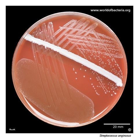
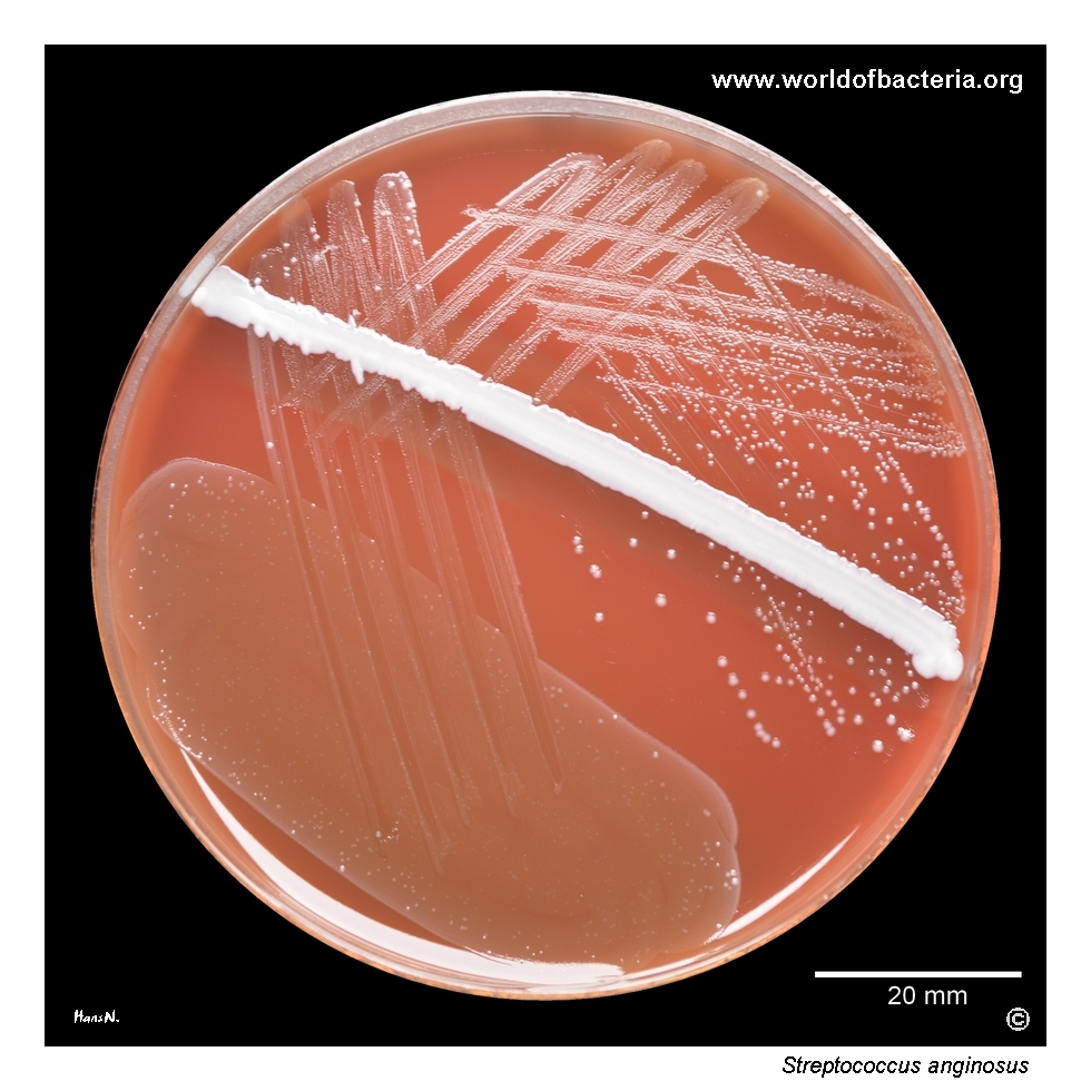
Streptococcus anginosus subcultured from enrichment broth onto blood agar with a staphylococcal streak (Staphylococcus aureus ATCC 25923). Culture conditions: 24 hours at 36 °C in 5% CO2. The specimen was pus from the gallbladder of a 72-year-old man treated in a surgical ward with the diagnosis K80.0, calculus of the gallbladder with acute cholecystitis. This type of purulent specimen fits well with the well-known tendency of S. anginosus group organisms to be associated with abscess formation.
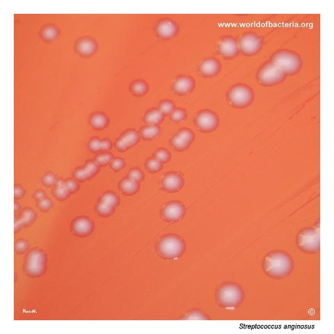
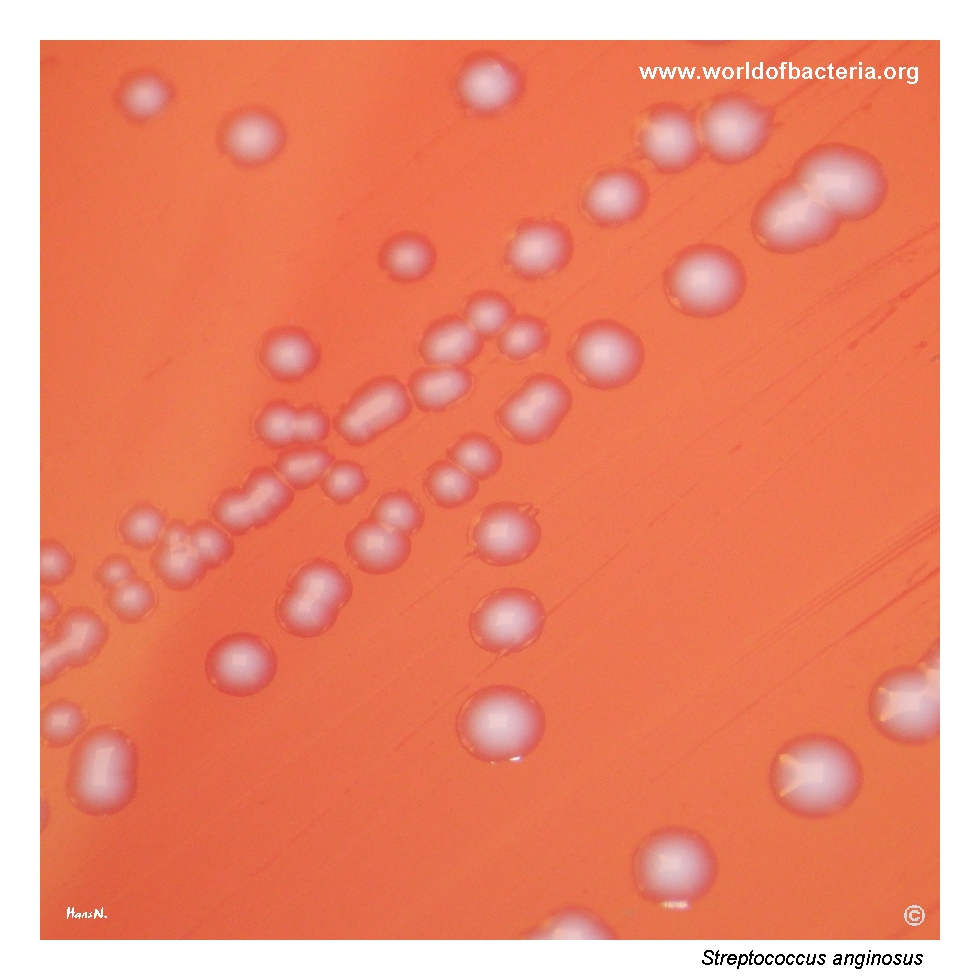
Colonies of Streptococcus anginosus on blood agar after 24 hours of incubation at 36 °C in 5% CO2. In this case, the colonies are small, convex, and non-haemolytic. However, isolates of S. anginosus may also show alpha-haemolysis or beta-haemolysis on blood agar. This variability is diagnostically important, because colony appearance alone does not reliably distinguish the species from other small streptococci encountered in routine clinical material.
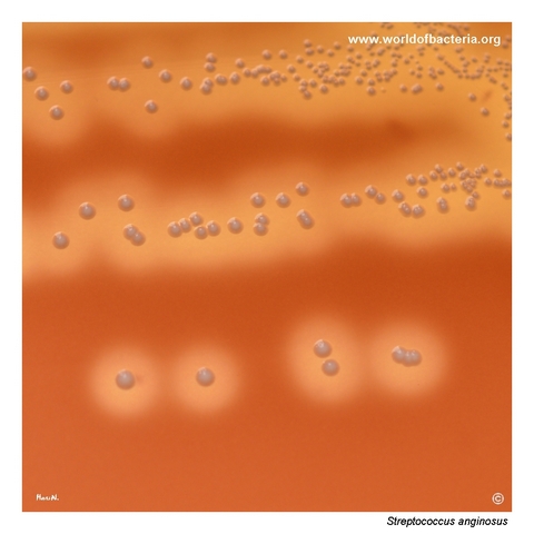
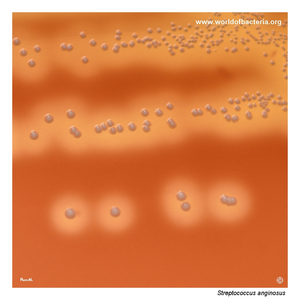
Close-up view of Streptococcus anginosus colonies on blood agar after 24 hours of incubation at 36 °C in 5% CO2. In this isolate, the organism produces beta-haemolysis around the colonies. The strain was recovered from the urogenital tract of a 33-year-old woman. This image is useful because it shows that S. anginosus may display a much more conspicuous haemolytic phenotype than the small non-haemolytic colonies often associated with the species.
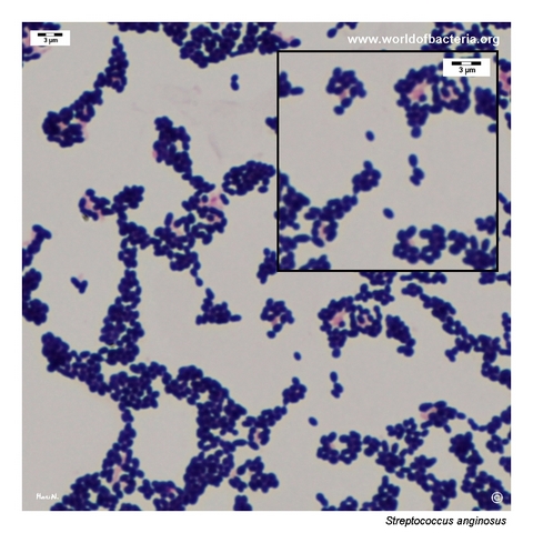
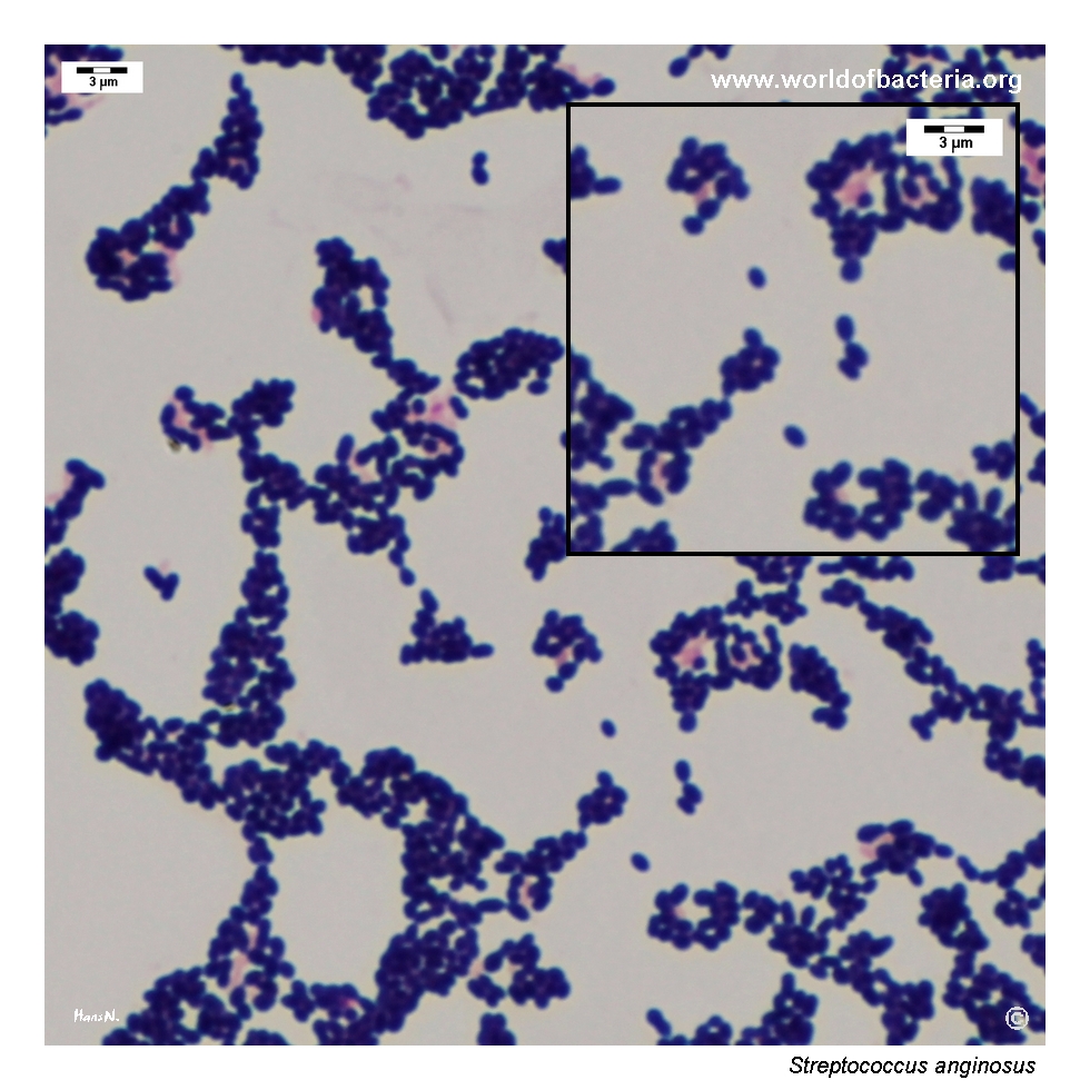
Gram-stained smear prepared from a colony of Streptococcus anginosus grown on blood agar. The organism appears as Gram-positive oval cocci occurring singly, in pairs, and in clusters. Although streptococci are more often associated with pairs and chains, members of the S. anginosus group may show somewhat variable microscopic arrangement. For that reason, Gram stain findings should always be interpreted together with colony morphology and the clinical source of the isolate.
Diagnostic and Clinical Notes
This page set is particularly useful because it demonstrates two visually different phenotypes of Streptococcus anginosus side by side: one isolate forming small non-haemolytic colonies and another producing clearly visible beta-haemolysis. This helps counter the common tendency to imagine the species as having only one routine plate appearance.
The gallbladder pus isolate fits well with the established clinical reputation of the anginosus group as organisms that frequently participate in purulent and abscess-associated infections. In such specimens, even relatively small colonies should not be dismissed too quickly as incidental flora.
The urogenital isolate adds a complementary teaching point: haemolysis can make S. anginosus look more conspicuous and potentially shift the initial differential diagnosis toward other streptococci. This is why haemolysis must be interpreted as a variable trait rather than as a definitive shortcut to identification.
The Gram-stained smear also illustrates that microscopic arrangement may be somewhat less stereotyped than expected for streptococci. Species identification should therefore rest on the whole picture: specimen type, culture morphology, and modern identification methods.
Laboratory Identification
Colony Morphology
On blood agar in 5% CO2, Streptococcus anginosus may present as small smooth convex colonies that remain non-haemolytic, but other isolates can produce alpha-haemolysis or even conspicuous beta-haemolysis. This page set deliberately shows both ends of that practical spectrum. The main teaching point is that the species cannot be reduced to a single visual stereotype on routine medium.
Microscopy
Gram staining shows small Gram-positive oval cocci, usually in pairs and short chains, but members of the anginosus group may also show less orderly clustering or irregular groupings in colony smears. The microscopic picture therefore supports the diagnosis of a streptococcal organism but is not sufficient to define the species on its own.
Key Identification Clues
Small streptococcal colonies with variable haemolysis on blood agar
Association with purulent or abscess-like clinical material
Possible recovery from intra-abdominal or urogenital specimens
Need to consider the broader anginosus group in the differential diagnosis
Requirement for reliable species-level confirmation by modern laboratory methods
Modern Identification Methods
Because haemolysis and colony size are variable and because overlap with other small streptococci is substantial, reliable identification is best achieved with modern laboratory methods such as MALDI-TOF MS supported by an adequate database. This is especially useful when the clinical specimen strongly suggests a clinically relevant anginosus-group organism but the plate phenotype remains ambiguous.
Antibiotic Characteristics
No simple therapeutic rule should be inferred from the photographed colonies alone. Although Streptococcus anginosus often remains broadly susceptible to standard anti-streptococcal therapy, treatment decisions must be based on the individual isolate and the clinical focus of infection.
For abscess-associated or deep purulent infections, the clinical management often depends not only on antimicrobial therapy but also on drainage of the infected focus. This is an important practical point whenever an anginosus-group organism is recovered from clearly suppurative material.
Note: In this page set, the major value of the images is diagnostic rather than therapeutic: they show how much the plate phenotype can vary within one clinically important species.