Streptococcus mitis is an alpha-haemolytic member of the mitis group and a common human commensal whose colonies may closely resemble pneumococci and other viridans streptococci on blood agar.
Basic Characteristics
Taxonomy
Domain: Bacteria
Phylum: Bacillota
Class: Bacilli
Order: Lactobacillales
Family: Streptococcaceae
Genus:Streptococcus
Species:Streptococcus mitis
Microscopy & Gram Stain
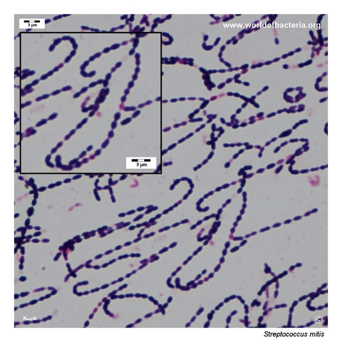
Gram-positive cocci that often appear as diplococci and short chains, with some cells showing a slightly lanceolate outline
In colony smears, the organism may look deceptively pneumococcus-like, especially when diplococci predominate
This page set demonstrates why morphology alone is not sufficient for species identification within the mitis group
The Gram stain remains valuable mainly for placing the isolate among alpha-haemolytic streptococci rather than for naming the species directly
Oxygen Relationship
Facultatively anaerobic streptococcus that grows readily on blood agar under capnophilic incubation
In this page set, all documented isolates formed visible colonies after 24 hours at 36 °C in 5% CO2
Routine blood agar therefore supports uncomplicated growth, even though the phenotype may overlap strongly with other viridans streptococci
Rapid Identification Tests
Catalase: negative
Haemolysis: typically alpha-haemolytic
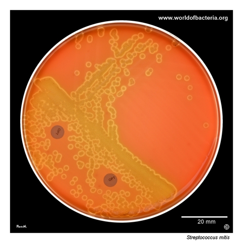
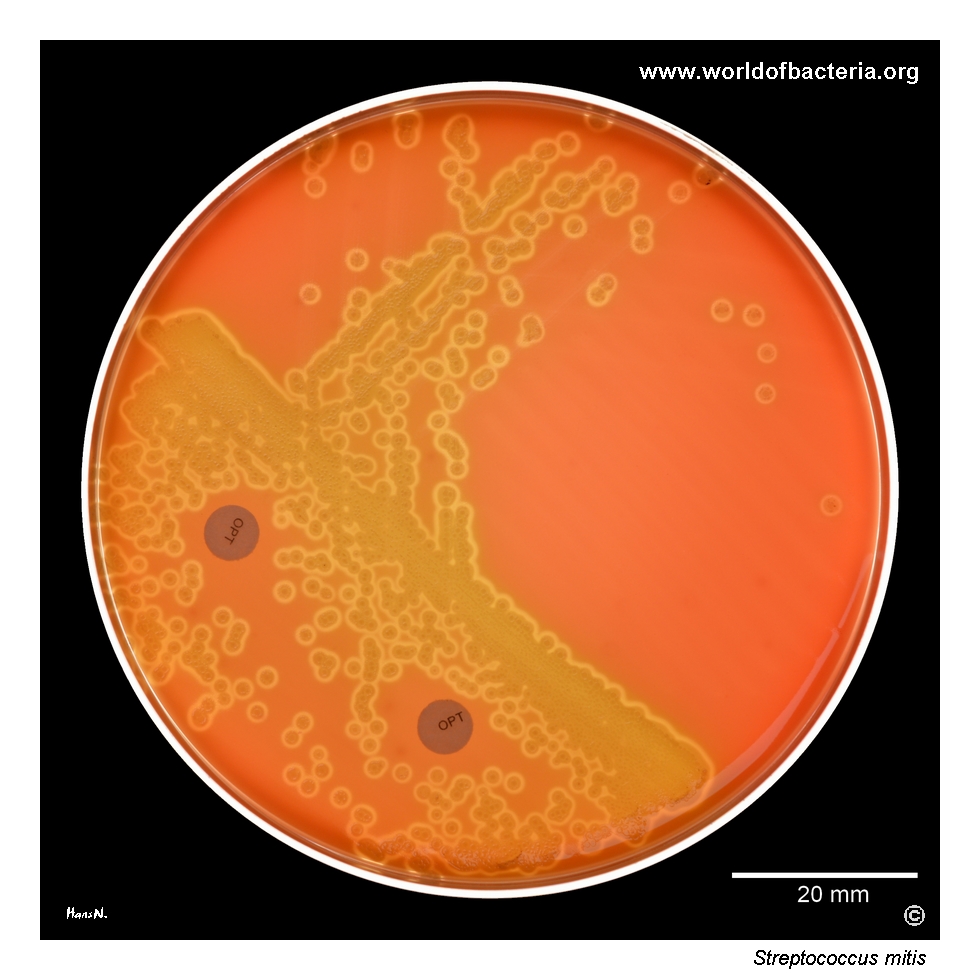
Optochin: usually resistant, which helps separate most isolates from Streptococcus pneumoniae
Bile solubility: typically negative
Key caution: members of the mitis group show major phenotypic overlap, so confirmatory identification is often needed
Ecology and Clinical Relevance
Natural Habitat
Streptococcus mitis is a common human commensal, especially in the oral cavity and upper respiratory tract
Its recovery from throat material therefore fits well with the expected ecology of the species
At the same time, similar mitis-group organisms may also appear in less typical sites, as illustrated here by an isolate from a urethral swab
Common Clinical Specimens
Throat and upper respiratory tract specimens
Oral and dental material
Blood cultures in invasive disease or endocarditis-related settings
Occasional urogenital or other mucosal specimens in which interpretation depends strongly on context
Clinical Significance
Streptococcus mitis is usually best known as a commensal of the mitis group, but it may also act as an opportunistic pathogen in susceptible patients
For routine diagnostic teaching, its greatest practical importance often lies in its close resemblance to other alpha-haemolytic streptococci, especially pneumococci and related mitis-group species
This makes the species highly relevant for differential diagnosis even when it is not the main pathogen in the specimen
Differential Considerations
Streptococcus pneumoniae, especially when the Gram stain shows lanceolate diplococci
Streptococcus pseudopneumoniae and other members of the mitis group
Other viridans streptococci with alpha-haemolytic colonies on blood agar
Mixed respiratory or mucosal cultures in which colony shape and haemolysis offer only limited discriminatory value
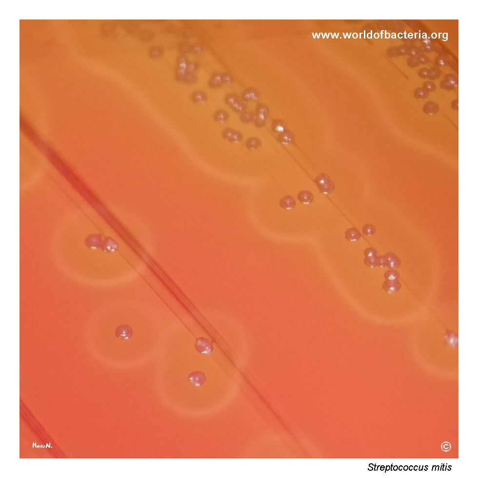
Streptococcus mitis isolate on blood agar after 24 hours of incubation at 36 °C in 5% CO2. Like most other alpha-haemolytic streptococci, strains of S. mitis are resistant to optochin, which helps distinguish them from most isolates of Streptococcus pneumoniae. This isolate originated from a urethral swab from a 62-year-old man. In routine diagnostics, this species is important mainly because of its close phenotypic resemblance to other members of the mitis group.
Colonies of Streptococcus mitis on blood agar. Some colonies are irregular in shape and often elongated along the direction of the inoculation streaks. The colonies are surrounded by a zone of alpha-haemolysis. This isolate originated from a throat swab and was cultured for 24 hours at 36 °C in 5% CO2. Such variable colony shape is common among viridans streptococci and may complicate visual differentiation from related alpha-haemolytic species.
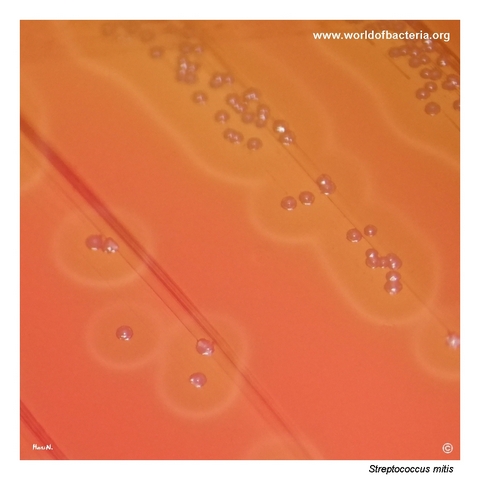
Close-up view of small colonies of Streptococcus mitis on blood agar with a broad zone of alpha-haemolysis surrounding the colonies. Colony morphology may vary considerably between different strains. In this example, the colonies are mostly small, round, and show a raised central area (umbonate). Culture conditions: 24 hours at 36 °C in 5% CO2.
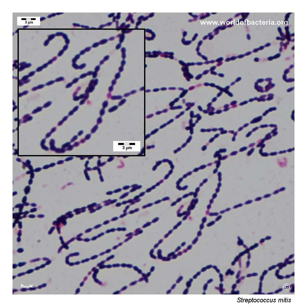
Gram-stained smear prepared from a colony of Streptococcus mitis grown on blood agar. The organism appears as Gram-positive diplococci, often with a lanceolate shape, arranged in chains. This microscopic appearance may resemble that of pneumococci, which is one of the reasons why members of the S. mitis group require careful interpretation in routine microbiology and should not be identified on morphology alone.
Diagnostic and Clinical Notes
This page set is didactically useful because it captures several reasons why Streptococcus mitis may be diagnostically frustrating: alpha-haemolysis, variable colony shape, occasional lanceolate diplococci, and strong overlap with other members of the mitis group.
The optochin-resistant blood agar isolate helps anchor one of the most practical routine distinctions from pneumococci. Even so, the page also shows why that distinction should not be reduced to one test result alone, because the overall appearance still remains highly pneumococcus-like.
The throat isolate demonstrates how irregular, elongated, or otherwise non-uniform colony shapes may appear within ordinary alpha-haemolytic growth. This variability is common enough to matter during plate reading but not specific enough to solve the identification problem by itself.
The Gram stain reinforces the central teaching point: members of the mitis group can look deceptively familiar. Species identification therefore depends on combining microscopy, haemolysis, screening tests such as optochin, and reliable modern identification methods.
Laboratory Identification
Colony Morphology
On blood agar, Streptococcus mitis typically forms small alpha-haemolytic colonies, but colony shape may vary considerably between isolates. Some colonies remain round and discrete, whereas others become irregular or elongated along the inoculation streak. In this page set, one isolate also shows a clearly raised central area. These features are useful descriptively, yet they overlap so extensively with other viridans streptococci that they cannot securely define the species.
Microscopy
Gram staining shows Gram-positive cocci, often as diplococci with a slightly lanceolate appearance, together with short chains. This is one reason why S. mitis can so easily be confused with pneumococci. The microscopic picture remains helpful for placing the isolate within the correct morphological group, but species-level interpretation requires correlation with culture phenotype and additional testing.
Key Identification Clues
Alpha-haemolytic growth on blood agar
Optochin resistance in routine testing
Considerable overlap with pneumococci and other mitis-group streptococci
Variable colony outline, including irregular or elongated colonies
Need for reliable species confirmation when the exact identification is clinically important
Modern Identification Methods
Because the mitis group contains several very closely related species, modern identification is often required for confident species assignment. MALDI-TOF MS can be very helpful, but interpretation still depends on database quality and local validation. When the distinction from pneumococcus or from another mitis-group species is clinically relevant, the result should be interpreted in the context of screening tests and the full laboratory picture.
Antibiotic Characteristics
No treatment recommendation should be inferred from the photographed colonies alone. Although Streptococcus mitis is often encountered primarily as a commensal or low-grade opportunist, clinically significant isolates require individual susceptibility testing and interpretation in context.
For this page, the main laboratory teaching point is not therapy but differential diagnosis: an alpha-haemolytic optochin-resistant isolate with a pneumococcus-like microscopic appearance may still belong to the mitis group rather than to Streptococcus pneumoniae.
Note: The educational value of this page lies mainly in recognizing a common diagnostic look-alike among alpha-haemolytic streptococci.