Streptococcus pneumoniae is a major human respiratory pathogen and a classical alpha-haemolytic streptococcus whose mature colonies often develop a centrally depressed appearance on blood agar; in this page set it is illustrated by the quality-control strain ATCC 49619 used in antimicrobial susceptibility testing.
Basic Characteristics
Taxonomy
Domain: Bacteria
Phylum: Bacillota
Class: Bacilli
Order: Lactobacillales
Family: Streptococcaceae
Genus:Streptococcus
Species:Streptococcus pneumoniae
Microscopy & Gram Stain
Gram-positive cocci that often appear as diplococci with a lanceolate or slightly flame-like outline
Pairs predominate, but short chains and more rounded forms may also be seen depending on the isolate and the age of the culture
In direct respiratory specimens, the classic diplococcal appearance may be striking, yet colony morphology and additional tests remain essential for secure identification
This page set illustrates the typical pneumococcal combination of alpha-haemolytic growth and Gram-positive diplococci rather than an unusual phenotypic variant
Oxygen Relationship
Facultatively anaerobic streptococcus that grows well under routine diagnostic conditions
Growth is usually enhanced in an atmosphere enriched with carbon dioxide, which is why pneumococci are commonly incubated on blood agar in 5% CO2
With prolonged incubation, older colonies undergo autolytic change and develop the classical centrally depressed appearance seen in mature pneumococcal cultures
Rapid Identification Tests
Catalase: negative
Haemolysis: alpha-haemolytic on blood agar
Optochin: usually susceptible
Bile solubility: typically positive
Important limitation: alpha-haemolysis alone does not distinguish pneumococci from all other viridans-group streptococci
Ecology and Clinical Relevance
Natural Habitat
Streptococcus pneumoniae colonizes the human nasopharynx and is transmitted between people, especially in close-contact settings
Carriage may be asymptomatic, but the same organism is also capable of causing major invasive and noninvasive disease
The strain shown on this page is ATCC 49619, a quality-control strain widely used in susceptibility testing rather than a current patient isolate from your laboratory
Common Clinical Specimens
Lower respiratory tract specimens such as sputum
Blood cultures and other normally sterile fluids in invasive disease
Cerebrospinal fluid in meningitis
Middle-ear and sinus material in localized infection
Clinical Significance
Streptococcus pneumoniae is one of the most important human bacterial pathogens and a major cause of pneumonia, otitis media, sinusitis, meningitis, and bacteraemia
In this page set, the organism is shown as a susceptibility-testing quality-control strain, which makes the images especially useful for teaching classical laboratory morphology
Because pneumococci share alpha-haemolysis with other streptococci, clinically relevant identification depends on combining plate appearance with confirmatory testing
Differential Considerations
Other alpha-haemolytic streptococci, especially members of the Streptococcus mitis group
Streptococcus pseudopneumoniae, which may resemble pneumococci but differs in key identification behaviour
Older pneumococcal colonies versus younger alpha-haemolytic streptococcal colonies that lack the same degree of central depression
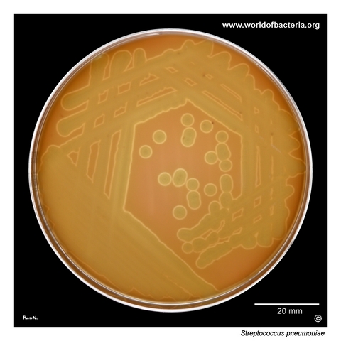
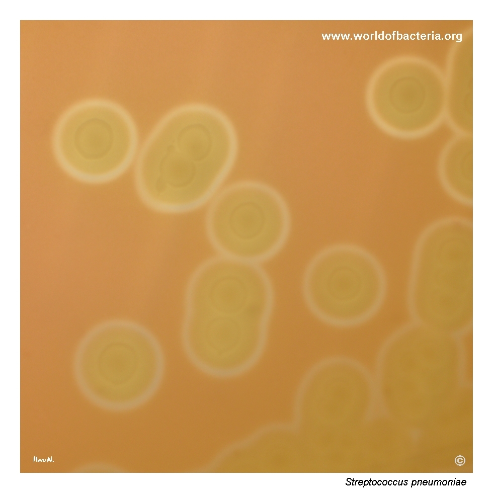
Streptococcus pneumoniae on blood agar. Strain ATCC 49619, incubated for 48 hours at 36 °C in 5% CO2. In diagnostic laboratories, this strain is widely used for quality control of antibiotic disks in susceptibility testing performed by the disk diffusion method. It was originally isolated from sputum from a 75-year-old patient in Phoenix, Arizona, USA. Quality-control strains such as this are essential for verifying the reliability of routine antimicrobial susceptibility testing.
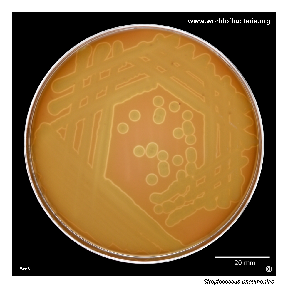
Colonies of Streptococcus pneumoniae ATCC 49619 on blood agar after 48 hours of incubation at 36 °C in 5% CO2. A broad zone of alpha-haemolysis surrounds the colonies. This greenish discolouration reflects partial alteration of erythrocytes in the agar and is one of the classic visual features associated with pneumococcal growth on blood agar, although other alpha-haemolytic streptococci may show a similar effect.
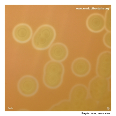
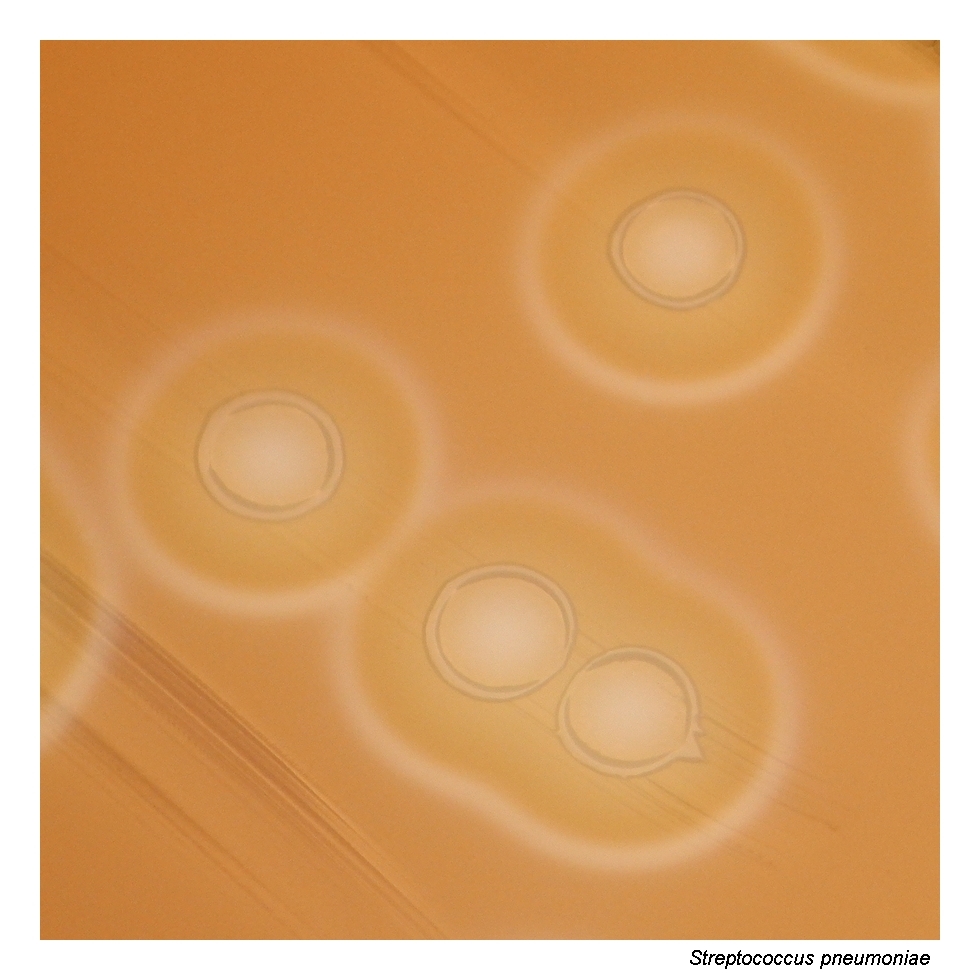
Close-up view of Streptococcus pneumoniae ATCC 49619 colonies on blood agar. The colonies are alpha-haemolytic and show the typical centrally depressed, dish-like appearance that becomes especially evident with further incubation. This umbilicated morphology is a classic feature of pneumococci and is related to autolytic changes in older colonies, making mature growth visually different from many other alpha-haemolytic streptococci.
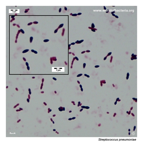
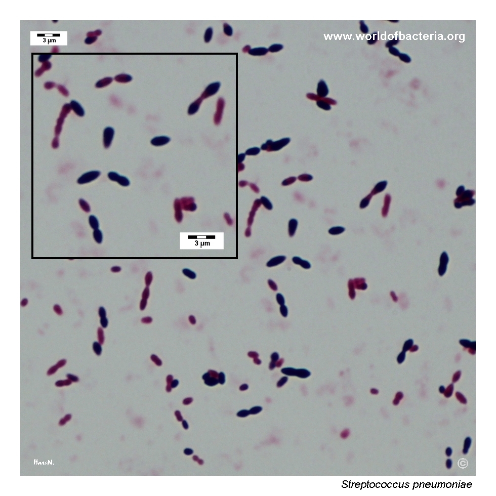
Gram-stained smear prepared from a colony of Streptococcus pneumoniae grown on blood agar. The Gram-positive cells often show a lanceolate, flame-like shape and commonly occur in pairs as diplococci. A similar appearance may sometimes be seen directly in lower respiratory tract specimens. In many isolates, however, the cells are more nearly spherical or only slightly elongated along the axis of division, so morphology alone should be interpreted with caution.
Diagnostic and Clinical Notes
This page is didactically useful because it presents the classical appearance of pneumococcus in a strain that many clinical laboratories know from susceptibility-testing quality control rather than from diagnostic recovery. Streptococcus pneumoniae ATCC 49619 is therefore not only a teaching strain but also a practical laboratory reference point.
The two most visually important culture features shown here are alpha-haemolysis and the centrally depressed, dish-like morphology of older colonies. Together, these findings create the familiar pneumococcal plate appearance, but neither feature is absolutely exclusive to the species and both must be interpreted in context.
The colony close-up nicely demonstrates the effect of autolysis in mature growth. This is one of the classical reasons why pneumococcal colonies often look flatter or umbilicated after further incubation than they do at an earlier reading time.
The Gram-stained smear adds the second major teaching point: the common pneumococcal impression of Gram-positive diplococci with a lanceolate outline. Even so, species identification should not rely on morphology alone, because alpha-haemolytic streptococci can overlap in both microscopic and colonial appearance.
Laboratory Identification
Colony Morphology
On blood agar incubated in 5% CO2, Streptococcus pneumoniae typically forms alpha-haemolytic colonies that become more distinctive with time. In this page set, the most characteristic finding is the mature centrally depressed or umbilicated appearance of the colonies, a classical pneumococcal feature linked to autolytic change. The broad greenish alpha-haemolytic zone is also visually prominent, but it must be interpreted cautiously because other alpha-haemolytic streptococci can produce a similar effect.
Microscopy
Gram staining shows Gram-positive cocci, often in pairs, with the familiar lanceolate outline of pneumococci. Depending on the preparation and the age of the culture, however, the cells may appear less elongate and more nearly spherical. The microscopic pattern is therefore strongly supportive but not sufficient on its own; it should always be correlated with colony morphology and confirmatory laboratory testing.
Key Identification Clues
Alpha-haemolytic growth on blood agar
Centrally depressed or umbilicated mature colonies
Gram-positive diplococci with a lanceolate tendency
Typical growth under capnophilic incubation conditions
Confirmatory differentiation from other alpha-haemolytic streptococci by optochin, bile solubility, or reliable modern identification methods
Modern Identification Methods
In modern routine practice, MALDI-TOF MS usually provides reliable species-level identification of pneumococci when the database is appropriately curated. Classical methods such as optochin susceptibility and bile solubility remain useful because they also reinforce the teaching distinction between S. pneumoniae and related alpha-haemolytic streptococci. When needed, molecular methods can further clarify difficult isolates within the mitis group.
Antibiotic Characteristics
This page is especially relevant to antimicrobial susceptibility testing because the illustrated strain, ATCC 49619, is widely used as a quality-control organism in disk diffusion and MIC procedures. Its practical role is to verify that the testing system is performing correctly, not to represent the susceptibility of a current clinical isolate.
For patient isolates of Streptococcus pneumoniae, susceptibility should always be determined individually. Resistance, particularly to beta-lactams, is commonly linked to altered penicillin-binding proteins rather than to a single simple colony-based clue that can be inferred from culture appearance.
The broader teaching point is that susceptibility interpretation in pneumococci is method-dependent and clinically important. A laboratory image of a classical alpha-haemolytic colony therefore connects directly to the larger discipline of standardized susceptibility testing and quality assurance.
Note: The strain shown here is valuable not only because it illustrates typical pneumococcal morphology, but also because it represents a standard reference organism in routine AST quality control.