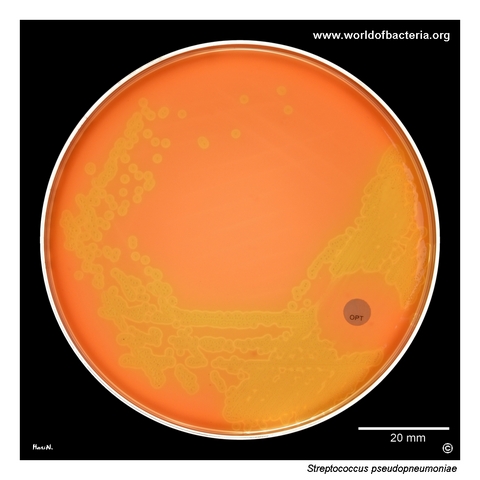
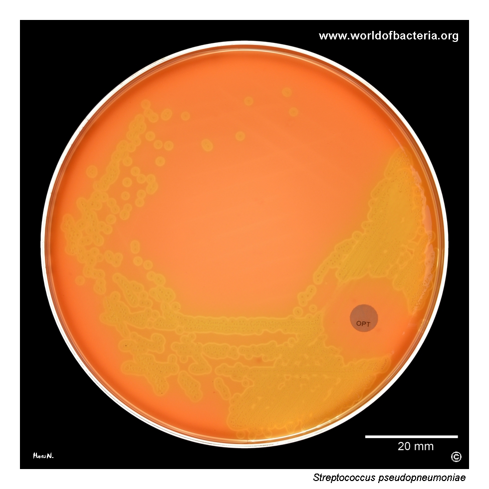
Streptococcus pseudopneumoniae is a member of the mitis group that may closely resemble pneumococci on blood agar, yet in this isolate the organism remained flatter than S. pneumoniae and showed a measurable optochin inhibition zone despite incubation in 5% CO2.
Basic Characteristics
Taxonomy
Domain: Bacteria
Phylum: Bacillota
Class: Bacilli
Order: Lactobacillales
Family: Streptococcaceae
Genus:Streptococcus
Species:Streptococcus pseudopneumoniae
Microscopy & Gram Stain
Gram-positive cocci that commonly occur in pairs and short chains and may resemble pneumococci on first inspection
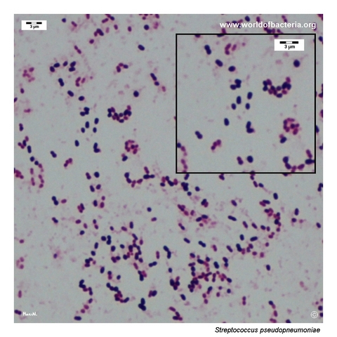
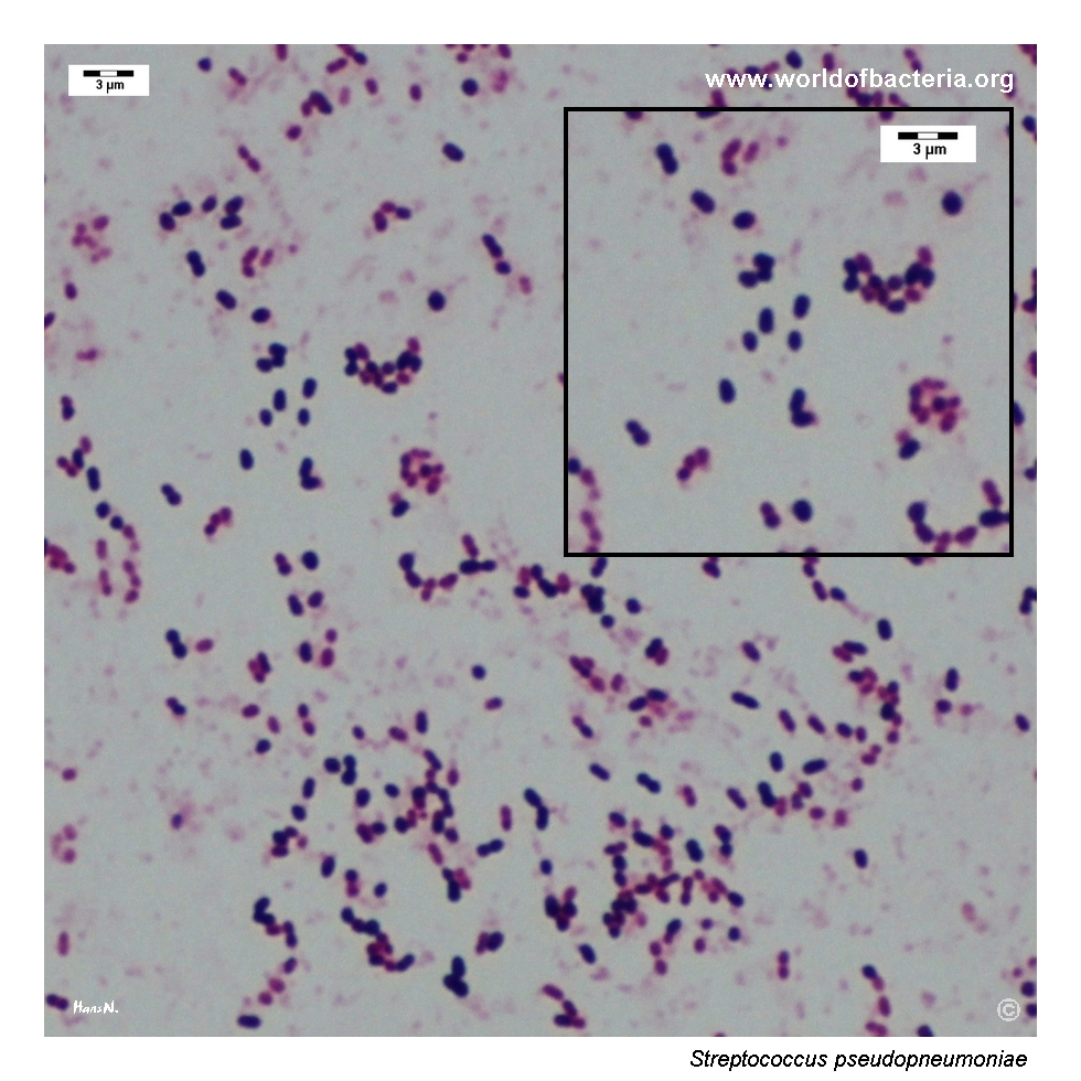
In this isolate, a colony smear prepared after about 20 hours showed partial Gram variability, with some cells staining more weakly than others
The overall morphology still fits the streptococcal group well, but microscopy alone is not sufficient for secure species identification
As with other members of the mitis group, colony appearance, haemolysis, and confirmatory identification methods remain essential
Oxygen Relationship
Facultatively anaerobic alpha-haemolytic streptococcus that grows readily under routine diagnostic conditions
Blood agar incubation in 5% CO2 supports visible growth and is entirely compatible with routine laboratory handling of respiratory isolates
In this page set, the capnophilic culture conditions are important because the isolate still showed a measurable optochin inhibition zone under those conditions
Rapid Identification Tests
Catalase: negative
Haemolysis: alpha-haemolytic on blood agar
Optochin: measurable inhibition zone in 5% CO2 in this isolate (17.7 mm on the photographed plate)
Key caution: this behaviour may invite confusion with Streptococcus pneumoniae, so identification should not rely on optochin alone
Practical implication: atypical phenotypes can occur and should be interpreted in the context of a pure culture and a complete identification work-up
Ecology and Clinical Relevance
Natural Habitat
Streptococcus pseudopneumoniae belongs to the mitis group and is mainly associated with the human respiratory tract
It is encountered particularly in respiratory material, where it may be recovered from patients with chronic lung disease or hospital-associated respiratory problems
In this page set, the organism was recovered in pure culture from an endotracheal aspirate from an intensive care unit patient with dyspnoea
Common Clinical Specimens
Lower respiratory tract material, including sputum and endotracheal aspirates
Respiratory specimens from hospitalized or chronically ill patients
Specimens in which differentiation from pneumococci and other alpha-haemolytic streptococci is diagnostically important
Clinical Significance
The clinical role of S. pseudopneumoniae is less clearly defined than that of pneumococci, but the species is regularly discussed in respiratory microbiology because of its close phenotypic resemblance to S. pneumoniae
In a pure culture from lower respiratory tract material, the isolate deserves more attention than an incidental alpha-haemolytic streptococcus in mixed oral flora
This page is especially instructive because it shows an isolate with a measurable optochin inhibition zone in 5% CO2, demonstrating that phenotypic overlap with pneumococci can be even greater than expected
Differential Considerations
Streptococcus pneumoniae, especially because of alpha-haemolysis and optochin inhibition
Other alpha-haemolytic streptococci of the mitis group
Flatter non-umbilicated colonies of S. pseudopneumoniae versus more classically umbilicated mature pneumococcal colonies
Atypical optochin behaviour that can blur the usual practical distinction between pseudopneumococci and true pneumococci
Streptococcus pseudopneumoniae in pure culture on blood agar after 24 hours of incubation at 36 °C in 5% CO2. The isolate was recovered from an endotracheal aspirate from a 78-year-old man hospitalized in the intensive care unit with the diagnosis R06.0, dyspnoea. The culture shows alpha-haemolytic streptococci with a measurable zone of growth inhibition around the optochin disk. In this case, the inhibition zone measured 17.7 mm despite incubation in 5% CO2, illustrating how closely this species may resemble pneumococci during routine culture reading.
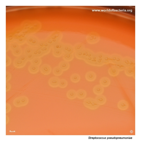
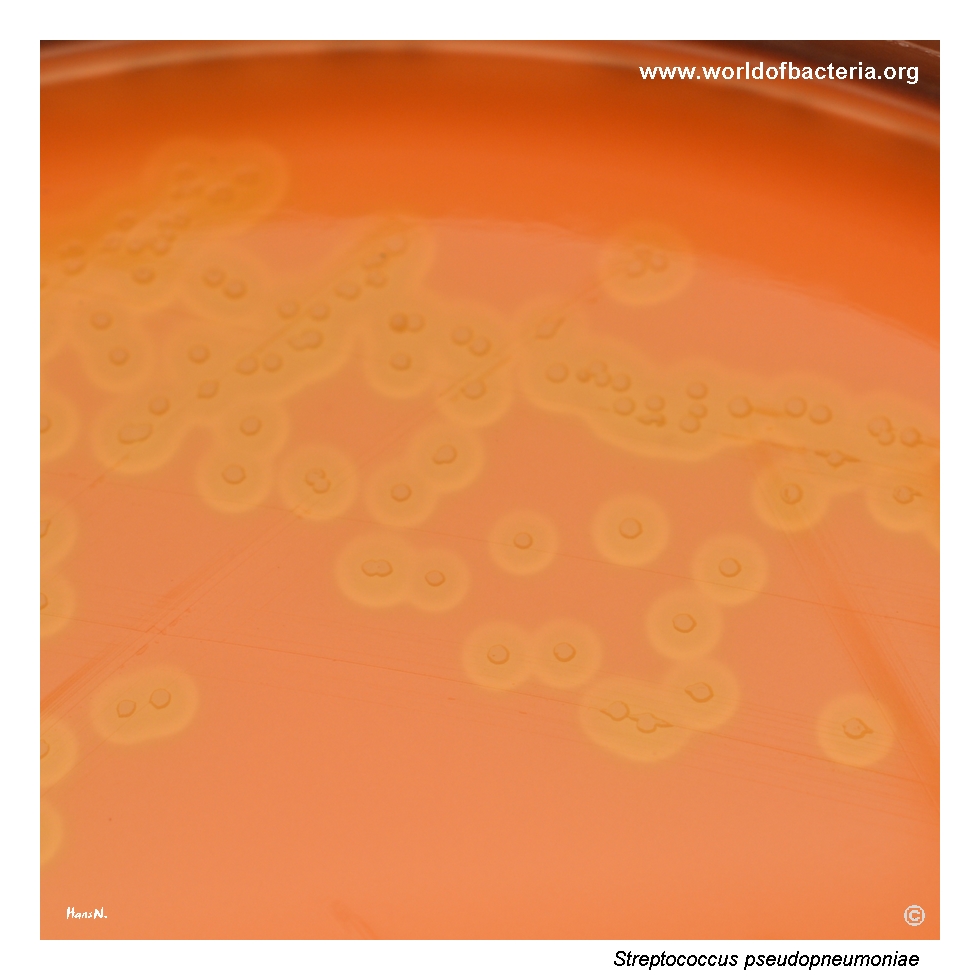
Colonies of Streptococcus pseudopneumoniae on blood agar resemble those of other alpha-haemolytic streptococci and may also suggest Streptococcus pneumoniae. A broad zone of alpha-haemolysis is visible around the individual colonies. Culture conditions: 24 hours at 36 °C in 5% CO2. This close visual similarity is diagnostically important, because species-level identification cannot be based on colony appearance alone.
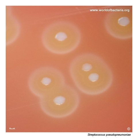
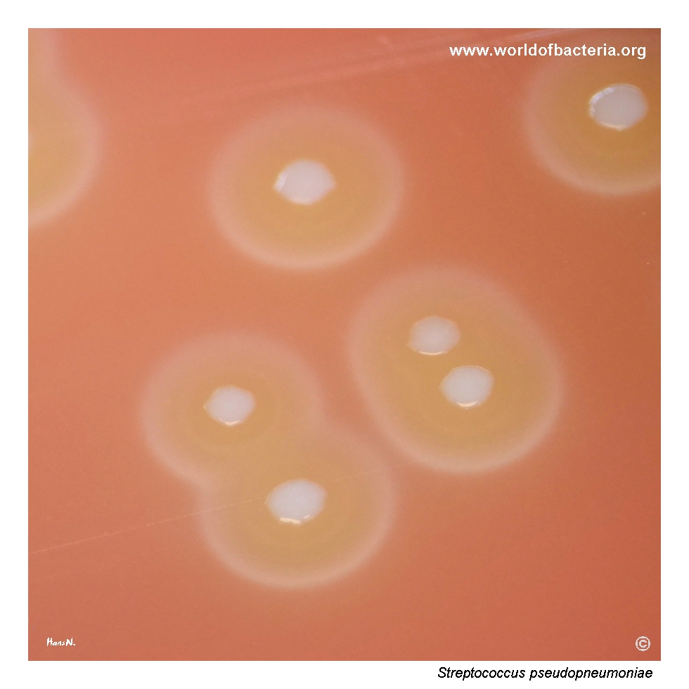
Close-up view of Streptococcus pseudopneumoniae colonies on blood agar with a conspicuous ring of alpha-haemolysis surrounding the growing colonies. Culture conditions: 48 hours at 36 °C in 5% CO2. In this isolate, the colonies remain flattened and do not show the central depression often seen in pneumococcal colonies after further incubation. This subtle difference can be useful during careful plate examination, although it is not fully reliable on its own.
Gram-stained smear prepared from a colony of Streptococcus pseudopneumoniae grown on blood agar. Cultures stained after about 20 hours of incubation may show partial Gram variability because of incomplete maturation of the cell wall. The organism appears as Gram-positive cocci and diplococci, although some cells may stain Gram-negative in places. This mixed appearance can complicate routine interpretation if the smear is evaluated without correlation to the culture plate.
Diagnostic and Clinical Notes
This page set is especially instructive because it documents a pure culture of Streptococcus pseudopneumoniae from endotracheal aspirate that visually approaches pneumococcus at several levels at once: alpha-haemolytic growth, respiratory origin, and measurable optochin inhibition on the photographed plate.
The most important interpretive point is that the optochin finding should be described exactly as observed. Here, the inhibition zone measured 17.7 mm after incubation in 5% CO2, so it is more accurate to present the isolate as optochin-susceptible under these conditions than to force the description toward a more typical textbook phenotype.
At the same time, the colony close-up still shows a useful difference from many pneumococci: the colonies remain relatively flat and do not develop the same degree of central depression or dish-like morphology that is often seen in older pneumococcal growth.
The Gram-stained smear adds another helpful teaching point. Partial Gram variability in younger cultures can complicate interpretation, especially if the observer is expecting a uniform Gram-positive diplococcal picture. For this reason, microscopy, colony morphology, and final identification should be interpreted together rather than in isolation.
Laboratory Identification
Colony Morphology
On blood agar in 5% CO2, this isolate formed alpha-haemolytic colonies that closely resembled other mitis-group streptococci and could easily suggest pneumococcus during routine reading. After further incubation, however, the colonies remained relatively flat rather than becoming classically umbilicated. This difference is subtle but didactically useful, especially when the plate also shows an optochin inhibition zone that might otherwise push the observer toward a pneumococcal interpretation.
Microscopy
Gram staining showed cocci and diplococci with partial Gram variability in younger culture. Although the general impression still fits an alpha-haemolytic streptococcus of the mitis group, the slide also illustrates how easily microscopic interpretation can become less straightforward when staining is performed before the culture has fully matured. Species identification therefore depends on combining microscopy with the culture plate and instrumental identification.
Key Identification Clues
Alpha-haemolytic respiratory isolate belonging to the mitis-group streptococci
Pure culture from endotracheal aspirate rather than mixed oral flora
Measurable optochin inhibition zone of 17.7 mm despite incubation in 5% CO2
Flatter colonies without the classical mature pneumococcal central depression
Need for species-level confirmation by reliable modern laboratory identification methods
Modern Identification Methods
Because phenotypic overlap with Streptococcus pneumoniae and other mitis-group streptococci may be substantial, reliable species identification is best secured by modern laboratory methods such as MALDI-TOF MS with an appropriate database and, when necessary, molecular clarification. This case illustrates why instrumental identification remains important even when classical plate reading seems to point in a familiar direction.
Antibiotic Characteristics
No simple therapeutic rule should be inferred from the photographed plate alone. Although the isolate showed a measurable optochin inhibition zone in 5% CO2, optochin is used here as an identification aid rather than as a guide to antimicrobial treatment.
For clinically relevant isolates of Streptococcus pseudopneumoniae, susceptibility testing should be performed directly on the individual strain. This is especially important because respiratory mitis-group streptococci may show variable susceptibility patterns and because phenotypic resemblance to pneumococci does not guarantee the same resistance profile.
Note: In this page set, the optochin finding is valuable mainly as a teaching point about atypical identification behaviour and the limits of rule-based presumptive identification.