Staphylococcus simulans is a coagulase-negative staphylococcal species that may be associated with animals but is also occasionally recovered from human clinical material, where visibly haemolytic isolates can resemble more familiar pathogenic staphylococci on blood agar.
Basic Characteristics
Taxonomy
Domain: Bacteria
Phylum: Bacillota (Firmicutes)
Class: Bacilli
Order: Bacillales
Family: Staphylococcaceae
Genus:Staphylococcus
Species:Staphylococcus simulans
Microscopy & Gram Stain
Gram-positive cocci
Cells occur singly, in pairs, and in irregular clusters
Microscopic morphology is typical of staphylococci but not species-specific
Oxygen Relationship
Facultatively anaerobic
Grows readily under routine aerobic blood agar conditions
Not regarded as a fastidious species in routine laboratory work
Rapid Identification Tests
Catalase: positive
Coagulase: negative
Colony pigmentation: usually absent or weak
Haemolysis on blood agar: variable, often weak or absent but occasionally conspicuous
Ecology and Clinical Relevance
Natural Habitat
Originally described from human skin
Also recognized as an animal-associated staphylococcal species, especially in veterinary microbiology
Human recovery appears less common than for major skin-colonizing coagulase-negative staphylococci
Common Clinical Specimens
Wound swabs and skin or soft tissue material
Occasional urine specimens
Blood culture and device-associated material in uncommon cases
Veterinary specimens, particularly those linked to livestock
Clinical Significance
Uncommon opportunistic pathogen in humans
Better known as an animal-associated species, including in bovine mastitis
May cause skin and soft tissue infection, urinary infection, or other invasive disease in selected cases
Can be overlooked or underestimated when isolated together with more familiar skin flora
Differential Considerations
Staphylococcus epidermidis, because both may occur in mixed skin-associated cultures
Staphylococcus haemolyticus, because both are coagulase-negative and may show haemolysis
Staphylococcus aureus, when haemolysis on blood agar is prominent
Species-level identification should not rely on morphology alone
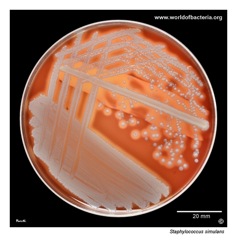
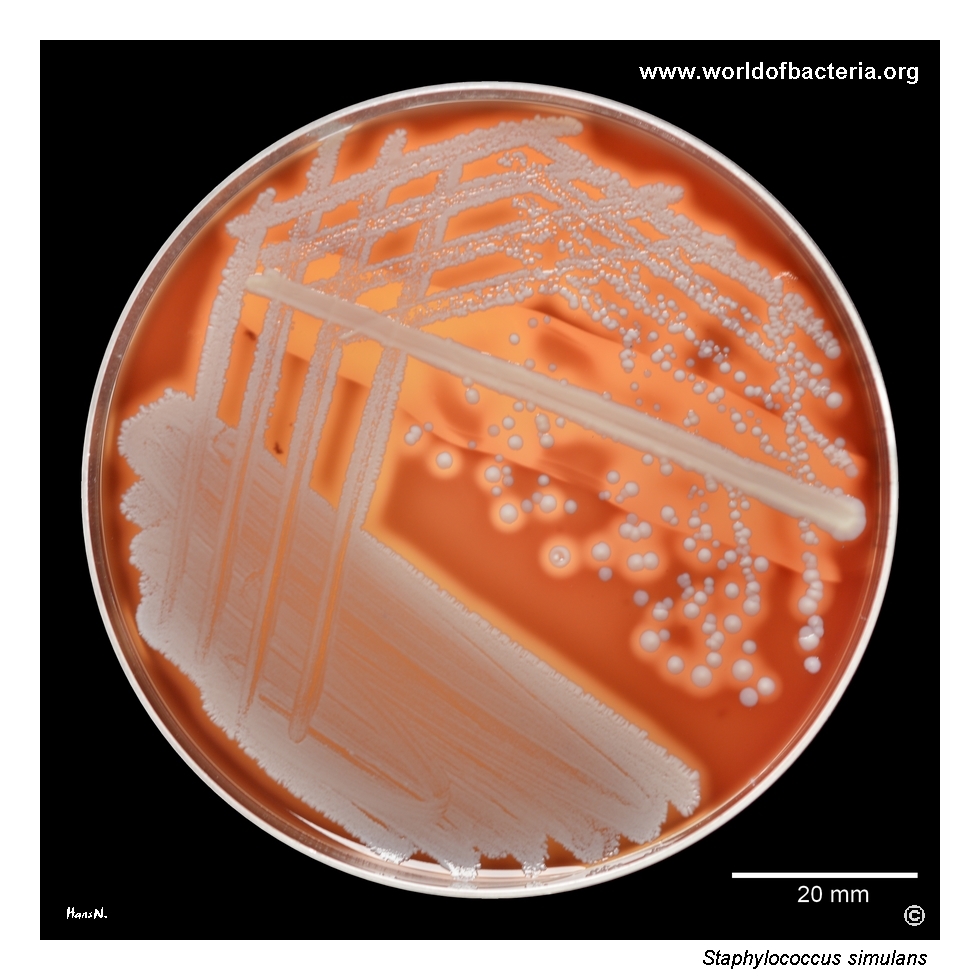
Subculture from enrichment broth after 24 hours of incubation. The isolate originated from a wound on the head of a 40-year-old man treated in a surgical intensive care unit. On blood agar, colonies of Staphylococcus simulans with marked haemolysis are visible together with non-haemolytic colonies of Staphylococcus epidermidis. A streak of Staphylococcus aureus ATCC 25923 was also inoculated on the plate. In the primary culture, S. simulans was present only sporadically.
Colonies of Staphylococcus simulans on blood agar after 24 hours at 36 °C in ambient air. This species more often shows weak haemolysis, or none at all, and the effect may become clearer only after prolonged incubation. In this isolate, however, haemolysis is already striking after 24 hours. The deformed colony in the lower part of the image reflects sampling for MALDI-TOF MS identification, which yielded a score of 2.54, supporting highly reliable species identification.
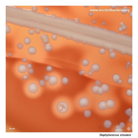
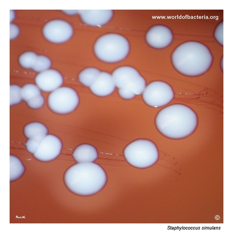
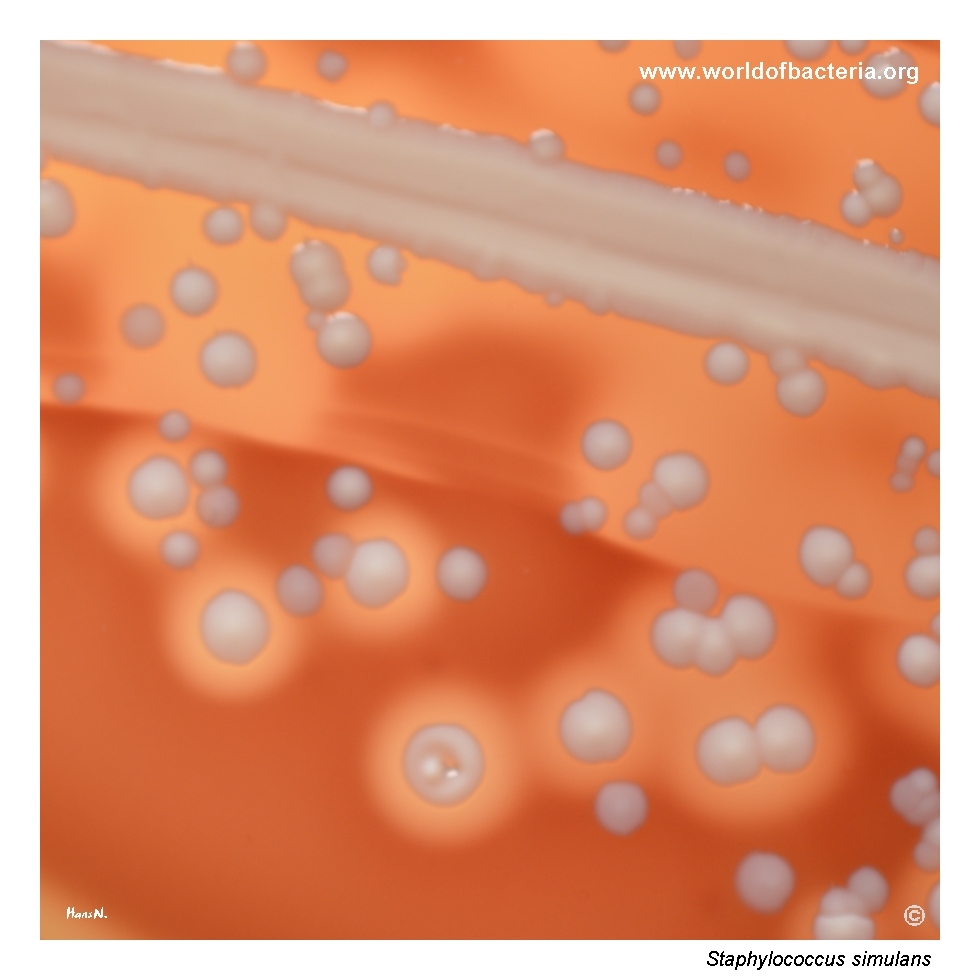
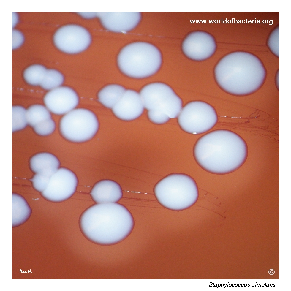
Close-up view of Staphylococcus simulans colonies after 24 hours of incubation at 36 °C in ambient air. The colonies are smooth, convex, non-pigmented, and round with entire margins, surrounded by a clear zone of beta-haemolysis. This is visually notable because S. simulans more often produces only weak haemolysis or may even appear non-haemolytic on blood agar, especially when compared with more strongly haemolytic staphylococci.
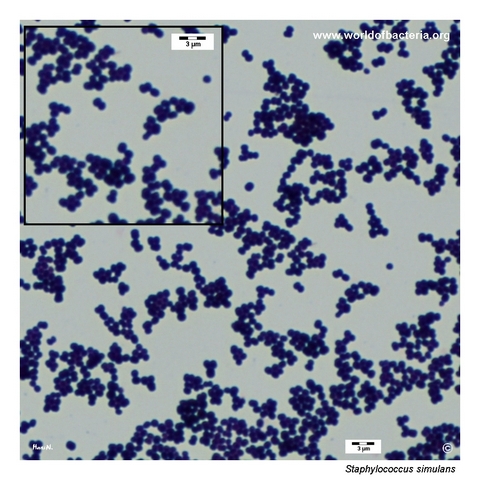
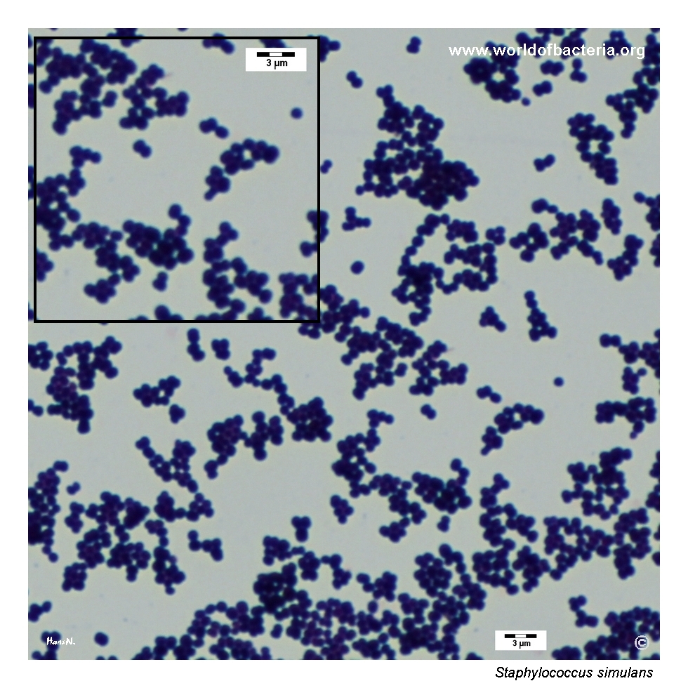
Gram-stained preparation of Staphylococcus simulans from a culture grown on blood agar. The organism appears as Gram-positive cocci occurring singly, in pairs, and in clusters. As in other staphylococci, this microscopic appearance is characteristic of the genus but not specific for the species. Final identification therefore depends on combining microscopic findings with colony morphology and modern laboratory identification methods.
Diagnostic and Clinical Notes
Staphylococcus simulans is an instructive species because the organism is usually discussed as an uncommon human pathogen, yet in some isolates the plate phenotype is unexpectedly striking. In this case, the isolate showed clear haemolysis already after 24 hours, which can easily draw attention toward more familiar haemolytic staphylococci.
The specimen context is also important. The isolate came from a wound in a patient treated in a surgical intensive care unit and became more evident only after subculture from enrichment broth. In the primary culture it was present only sporadically, reminding us that potentially relevant organisms may be underrepresented at first reading.
This culture is visually mixed, with haemolytic colonies of S. simulans present together with non-haemolytic colonies of Staphylococcus epidermidis. Such combinations are diagnostically useful because they highlight how strongly haemolysis can influence the visual impression of a mixed plate.
Although S. simulans is best known from veterinary microbiology, especially in animal-associated infections, it should not be dismissed automatically when recovered from human clinical material. Human infections are uncommon but well documented, including skin and soft tissue infection and occasional invasive disease.
The MALDI-TOF MS score of 2.54 is a particularly strong point in this case. For less familiar coagulase-negative staphylococci, reliable modern identification is essential because morphology alone does not securely separate them from better known species.
Laboratory Identification
Colony Morphology
On blood agar, Staphylococcus simulans forms smooth, convex, round colonies with entire margins. The colonies in this page set are non-pigmented but surrounded by a conspicuous zone of beta-haemolysis. This is diagnostically noteworthy because many isolates of S. simulans show only weak haemolysis or none at all, and stronger haemolysis may therefore create a misleading first impression.
Microscopy
Gram staining shows Gram-positive cocci occurring singly, in pairs, and in clusters. This microscopic pattern is entirely compatible with the genus Staphylococcus but does not allow reliable distinction of S. simulans from other coagulase-negative or even more familiar haemolytic staphylococci.
Key Identification Clues
Gram-positive cocci in clusters
Catalase positive
Coagulase negative
Usually non-pigmented colonies
Haemolysis may be weak, absent, or occasionally pronounced
May occur in mixed cultures and be overlooked in primary material
MALDI-TOF MS is highly useful for reliable species-level identification
Modern Identification Methods
Species-level identification is best supported by MALDI-TOF mass spectrometry with an up-to-date database, or by molecular methods if needed. In this isolate, MALDI-TOF MS yielded a score of 2.54, supporting highly reliable identification. This is especially valuable for S. simulans because phenotypic overlap with other coagulase-negative staphylococci can be substantial.
Antibiotic Characteristics
Staphylococcus simulans should not be assigned a susceptibility pattern on the basis of species name or colony appearance alone. As with other coagulase-negative staphylococci, antimicrobial susceptibility may vary from isolate to isolate.
When the species is recovered from human clinical material, susceptibility testing should be performed on the individual isolate and interpreted together with the specimen type, the amount of growth, and the broader clinical context.
In mixed cultures, therapeutic interpretation is especially dependent on deciding whether S. simulans is acting as a true pathogen, a copathogen, or a less significant accompanying organism.
Note: Antimicrobial therapy should always be guided by laboratory susceptibility testing, specimen context, and overall clinical evaluation.