Staphylococcus pseudintermedius is a Gram-positive, coagulase-positive staphylococcal species associated primarily with dogs and other companion animals, but occasionally recovered from human clinical material as a zoonotic or opportunistic pathogen.
Basic Characteristics
Taxonomy
Domain: Bacteria
Phylum: Bacillota (Firmicutes)
Class: Bacilli
Order: Bacillales
Family: Staphylococcaceae
Genus:Staphylococcus
Species:Staphylococcus pseudintermedius
Microscopy & Gram Stain
Gram-positive cocci
Cells occur singly, in pairs, and in irregular clusters
Microscopic morphology is typical of staphylococci but not species-specific
Oxygen Relationship
Facultatively anaerobic
Grows readily under routine blood agar conditions
Rapid Identification Tests
Catalase: positive
Coagulase: positive
Colony pigmentation: often absent or weak
Haemolysis on blood agar: may be present and should be assessed carefully
Ecology and Clinical Relevance
Natural Habitat
Primarily associated with dogs and other companion animals
Part of the normal microbiota of animal skin and mucous membranes
Frequently discussed in relation to canine skin disease and otitis
Common Clinical Specimens
Ear swabs
Skin and soft tissue specimens
Wound swabs and abscess material
Veterinary pyoderma-associated material
Occasionally human wound, device-associated, or other zoonotic specimens
Clinical Significance
Important veterinary pathogen, especially in dogs
Can cause otitis, skin and soft tissue infection, and other opportunistic infections
Occasional zoonotic or opportunistic human pathogen
May be underrecognized in human microbiology because of misidentification
Differential Considerations
Staphylococcus aureus, because both species are coagulase positive and may be haemolytic
Staphylococcus intermedius and other members of the Staphylococcus intermedius group
Non-pigmented coagulase-positive staphylococci in mixed clinical material
Species-level identification should not rely on morphology alone
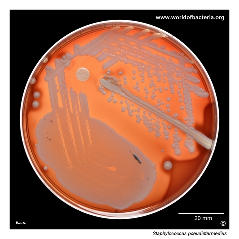
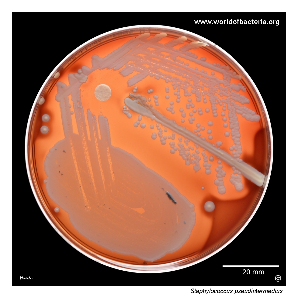
Ear canal swab from a 50-year-old woman with otitis externa. A streak of Staphylococcus aureus ATCC 25923 and a bacitracin disk were placed on the blood agar plate. The culture was incubated for 48 hours at 36 °C in an atmosphere enriched with 5% carbon dioxide. Candida parapsilosis was also present in significant quantity and grew on Sabouraud agar, whereas its growth on blood agar was suppressed by the more rapidly growing staphylococcal isolate.
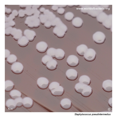
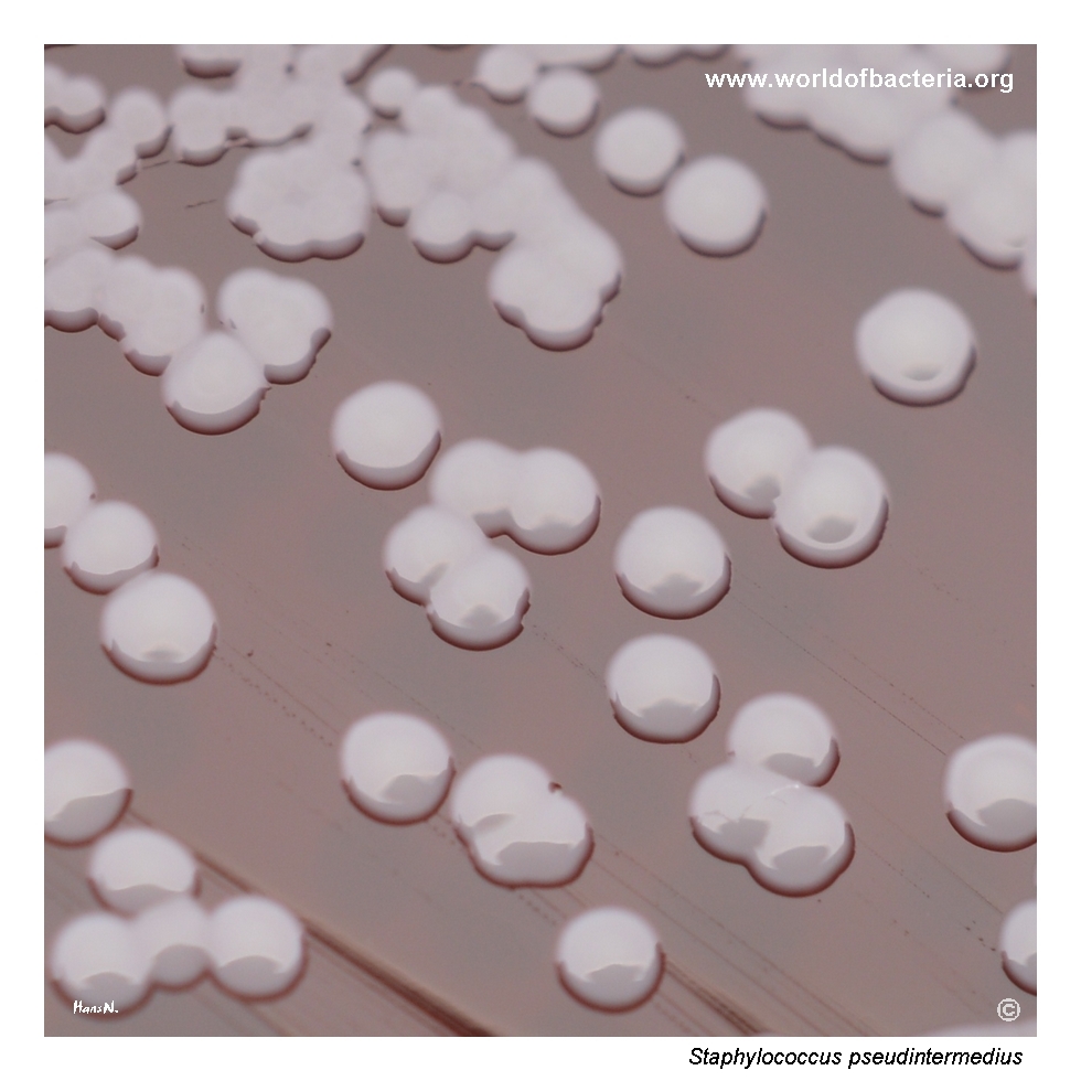
Colonies of Staphylococcus pseudintermedius on blood agar after 48 hours of incubation at 36 °C in 5% CO2. The colonies are smooth, convex, and round with entire margins and show no yellow pigment production. In routine culture, such non-pigmented colonies may appear relatively inconspicuous, which makes careful evaluation of colony shape, haemolysis, and growth conditions important when reading mixed specimens.
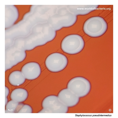
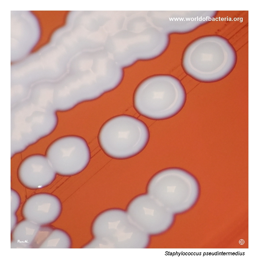
Close-up view of Staphylococcus pseudintermedius colonies on blood agar after 48 hours at 36 °C in 5% CO2. The colonies are smooth and convex with entire margins, and some show a distinctly raised centre. In English colony morphology, this appearance is described as umbonate. Such subtle surface features can be useful in detailed colony description, although they are not sufficient on their own for definitive identification.
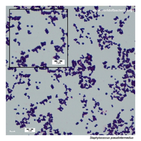
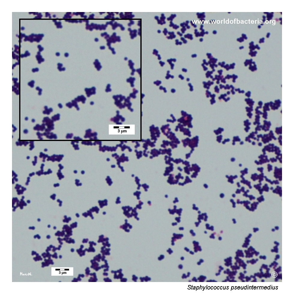
Gram-stained smear prepared from a colony of Staphylococcus pseudintermedius grown on blood agar. The organism appears as relatively small Gram-positive cocci occurring singly, in pairs, and in clusters. This morphology is consistent with the genus Staphylococcus, but it does not by itself distinguish S. pseudintermedius from other related species and must be interpreted together with colony characteristics and identification testing.
Diagnostic and Clinical Notes
Staphylococcus pseudintermedius should be regarded primarily as an animal-associated species, especially a canine pathogen, even when it is recovered from human clinical material. Its presence in human specimens therefore raises the possibility of zoonotic acquisition or at least an epidemiological link to companion animals.
In veterinary medicine, the species is well known as an important cause of skin and ear infections in dogs. In human microbiology, by contrast, it is still comparatively unfamiliar and may be underrecognized because many laboratories historically grouped similar isolates under S. intermedius or confused them with S. aureus.
This case is visually instructive because the colonies are non-pigmented and relatively inconspicuous, which reduces the immediate visual impact that often draws attention to more familiar yellow-pigmented staphylococci. Careful reading of haemolysis, colony form, and specimen context is therefore important.
The mixed specimen context is also notable. Candida parapsilosis was recovered in significant quantity on Sabouraud agar, while the rapidly growing staphylococcal isolate dominated the blood agar culture. Such interactions can affect the apparent importance of individual organisms when a plate is interpreted only at first glance.
Because S. pseudintermedius belongs to the Staphylococcus intermedius group, species-level identification is best supported by updated MALDI-TOF MS or molecular methods rather than by routine colony morphology alone.
Laboratory Identification
Colony Morphology
On blood agar after 48 hours of incubation at 35–37 °C, Staphylococcus pseudintermedius forms smooth, convex, round colonies with entire margins. In this isolate the colonies are non-pigmented and therefore relatively inconspicuous, although close inspection shows that some colonies have a distinctly raised, umbonate centre. Haemolysis may be present, but its extent should be interpreted together with incubation conditions and the overall culture context.
Microscopy
Gram staining shows relatively small Gram-positive cocci occurring singly, in pairs, and in clusters. This appearance is fully compatible with the genus Staphylococcus but does not distinguish S. pseudintermedius from S. aureus, S. intermedius, or other related staphylococci.
Key Identification Clues
Gram-positive cocci in clusters
Catalase positive
Coagulase positive
Usually animal-associated, especially linked to dogs
May appear non-pigmented and less conspicuous than many S. aureus isolates
Belongs to the Staphylococcus intermedius group and may be misidentified without modern methods
Modern Identification Methods
Reliable species-level identification is best achieved with MALDI-TOF mass spectrometry when the reference database is up to date, or with molecular methods such as species-specific PCR or sequencing. This is particularly important for S. pseudintermedius because older phenotypic workflows often did not separate it reliably from S. intermedius or other coagulase-positive staphylococci.
Antibiotic Characteristics
Staphylococcus pseudintermedius may show variable antimicrobial susceptibility, and clinically significant isolates should always be tested individually. Appearance on culture media should never be used to infer susceptibility.
Methicillin-resistant S. pseudintermedius (MRSP) is of particular practical importance, especially in veterinary medicine, because it can markedly restrict treatment options and may also appear in zoonotic contexts.
When the organism is recovered from mixed material, treatment decisions should take into account the overall specimen interpretation, the possibility of copathogens, and the clinical setting in which the isolate was obtained.
Note: Antimicrobial therapy should always be guided by laboratory susceptibility testing, specimen context, and overall clinical evaluation.