Neisseria meningitidis is an oxidase-positive Gram-negative diplococcus that commonly colonizes the human nasopharynx but can occasionally invade the bloodstream or meninges and cause rapidly progressive meningococcal disease.
Basic Characteristics
Taxonomy
Domain: Bacteria
Phylum: Pseudomonadota (Proteobacteria)
Class: Betaproteobacteria
Order: Neisseriales
Family: Neisseriaceae
Genus:Neisseria
Species:Neisseria meningitidis
Microscopy & Gram Stain
Gram-negative cocci occurring predominantly in pairs as diplococci
Adjacent sides of paired cells may appear flattened
A polysaccharide capsule is present in many invasive strains but is not reliably demonstrated by routine Gram staining
Oxygen Relationship
Strictly aerobic
Capnophilic; primary isolation is enhanced by incubation in approximately 5% CO2
Rapid Identification Tests
Oxidase: positive
Catalase: positive
Acid production: glucose and maltose positive
Lactose and sucrose: negative
Growth on routine MacConkey agar: absent
Ecology and Clinical Relevance
Natural Habitat
Human nasopharynx and upper respiratory tract
Transient asymptomatic carriage in healthy individuals
Humans are the only established natural reservoir
Common Clinical Specimens
Cerebrospinal fluid
Blood cultures
Petechial or purpuric skin-lesion material in selected cases
Lower respiratory tract specimens
Nasopharyngeal or throat specimens for carriage studies and epidemiological investigations
Clinical Significance
Cause of meningococcal meningitis and meningococcaemia
May produce fulminant septic shock, disseminated intravascular coagulation, and purpura fulminans
Can also cause pneumonia, septic arthritis, pericarditis, and other focal invasive infections
Recovery from a non-sterile respiratory specimen may represent carriage rather than invasive disease
Differential Considerations
Neisseria gonorrhoeae
Commensal Neisseria species such as N. subflava
Moraxella catarrhalis
Other causes of bacterial meningitis, especially Streptococcus pneumoniae and Haemophilus influenzae
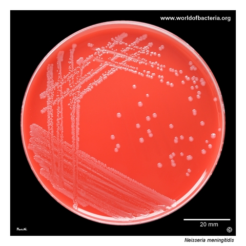
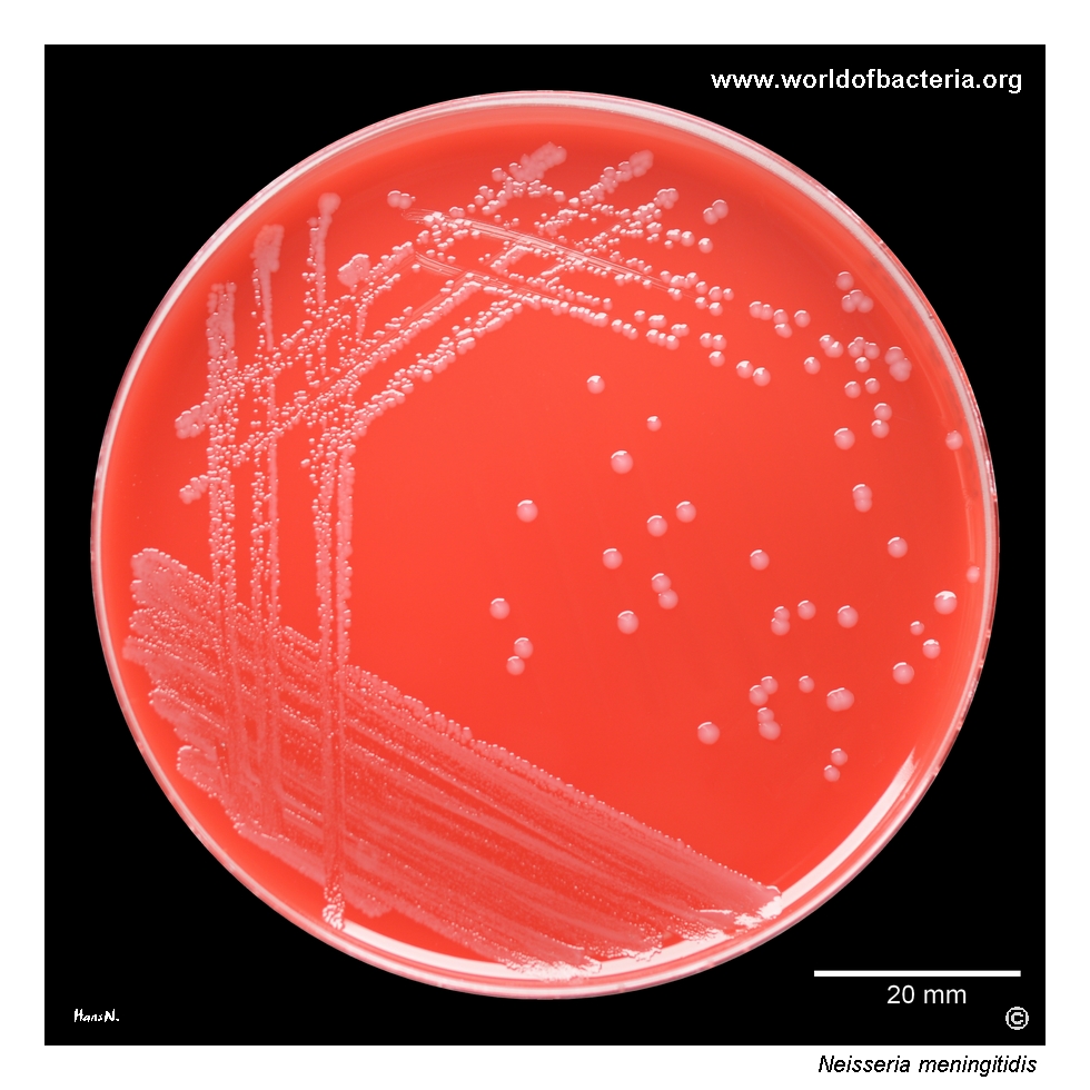
Neisseria meningitidis on blood agar after 32 hours of incubation at 36 °C in 5% CO2. The small, non-haemolytic colonies resemble those of other non-pigmented Neisseria species. The strain was recovered from sputum at a dilution of 10-5 together with quantitatively predominant Streptococcus pneumoniae. Because meningococci commonly colonize the nasopharynx, recovery from a respiratory specimen may reflect upper-airway carriage rather than invasive disease.
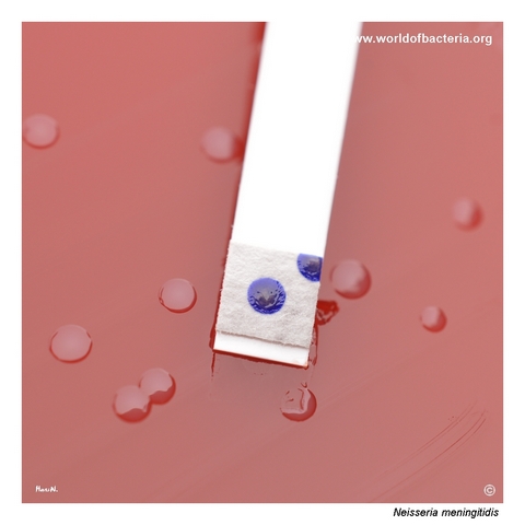
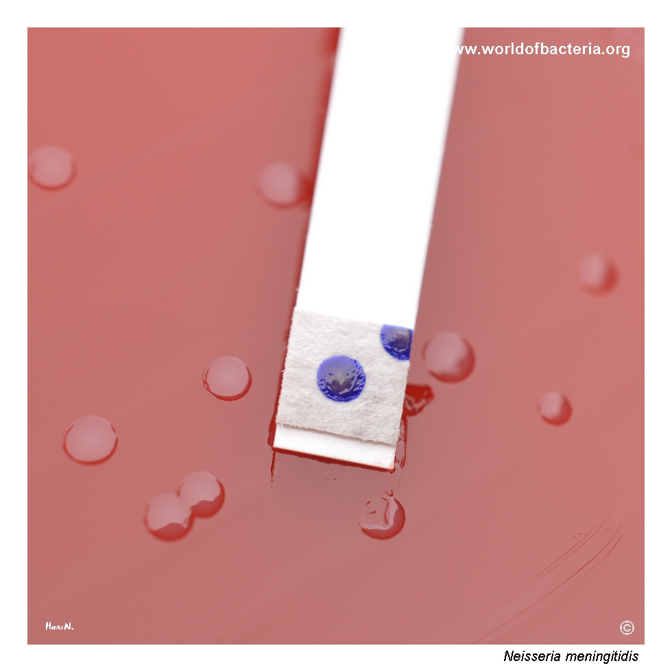
Positive oxidase reaction produced by Neisseria meningitidis. Both clinically important pathogenic species, N. meningitidis and N. gonorrhoeae, are oxidase-positive, as are other members of the genus Neisseria. Rapid development of a purple or blue colour after application of the oxidase reagent confirms cytochrome c oxidase activity. The reaction is a valuable preliminary clue but does not provide definitive species identification.
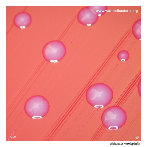
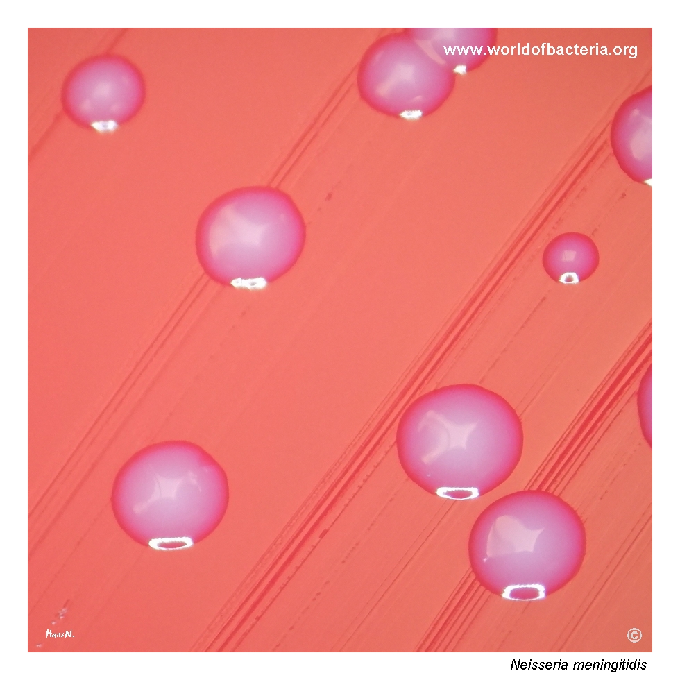
Colonies of Neisseria meningitidis may have a relatively characteristic appearance, although it depends strongly on illumination and can be difficult to reproduce in photographs. In this image, the colonies are semi-transparent and show a subtle pearly or opalescent sheen. Culture conditions: 24 hours at 36 °C in ambient air. This delicate appearance may support preliminary recognition but is not sufficiently specific to distinguish meningococci from related Neisseria species.
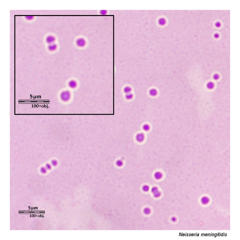
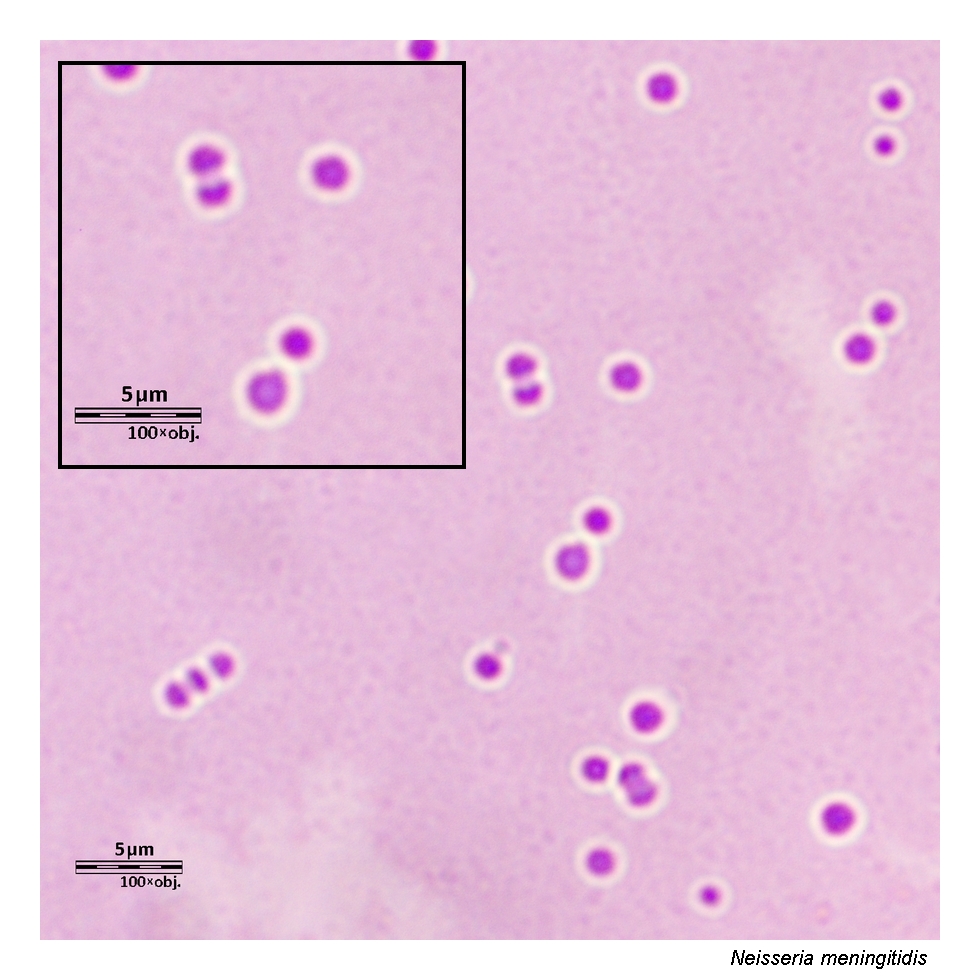
Gram-stained smear prepared from a colony of Neisseria meningitidis grown on blood agar. The organism appears as Gram-negative cocci, predominantly arranged in pairs as diplococci. Clear halos are visible around some cells, consistent with unstained capsular material, although the capsule cannot be confirmed reliably by routine Gram staining alone. Microscopic morphology supports presumptive recognition, while definitive identification requires culture or molecular testing.
Diagnostic and Clinical Notes
Neisseria meningitidis commonly colonizes the nasopharynx without causing symptoms. Detection in sputum or another non-sterile respiratory specimen must therefore be interpreted differently from recovery from blood, cerebrospinal fluid, or another normally sterile site.
The organism may invade rapidly and cause meningitis or meningococcaemia with shock, disseminated intravascular coagulation, and purpura fulminans. Suspected invasive meningococcal disease is a medical emergency, and antimicrobial treatment should not be delayed while awaiting final laboratory confirmation.
Primary isolation is supported by enriched blood or chocolate agar incubated promptly in a humid atmosphere containing approximately 5% CO2. Selective chocolate-based media may be useful for specimens containing abundant competing flora.
Oxidase positivity and Gram-negative diplococcal morphology provide important preliminary clues, but neither is species-specific. Traditional differentiation includes acid production from glucose and maltose, while modern confirmation commonly uses MALDI-TOF mass spectrometry or a validated nucleic acid amplification assay.
Serogroup determination is important for surveillance, outbreak recognition, vaccination strategy, and public-health response. Culture remains valuable because it permits antimicrobial susceptibility testing and detailed characterization, while PCR can remain positive after antimicrobial treatment has reduced culture yield.
Laboratory Identification
Colony Morphology
On enriched blood or chocolate agar incubated at approximately 35–37 °C, preferably with 5% CO2, Neisseria meningitidis forms small to medium-sized, smooth, convex, non-haemolytic colonies. Colonies may be grey, semi-transparent, pearly, or subtly opalescent, but morphology varies and overlaps substantially with that of other Neisseria species.
Microscopy
Gram staining typically reveals Gram-negative diplococci, sometimes intracellular in polymorphonuclear leukocytes in direct specimens from invasive disease. Pale halos may suggest capsular material, but routine Gram staining cannot confirm encapsulation or determine the serogroup.
Key Identification Clues
Gram-negative diplococci
Oxidase positive and catalase positive
Growth on enriched blood or chocolate agar, enhanced by CO2
No growth on routine MacConkey agar
Acid production from glucose and maltose but not lactose or sucrose
Potential polysaccharide capsule in invasive strains
Definitive confirmation by MALDI-TOF, validated NAAT, or other reference methods
Modern Identification Methods
Culture and validated molecular testing from a normally sterile site are central to confirmation of invasive meningococcal disease. MALDI-TOF mass spectrometry can identify cultured isolates when the reference database is appropriate. Slide agglutination or serogroup-specific PCR may determine the capsular serogroup, while whole-genome sequencing provides high-resolution characterization for surveillance and outbreak investigation.
Antibiotic Characteristics
Suspected invasive meningococcal disease requires immediate effective therapy. Extended-spectrum cephalosporins such as ceftriaxone or cefotaxime are commonly used for empirical treatment.
After microbiological confirmation, penicillin G or ampicillin may be considered only when susceptibility of the isolate has been demonstrated. Reduced penicillin susceptibility and acquired resistance mechanisms occur and should not be excluded by species identification alone.
Antimicrobial susceptibility testing is particularly relevant for invasive isolates and for public-health decisions where resistance to agents used for chemoprophylaxis is a concern.
Note: Close contacts of patients with invasive meningococcal disease require prompt public-health assessment and appropriate chemoprophylaxis. Selection of the prophylactic agent should follow current local guidance and resistance information.
For broader information about Neisseria meningitidis, meningococcal disease, laboratory confirmation, treatment, prevention, and current taxonomy, see: