Neisseria subflava is an oxidase-positive, yellow-pigmented Gram-negative coccus that commonly forms part of the human upper respiratory tract microbiota but may occasionally act as an opportunistic pathogen.
Basic Characteristics
Taxonomy
Domain: Bacteria
Phylum: Pseudomonadota (Proteobacteria)
Class: Betaproteobacteria
Order: Neisseriales
Family: Neisseriaceae
Genus:Neisseria
Species:Neisseria subflava
Microscopy & Gram Stain
Gram-negative cocci occurring singly, in pairs as diplococci, and occasionally in tetrads
Some cells may resist decolourization and appear Gram-variable in older cultures
Non-motile and non-spore-forming
Oxygen Relationship
Aerobic
Growth is generally enhanced by a humid atmosphere containing approximately 5% CO2
Rapid Identification Tests
Oxidase: positive
Catalase: usually positive
Acid production: glucose and maltose positive
Fructose and sucrose: variable
Lactose: negative
Nitrite reduction: positive
Nitrate reduction: negative
Ecology and Clinical Relevance
Natural Habitat
Human oropharynx and nasopharynx
Oral cavity and upper respiratory tract
Usually present as part of the commensal microbiota
Common Clinical Specimens
Throat and nasopharyngeal swabs
Sputum and other respiratory specimens
Oral specimens
Blood, cerebrospinal fluid, joint fluid, or other normally sterile material in rare invasive infections
Clinical Significance
Usually a non-pathogenic upper respiratory tract commensal
Recovery from non-sterile respiratory material most often reflects colonization
Rarely associated with endocarditis, meningitis, bacteraemia, septic arthritis, otitis, or other opportunistic infections
Clinical significance is greatest when repeatedly or purely isolated from a normally sterile site
Differential Considerations
Neisseria meningitidis
Neisseria gonorrhoeae
Other pigmented commensal Neisseria species, including N. flava, N. flavescens, N. sicca, and N. mucosa
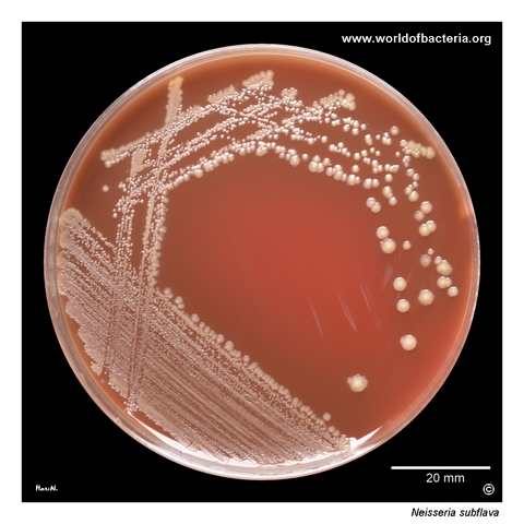
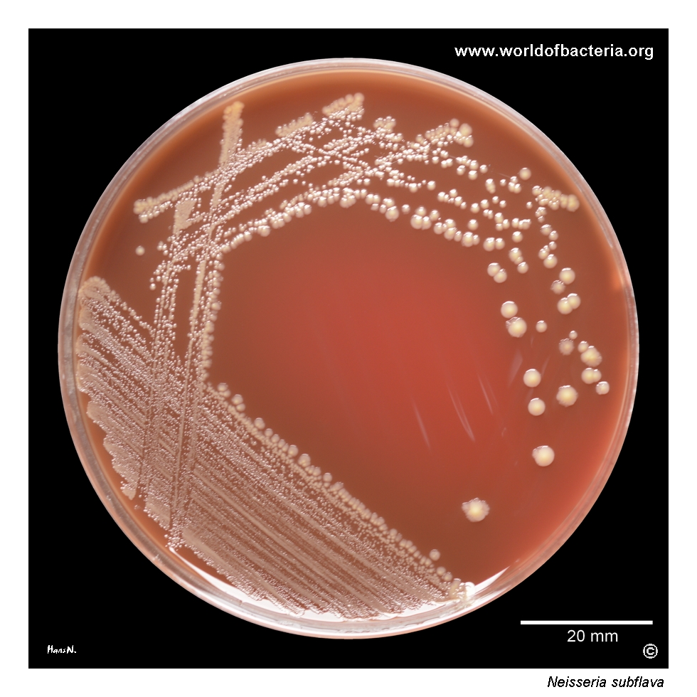
Neisseria subflava on blood agar after 48 hours of incubation at 36 °C in 5% CO2. This species belongs to the pigmented neisseriae and may produce yellow coloration of varying intensity depending on the strain and culture conditions. N. subflava is usually a non-pathogenic member of the upper respiratory tract microbiota, although its recovery from clinical material should always be interpreted in relation to the specimen source.
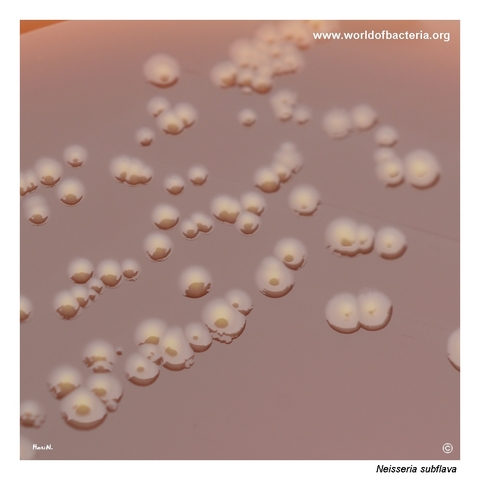
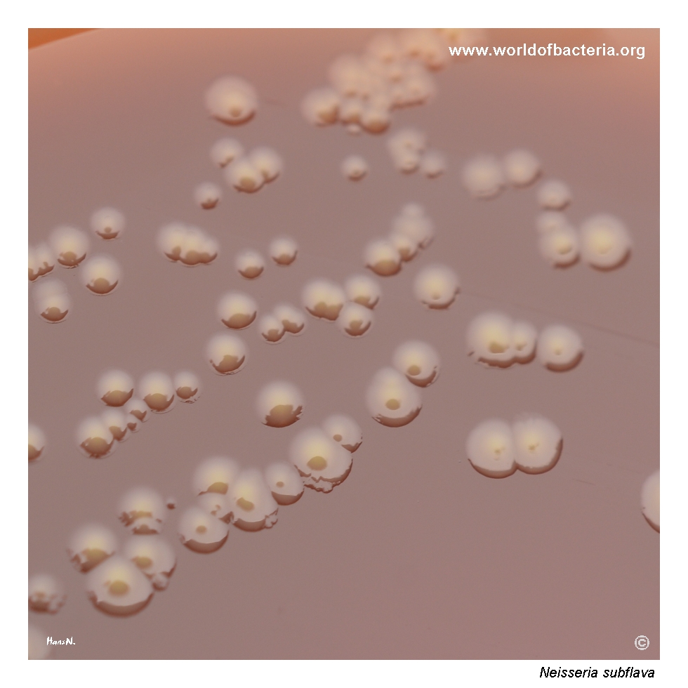
Colonies of Neisseria subflava on blood agar after 48 hours at 36 °C in 5% CO2. The colonies are yellow-pigmented and range from convex to relatively flat, with some showing a distinctly raised central area. Pigment intensity may vary between individual colonies and isolates. This coloration provides a useful preliminary clue but is not sufficiently specific for identification without additional laboratory testing.
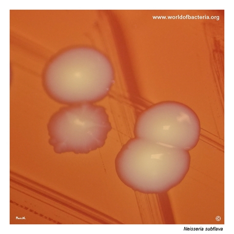
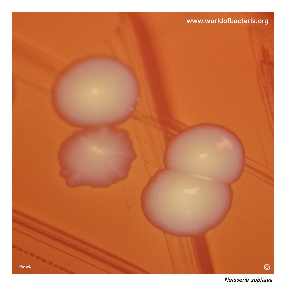
Close-up view of Neisseria subflava colonies on blood agar. In addition to smooth, convex colonies, some isolates may form colonies with conspicuous radial ridges extending from the centre towards the periphery. The colony shown here also has an irregular margin. Culture conditions: 48 hours at 36 °C in 5% CO2. This unusual surface structure illustrates the morphological variability that may occur within the species.
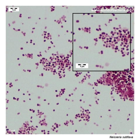
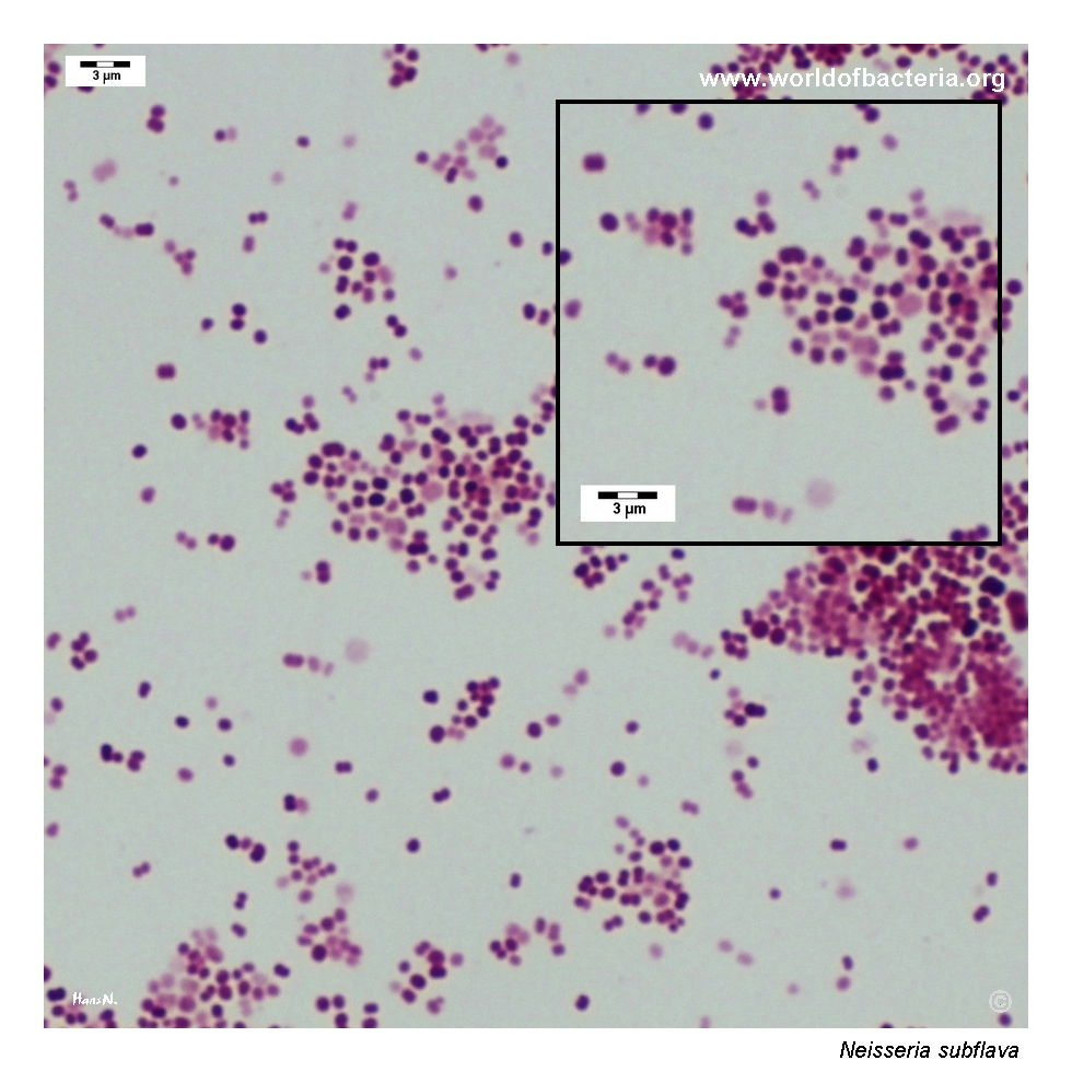
Gram-stained smear prepared from a colony of Neisseria subflava grown on blood agar. The organism appears as Gram-negative cocci occurring singly and predominantly in pairs as diplococci. This microscopic appearance is typical of neisseriae but does not distinguish N. subflava from pathogenic or other commensal members of the genus. Reliable identification therefore requires correlation with pigmentation, culture characteristics, and confirmatory testing.
Diagnostic and Clinical Notes
Neisseria subflava is usually part of the normal microbiota of the oral cavity, nasopharynx, and upper respiratory tract. Its detection in sputum, throat, or other non-sterile respiratory material generally represents colonization unless supported by the clinical picture and quantitative or repeated recovery.
Yellow pigmentation is a useful descriptive feature and explains the species epithet, which means yellowish. Pigment intensity varies with the strain, medium, incubation time, and illumination, and cannot provide definitive species identification.
The organism is oxidase positive and typically produces acid from glucose and maltose. Reactions with fructose and sucrose are variable, and the biochemical overlap among commensal neisseriae may limit the reliability of older identification schemes.
Rare invasive infections caused by commensal Neisseria species have been reported, particularly in patients with disrupted mucosal barriers, underlying disease, prosthetic material, or immunocompromise. Recovery from blood, cerebrospinal fluid, synovial fluid, or another normally sterile site should therefore not be dismissed automatically as contamination.
Accurate identification may require MALDI-TOF mass spectrometry with an appropriate reference database or molecular methods. Misidentification with other pigmented Neisseria species is possible when identification relies only on colony colour and carbohydrate reactions.
Laboratory Identification
Colony Morphology
On blood or chocolate agar incubated at approximately 35–37 °C, usually with added CO2, Neisseria subflava produces smooth, transparent to opaque, non-haemolytic colonies with yellow pigmentation of variable intensity. Colonies may be convex, flat, centrally raised, adherent, or occasionally irregular and radially ridged.
Microscopy
Gram staining shows cocci occurring singly, in pairs, and sometimes in tetrads. Some cells may retain crystal violet more strongly than expected and appear Gram-variable. The microscopic appearance confirms a neisserial pattern but does not distinguish pathogenic from commensal species.
Key Identification Clues
Yellow-pigmented, non-haemolytic colonies
Gram-negative cocci, commonly diplococci
Oxidase positive and usually catalase positive
Acid production from glucose and maltose
Variable acid production from fructose and sucrose
Lactose negative
Nitrite reduction positive and nitrate reduction negative
Confirmation by MALDI-TOF or molecular methods when clinically important
Modern Identification Methods
MALDI-TOF mass spectrometry can provide rapid identification when the database contains well-characterized reference spectra for commensal Neisseria. Difficult or clinically significant isolates may require 16S rRNA gene sequencing, other target-gene sequencing, or whole-genome analysis. Results should be interpreted with awareness of the close genetic and phenotypic relationships among pigmented neisseriae.
Antibiotic Characteristics
Colonization by Neisseria subflava does not require antimicrobial treatment. Therapy is considered only when the organism is judged to be the cause of a clinically significant infection.
There is no single predictable susceptibility profile for rare invasive isolates. Commensal neisseriae may carry altered penicillin-binding proteins, beta-lactamase genes, macrolide resistance determinants, or other resistance mechanisms.
A clinically significant isolate from a normally sterile site should undergo antimicrobial susceptibility testing using an appropriate validated method, with treatment selected according to the complete result and the site and severity of infection.
Note: Commensal oropharyngeal Neisseria species can serve as reservoirs of resistance determinants that may be exchanged with pathogenic neisseriae. This ecological role does not mean that every colonizing isolate is itself clinically resistant or requires treatment.
For broader information about Neisseria subflava, commensal neisseriae, occasional opportunistic infection, laboratory identification, and current taxonomy, see: