Neisseria gonorrhoeae is a fastidious, oxidase-positive Gram-negative diplococcus adapted to human mucosal surfaces and responsible for gonorrhoea, pelvic inflammatory disease, neonatal conjunctivitis, and occasional disseminated infection.
Basic Characteristics
Taxonomy
Domain: Bacteria
Phylum: Pseudomonadota (Proteobacteria)
Class: Betaproteobacteria
Order: Neisseriales
Family: Neisseriaceae
Genus:Neisseria
Species:Neisseria gonorrhoeae
Microscopy & Gram Stain
Gram-negative cocci occurring predominantly as diplococci
Adjacent sides of paired cells may appear flattened or slightly concave
Intracellular diplococci may be visible within polymorphonuclear leukocytes in direct genital specimens
Oxygen Relationship
Strictly aerobic
Capnophilic; primary isolation is enhanced by a humid atmosphere containing approximately 5% CO2
Rapid Identification Tests
Oxidase: positive
Catalase: positive
Acid production: glucose positive
Maltose, lactose, and sucrose: negative
Growth on routine MacConkey agar: absent
Ecology and Clinical Relevance
Natural Habitat
Human genital mucosa
Rectal and pharyngeal mucosal surfaces
Conjunctiva, particularly in neonatal exposure
Humans are the only established natural reservoir
Common Clinical Specimens
Vaginal and endocervical swabs
Urethral swabs and first-catch urine
Rectal and pharyngeal swabs
Conjunctival specimens
Blood, synovial fluid, or skin-lesion material in disseminated infection
Clinical Significance
Cause of gonorrhoea and mucosal infection of the genital, rectal, pharyngeal, and conjunctival sites
May ascend to cause pelvic inflammatory disease, epididymitis, infertility, and ectopic pregnancy
Can cause neonatal ophthalmia following exposure during delivery
Occasionally causes disseminated gonococcal infection with dermatitis, tenosynovitis, septic arthritis, or bacteraemia
Differential Considerations
Neisseria meningitidis
Commensal Neisseria species, especially in pharyngeal specimens
Moraxella catarrhalis
Other causes of urethritis, cervicitis, proctitis, and pelvic inflammatory disease
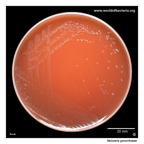
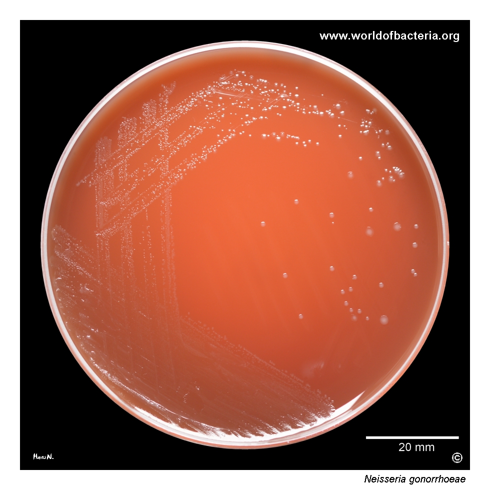
Colonies of Neisseria gonorrhoeae on selective agar for the primary isolation of gonococci from clinical material. The strain was recovered from a vaginal swab. After 48 hours of incubation at 36 °C in 5% CO2, the colonies show a semi-transparent appearance with a subtle pearly or opalescent sheen. This delicate visual character may support preliminary recognition, although it is strongly influenced by illumination and is not specific on its own.
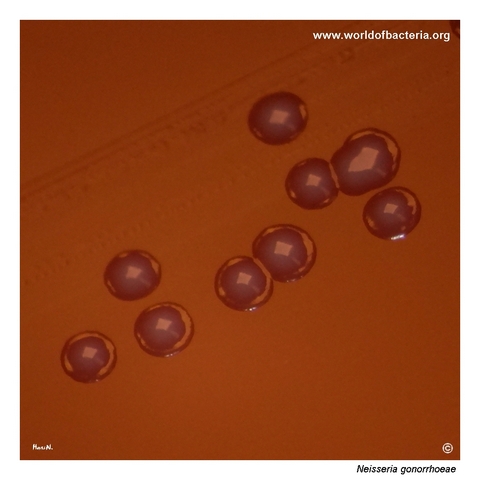
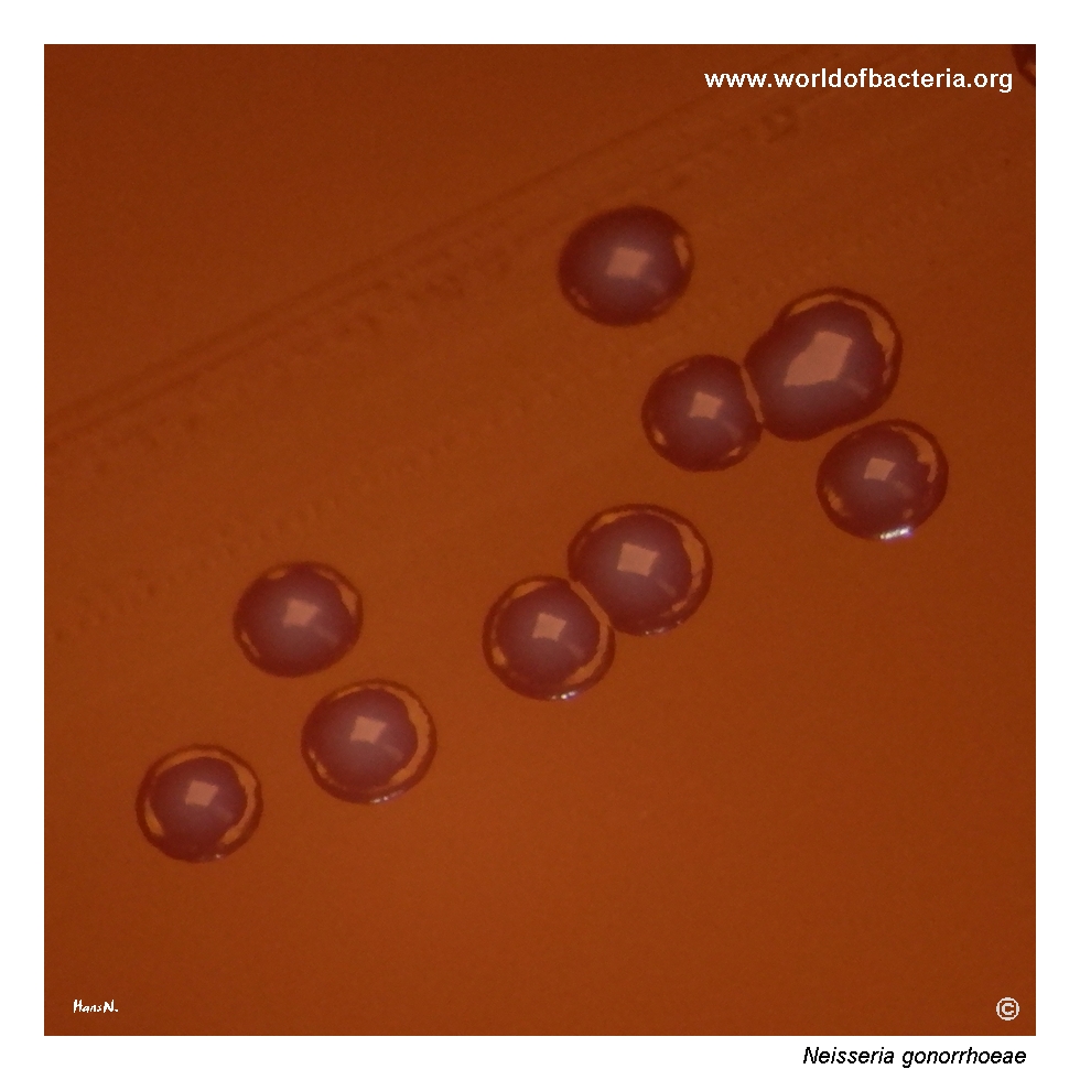
Colonies of Neisseria gonorrhoeae on selective agar after 48 hours at 36 °C in 5% CO2. The colonies remain small, measuring approximately 2 mm in diameter. At higher magnification, some show slightly projecting or irregular margins, a feature that may also occur in other bacterial species. More useful clues are the relatively slow growth and small colony size. Many strains grow best in an atmosphere enriched with carbon dioxide.
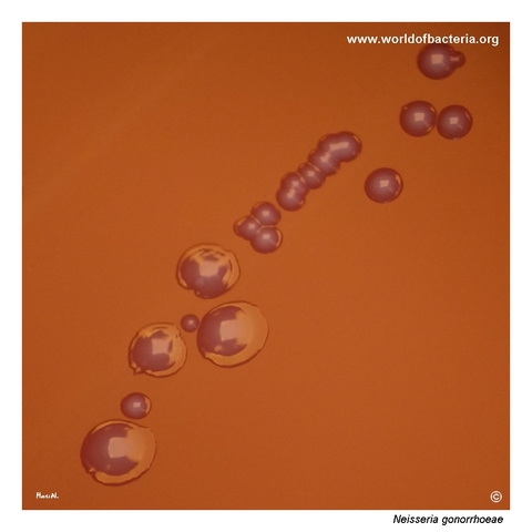
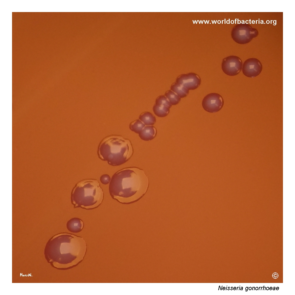
Close-up view of Neisseria gonorrhoeae colonies. A single strain may produce a heterogeneous population of colonies differing in size and morphology, as demonstrated in this image. Historically, smaller colony types were sometimes associated with greater virulence, but colony size alone is not a reliable measure of pathogenic potential. Culture conditions: 24 hours at 36 °C in ambient air.
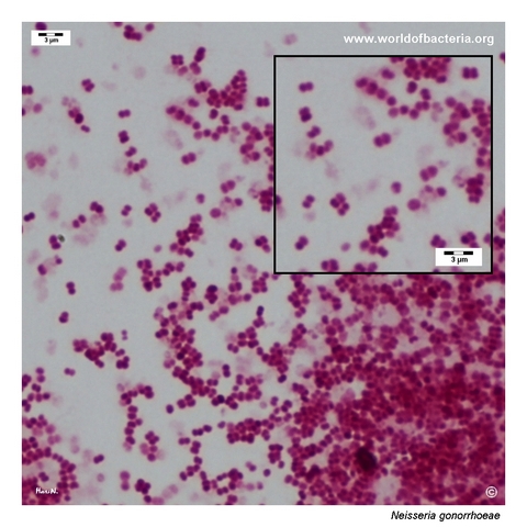
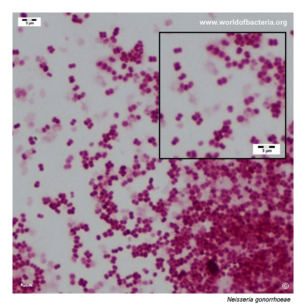
Gram-stained smear prepared from a colony of Neisseria gonorrhoeae grown on selective agar. The organism appears as Gram-negative cocci, predominantly arranged in pairs as diplococci. This morphology is characteristic of gonococci but is not sufficient for definitive identification when observed in a cultured isolate. Interpretation should therefore be combined with colony characteristics and appropriate confirmatory testing.
Diagnostic and Clinical Notes
Neisseria gonorrhoeae is highly adapted to human mucosal surfaces and is transmitted primarily through sexual contact or from mother to infant during delivery. Infection may be symptomatic or asymptomatic, particularly at cervical, rectal, and pharyngeal sites.
The organism is fragile outside the host and is sensitive to drying, temperature change, and delays in transport. Successful culture therefore depends on appropriate specimen collection, rapid inoculation or transport, enriched selective media, high humidity, and incubation in carbon dioxide.
Selective chocolate-based media suppress much of the accompanying genital or pharyngeal flora while supporting gonococcal growth. Small translucent, pearly, or opalescent colonies may suggest the organism, but morphology alone cannot distinguish it reliably from related species.
Nucleic acid amplification tests are the principal diagnostic method for most uncomplicated infections because of their high sensitivity and ability to test urine and specimens from multiple anatomical sites. Culture remains essential when antimicrobial susceptibility testing is required, particularly after suspected treatment failure or when reduced cephalosporin susceptibility is a concern.
In a direct urethral specimen from a symptomatic man, intracellular Gram-negative diplococci provide strong presumptive evidence. The diagnostic value of Gram staining is lower in cervical, vaginal, rectal, and pharyngeal specimens and in asymptomatic infection.
Laboratory Identification
Colony Morphology
On enriched selective agar incubated at approximately 35–37 °C in a humid atmosphere containing about 5% CO2, Neisseria gonorrhoeae forms small, smooth to subtly irregular, translucent, grey-white, pearly, or opalescent colonies. Colony size and morphology may vary within a single strain and do not provide a reliable measure of virulence.
Microscopy
Gram staining reveals Gram-negative diplococci with adjacent flattened surfaces. Intracellular organisms within polymorphonuclear leukocytes may be seen in direct genital specimens, whereas smears from cultured colonies lack the clinical and cellular context needed for a definitive species-level diagnosis.
Key Identification Clues
Fastidious Gram-negative diplococcus
Oxidase positive and catalase positive
Growth on enriched selective agar, enhanced by CO2 and humidity
No growth on routine MacConkey agar
Acid production from glucose but not maltose, lactose, or sucrose
Confirmation by validated NAAT or identification of a cultured isolate
Culture required for phenotypic antimicrobial susceptibility testing
Modern Identification Methods
Validated nucleic acid amplification tests are widely used for direct detection from urine and genital or extragenital specimens. Cultured isolates may be confirmed using MALDI-TOF mass spectrometry, biochemical or enzymatic systems, and molecular assays. Care is required with pharyngeal specimens because commensal Neisseria species may affect the specificity of some assays. Whole-genome sequencing provides high-resolution information for resistance surveillance and outbreak investigation.
Antibiotic Characteristics
Neisseria gonorrhoeae has developed resistance to nearly every antimicrobial class previously used routinely for gonorrhoea, including sulfonamides, penicillins, tetracyclines, macrolides, and fluoroquinolones.
Ceftriaxone remains the principal empirical treatment in many current guidelines. Selection of the regimen should follow the current national or international recommendation and should account for infection site, allergy history, pregnancy, and whether concomitant Chlamydia trachomatis infection has been excluded.
Suspected treatment failure requires culture from relevant anatomical sites and antimicrobial susceptibility testing before additional treatment whenever possible. Isolates with reduced cephalosporin susceptibility require prompt communication with public-health or reference laboratories.
Note: Gonococcal resistance patterns evolve rapidly. Species identification alone cannot predict susceptibility, and treatment guidance should always be checked against the most recent recommendations.