Listeria monocytogenes is a small Gram-positive foodborne pathogen of major clinical importance because it can cause invasive infection, especially bacteraemia, meningitis, and severe disease in pregnancy, while on blood agar its colonies may initially appear deceptively inconspicuous.
Basic Characteristics
Taxonomy
Domain: Bacteria
Phylum: Bacillota
Class: Bacilli
Order: Bacillales
Family: Listeriaceae
Genus:Listeria
Species:Listeria monocytogenes
Microscopy & Gram Stain
Small Gram-positive rods that may appear singly, in pairs, and in short chains or small irregular groups
In direct blood-culture microscopy, the cells may easily be mistaken for less significant small Gram-positive rods unless interpreted carefully
The organism is not coryneform in the classical diphtheroid sense, yet in practice the microscopic impression may still overlap with other small Gram-positive bacilli
For this reason, microscopy is highly useful as an early clue but remains insufficient on its own for final species identification
Oxygen Relationship
Facultatively anaerobic organism that grows well under routine laboratory conditions
Good growth is obtained on blood agar, often in a carbon-dioxide-enriched atmosphere, although the species is not fastidious in the strict sense
A practical diagnostic difficulty is that haemolysis may remain weak or inconspicuous after the first day of incubation and become clearer only with further incubation
Rapid Identification Tests
Catalase: positive
Haemolysis: usually beta-haemolytic, but often only weakly so on sheep blood agar
Motility: classically tumbling at room temperature, although this is not the main focus of the present image set
Important caveat: small grey-white colonies on blood agar may resemble enterococci or other Gram-positive bacteria if the subtle haemolysis is overlooked
Practical warning: a small Gram-positive rod in blood culture should not automatically be dismissed as contamination when the clinical setting fits listeriosis
Ecology and Clinical Relevance
Natural Habitat
Listeria monocytogenes is widely distributed in the environment and is classically associated with foodborne transmission to humans
Unlike many low-significance Gram-positive rods, it is a true invasive pathogen rather than a routine skin commensal
Its clinical importance is greatest in older adults, immunocompromised patients, pregnant women, fetuses, and neonates
Common Clinical Specimens
Blood cultures in invasive listeriosis
Cerebrospinal fluid in meningitis or meningoencephalitis
Placental, fetal, or neonatal material in pregnancy-associated disease
Less commonly, other sterile-site material depending on the focus of infection
Clinical Significance
Listeria monocytogenes is a major invasive foodborne pathogen capable of causing sepsis, meningitis, meningoencephalitis, and severe infection in pregnancy and the neonatal period
The species is diagnostically important because its colonies may look surprisingly ordinary on blood agar despite the seriousness of the diseases it can cause
In blood-culture work especially, correct recognition has direct clinical importance because delayed interpretation may postpone appropriate targeted therapy
Differential Considerations
Enterococci when only small grey-white colonies on blood agar are considered
Small coryneform or diphtheroid-like Gram-positive rods that may be dismissed as contaminants
Other Gram-positive rods recovered from blood culture, including less clinically important aerobic species
Beta-haemolytic organisms in which the haemolytic pattern is more obvious than the often subtle haemolysis produced by L. monocytogenes
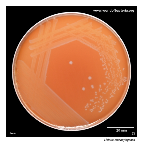
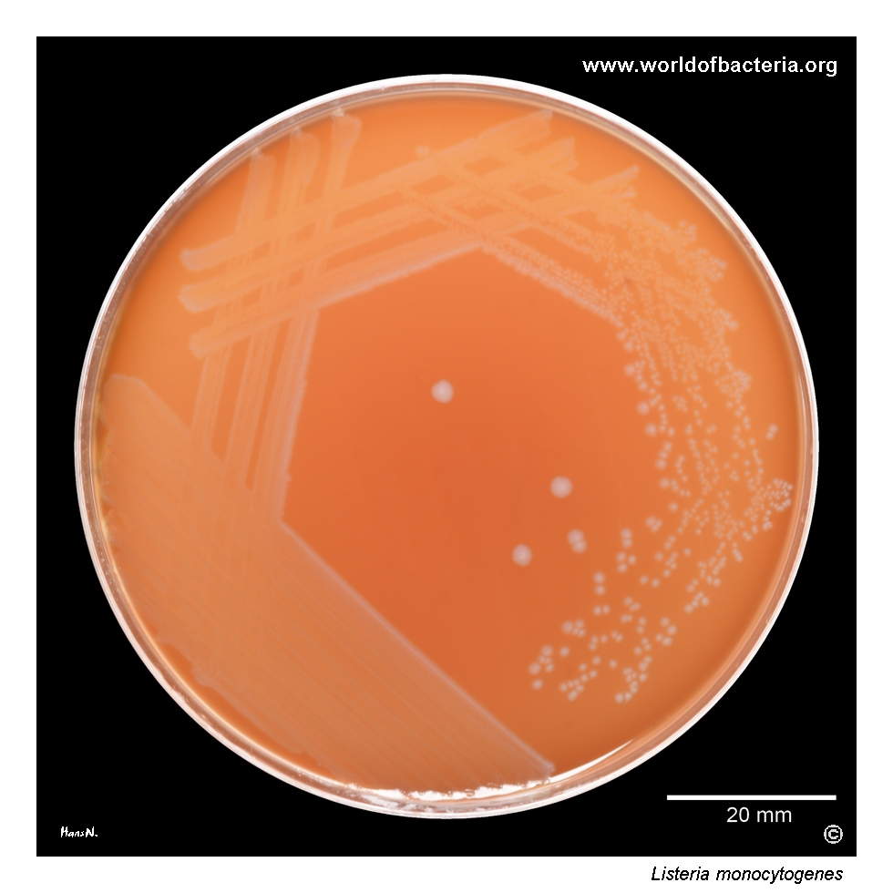
Listeria monocytogenes is classically described as a beta-haemolytic organism on blood agar. In many isolates, however, the haemolysis may be very subtle after 24 hours of incubation and becomes more apparent only after two or more days, especially in areas of confluent growth. Culture conditions: 48 hours at 36 °C in 5% CO2. This delayed and often inconspicuous haemolysis is one reason why listerial colonies may initially be underestimated during routine plate reading.
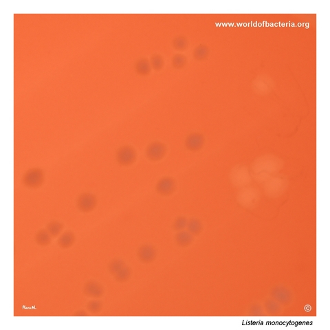
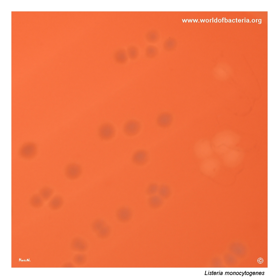
Colonies of Listeria monocytogenes on blood agar, photographed in transmitted light. Part of the colony growth was removed from the agar, and the resulting clearing of the medium indicates haemolysis of sheep erythrocytes, consistent with beta-haemolysis. Around the colonies that remained on the plate, haemolysis is not clearly visible. This image nicely illustrates how the haemolytic effect may be easier to appreciate after removal of the colony mass than around intact colonies themselves.
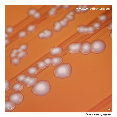
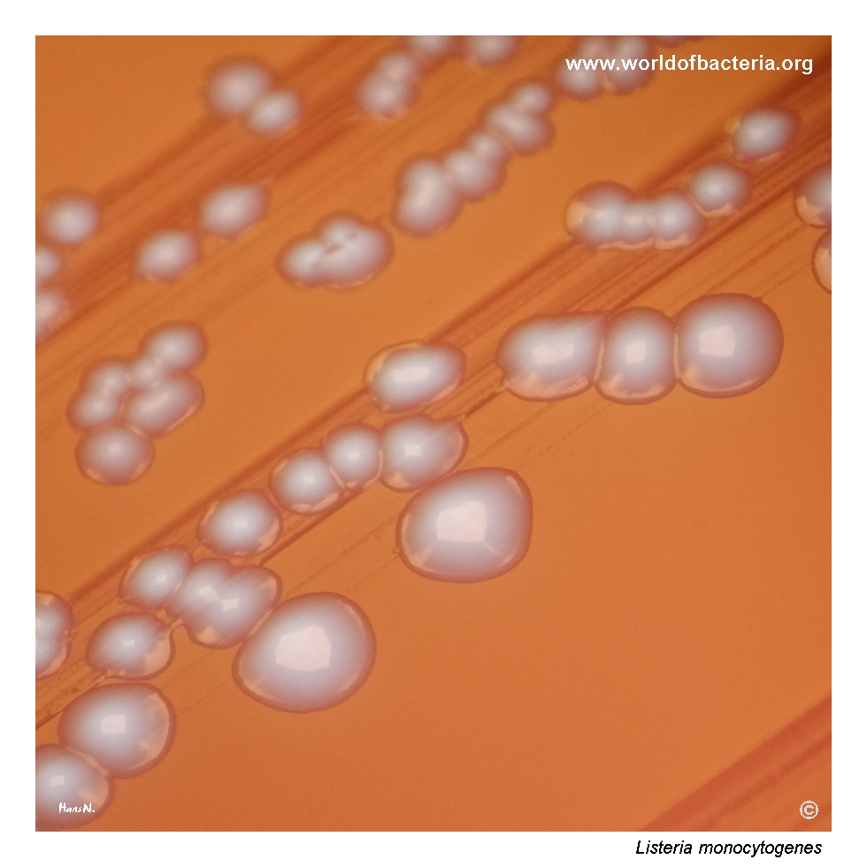
Close-up view of Listeria monocytogenes colonies after 48 hours of incubation at 36 °C in 5% CO2. Listerial colonies are sometimes compared in appearance to those of enterococci, partly because more obvious haemolysis is often absent or only weakly expressed. This visual similarity is diagnostically important, as small grey-white colonies on blood agar may easily be misinterpreted unless the characteristic microscopic morphology is also taken into account.
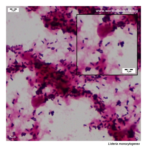
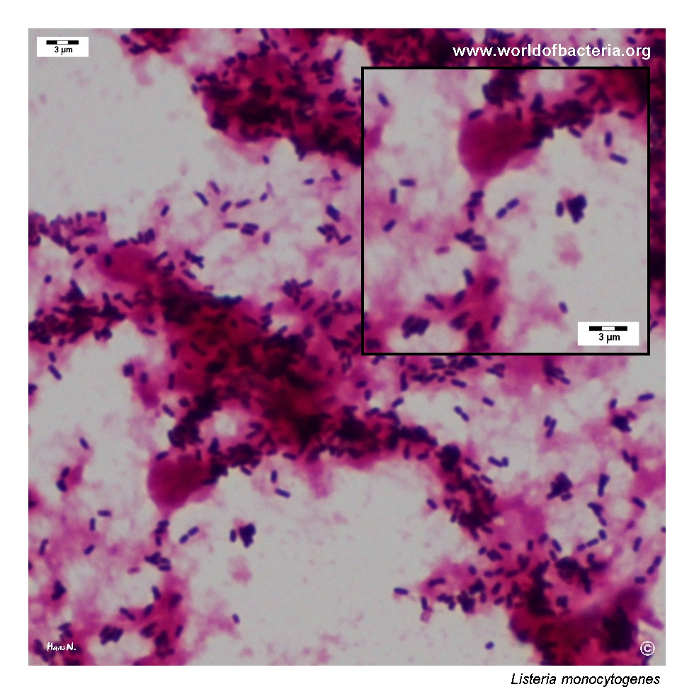
Listeria monocytogenes in a positive blood culture, Gram stained. The organism appears as small Gram-positive rods occurring singly, in pairs, and in small clusters. In blood culture microscopy, this appearance is a useful clue, but it still requires careful interpretation because small Gram-positive rods may be mistaken for less significant contaminants unless correlated with the clinical picture and the culture findings.
Diagnostic and Clinical Notes
This page is especially instructive because it shows how misleadingly ordinary Listeria monocytogenes may look on blood agar. The species is classically beta-haemolytic, yet the haemolysis can be very faint after the first day of incubation and may remain easier to appreciate after colony removal or in confluent growth than around intact colonies themselves.
That subtlety matters in routine practice. Small grey-white colonies with only weak haemolysis can be underestimated or confused with enterococci or other Gram-positive bacteria, particularly if the plate is read quickly or without close attention to the accompanying microscopic findings.
The blood-culture image adds the more clinically urgent lesson. A small Gram-positive rod in a positive blood culture is not always a trivial contaminant. In the correct clinical setting, such a finding may represent invasive listeriosis, and delayed recognition can have major consequences for patient management.
Together, the images therefore teach a valuable contrast: the plate appearance may seem modest, but the clinical significance of the species is potentially very high. This discrepancy is one reason why L. monocytogenes remains such an important organism in diagnostic microbiology.
Laboratory Identification
Colony Morphology
On blood agar, Listeria monocytogenes usually forms small grey-white colonies with a smooth to slightly matt surface. In many isolates, the beta-haemolysis is only weakly expressed after 24 hours and becomes more apparent after prolonged incubation or after close inspection of areas where colony mass has been removed. This often modest appearance is a key practical point, because the species can initially resemble less significant organisms despite its major clinical importance.
Microscopy
Gram staining shows small Gram-positive rods occurring singly, in pairs, and in short groupings. In blood cultures, the microscopic image may be deceptively understated and can overlap with other small Gram-positive bacilli. The finding is therefore strongly suggestive but must always be interpreted together with culture morphology and the clinical situation.
Key Identification Clues
Small Gram-positive rods in blood culture or colony smear
Small grey-white colonies on blood agar
Usually beta-haemolytic, but often only subtly so
Catalase positive
Characteristic diagnostic importance in invasive disease and pregnancy-associated infection
Need to distinguish the species from enterococci and low-significance Gram-positive rods
Modern Identification Methods
In current routine practice, MALDI-TOF MS usually provides reliable species identification when database coverage is adequate. Classical orientation tests such as catalase and motility can still be useful, but modern laboratory workflows generally depend on instrumental identification combined with careful interpretation of blood-culture microscopy and colony morphology. When clinically indicated, the laboratory result must then be integrated rapidly with the broader picture of invasive listeriosis.
Antibiotic Characteristics
For Listeria monocytogenes, culture appearance alone gives no dependable information about susceptibility. The important laboratory task is first to recognize the species correctly and then to perform or interpret susceptibility testing within the clinical context of invasive infection.
In practical diagnostics, the real antibiotic lesson is that the seriousness of listeriosis should not be underestimated just because the colonies appear small or only weakly haemolytic. Prompt species recognition matters more than any visual impression of colony vigour.
Because invasive listeriosis often affects older adults, pregnant patients, neonates, and people with impaired immunity, laboratory communication is clinically important once the species has been identified.
Note: therapeutic decisions should always be guided by the confirmed identification, the infection syndrome, and formal susceptibility interpretation rather than by colony morphology alone.