Corynebacterium pseudodiphtheriticum is a non-lipophilic respiratory-associated coryneform species that commonly colonizes the upper airway and usually forms white non-haemolytic colonies on blood agar.
Basic Characteristics
Taxonomy
Domain: Bacteria
Phylum: Actinomycetota
Class: Actinomycetes
Order: Corynebacteriales
Family: Corynebacteriaceae
Genus:Corynebacterium
Species:Corynebacterium pseudodiphtheriticum
Microscopy & Gram Stain
Gram-positive coryneform rods
Pleomorphic bacilli with the usual diphtheroid appearance
Cells may occur singly, in pairs, in palisades, or in angular V-like arrangements
Microscopy supports recognition of a coryneform isolate but does not by itself determine the species
Oxygen Relationship
Aerobic to aerotolerant coryneform organism that grows well under routine culture conditions
In practical diagnostics it grows readily on blood agar and also grows well in 5% CO2
The species is not notable for fastidiousness but may still be overlooked because its colonies can resemble ordinary skin flora
Rapid Identification Tests
Catalase: positive
Oxidase: negative
Microscopy: Gram-positive coryneform rod
Practical clue: white, smooth, non-haemolytic colonies that may resemble coagulase-negative staphylococci on blood agar
Important caveat: reliable species identification usually requires MALDI-TOF MS or another robust species-level method
Ecology and Clinical Relevance
Natural Habitat
The species is strongly associated with the human upper respiratory tract, especially the nasal cavity and pharyngeal mucosa
In many specimens it behaves as ordinary colonizing flora rather than as a true pathogen
Its recovery from nasal material therefore requires careful clinical interpretation
Common Clinical Specimens
Nasal swabs
Throat or upper respiratory tract specimens
Respiratory samples from lower airway infection in selected patients
Occasionally other mucosal or mixed clinical samples
Clinical Significance
Often a commensal or colonizing corynebacterial species of the upper airway
Can occasionally act as an opportunistic respiratory pathogen, especially in predisposed patients
In mixed cultures its presence may simply reflect colonization, and another organism may be the more likely cause of symptoms
Differential Considerations
Corynebacterium diphtheriae and other respiratory corynebacteria
Corynebacterium propinquum and related upper-airway diphtheroids
Corynebacterium macginleyi in small-colony mixed cultures when ocular material is involved
Non-haemolytic coagulase-negative staphylococci or enterococci when only colony appearance on blood agar is considered
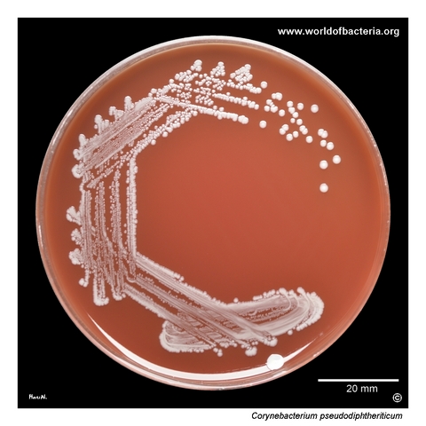
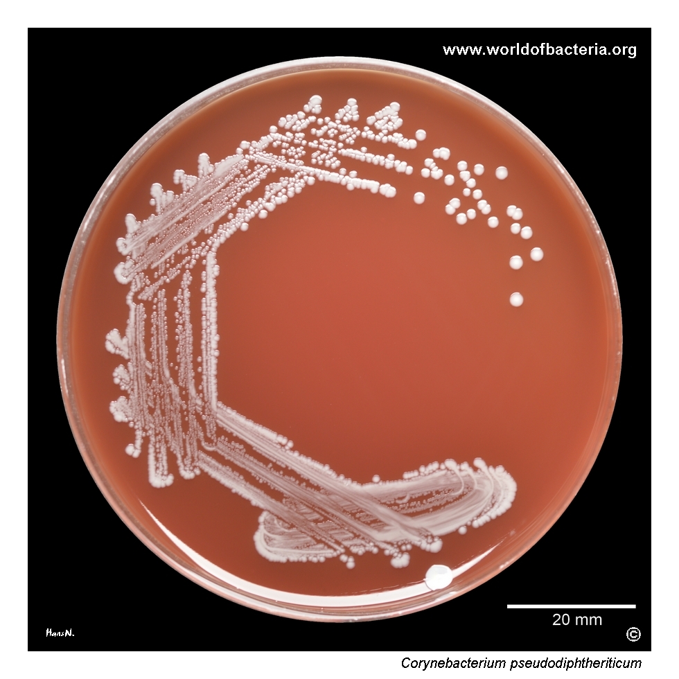
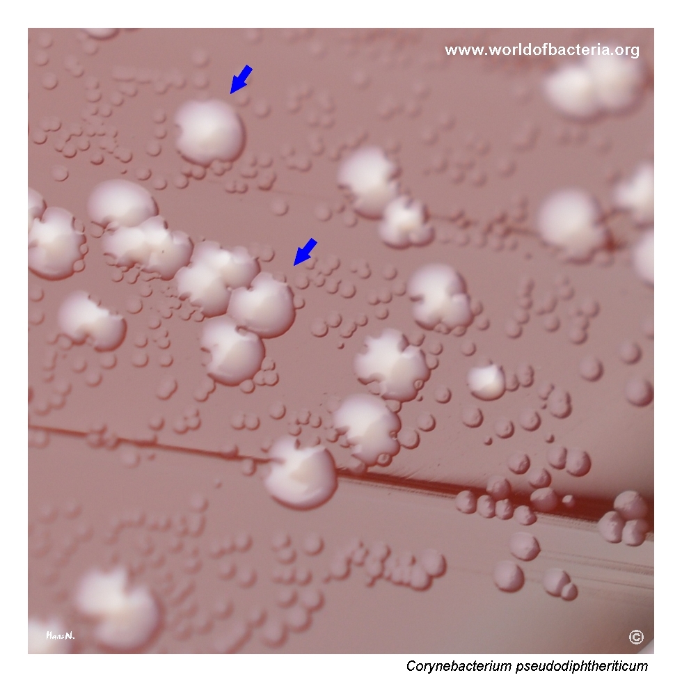
Blood agar culture of Corynebacterium pseudodiphtheriticum. Incubation: 48 hours at 36 °C in 5% CO2. The strain was isolated from a nasal swab from a 4-year-old boy with diagnosis R05, cough. Corynebacteria may occur on the nasal mucosa as ordinary colonizing flora without pathogenic significance. In this specimen, Streptococcus pneumoniae was the more likely cause of the patient’s symptoms.
White colonies of Corynebacterium pseudodiphtheriticum together with tiny colonies of C. macginleyi. Incubation: 48 hours at 36 °C in 5% CO2. Patient: 97-year-old woman, diagnosis H100, mucopurulent conjunctivitis.
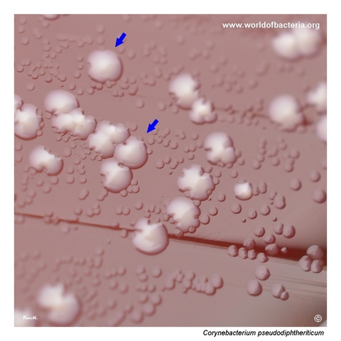
Close-up view of Corynebacterium pseudodiphtheriticum colonies on blood agar. The colonies are white, convex, smooth, and have entire margins, and they can resemble colonies of coagulase-negative staphylococci. Incubation: 48 hours at 36 °C in 5% CO2.
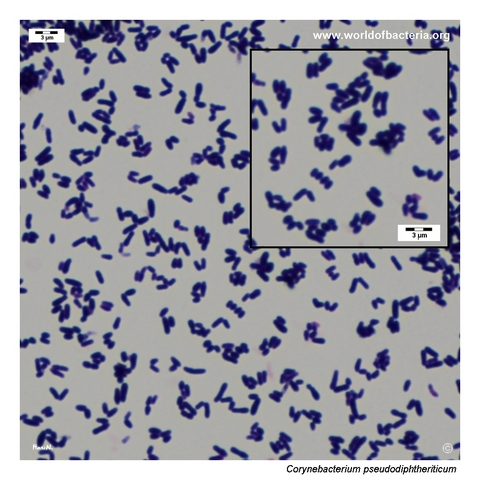
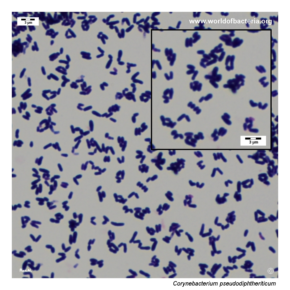
Smear prepared from a colony of Corynebacterium pseudodiphtheriticum grown on blood agar and stained by the Gram method. Gram-positive coryneform rods are visible, and local palisade-like arrangement can be seen in some parts of the field.
Diagnostic and Clinical Notes
This species is diagnostically instructive because it often looks deceptively ordinary on blood agar. The colonies are white, smooth, and non-haemolytic and may therefore be mistaken for coagulase-negative staphylococci or dismissed as insignificant diphtheroid flora unless the plate is read carefully.
The nasal specimen shown here illustrates a common interpretive problem: a respiratory corynebacterium may be recovered from symptomatic patients without necessarily being the real cause of disease. In this case, Streptococcus pneumoniae present in the same specimen was the more likely pathogen, while C. pseudodiphtheriticum was probably part of the background flora.
The comparison with Corynebacterium macginleyi is also useful. In mixed culture, C. pseudodiphtheriticum forms larger, more conspicuous white colonies, whereas C. macginleyi remains tiny and much easier to overlook.
Microscopy confirms a coryneform organism and may show palisades or angular arrangements, but species identification still depends on the total culture picture and reliable instrumental methods.
Laboratory Identification
Colony Morphology
On blood agar, Corynebacterium pseudodiphtheriticum forms white to grey-white, smooth, convex, non-haemolytic colonies with entire margins. After 48 hours the colonies are usually visually more obvious than those of very small ocular corynebacteria such as C. macginleyi, but they can still resemble non-haemolytic coagulase-negative staphylococci at first glance.
Microscopy
Gram staining shows Gram-positive coryneform rods with the usual diphtheroid appearance. Cells may occur singly, in pairs, in short palisades, or in angular arrangements. This supports assignment to the coryneform group but is not sufficient for definitive species identification.
Key Identification Clues
White non-haemolytic colonies on blood agar
Colony appearance that may mimic coagulase-negative staphylococci
Gram-positive coryneform rods with occasional palisade formation
Strong association with nasal and upper respiratory tract specimens
Frequent role as colonizing flora rather than a proven pathogen
Need for MALDI-TOF MS or another reliable identification method when precise species assignment matters
Modern Identification Methods
Reliable species identification is best achieved by MALDI-TOF mass spectrometry when the database is sufficiently complete. In problematic isolates, sequencing-based methods may still be helpful. This is particularly relevant because the organism is common enough to be encountered in routine work, yet visually nonspecific enough to be reported only generically as “diphtheroids” if instrumental identification is not pursued.
Antibiotic Characteristics
No simple species-level antimicrobial rule should be assumed from colony morphology alone for Corynebacterium pseudodiphtheriticum. Interpretation should be based on the actual clinical setting and, where clinically relevant, on isolate-specific susceptibility testing.
Because this species is often recovered as colonizing flora from the upper airway, the main practical issue is not only which antibiotic might work, but first whether treatment is indicated at all.
When the organism is judged clinically relevant, laboratory susceptibility testing is more informative than assumptions based on its coryneform appearance or on its resemblance to other nondiphtheriae corynebacteria.
Note: Antimicrobial decisions should follow the clinical syndrome, specimen context, and actual susceptibility results of the isolate.