Enterococcus faecalis is the most familiar enterococcal species in routine clinical microbiology, commonly encountered in urine culture and notable for its PYR positivity, environmental hardiness, and ability to cause both localized and invasive infection.
Basic Characteristics
Taxonomy
Domain: Bacteria
Phylum: Bacillota
Class: Bacilli
Order: Lactobacillales
Family: Enterococcaceae
Genus:Enterococcus
Species:Enterococcus faecalis
Microscopy & Gram Stain
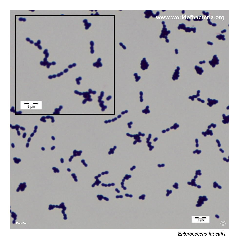
Gram-positive cocci usually arranged in pairs and short chains, with occasional small clusters
In this page set, the colony Gram stain shows a pattern fully compatible with enterococci
Microscopy is useful for placing the isolate among Gram-positive cocci, but it does not by itself distinguish E. faecalis from E. faecium or from some streptococci
Correlation with colony morphology and simple biochemical tests remains important in routine practice
Oxygen Relationship
Facultatively anaerobic organism that grows well under standard aerobic incubation
In this page set, typical colony growth was documented after 24 hours at 37 °C in ambient air
Like other enterococci, the species is hardy rather than fastidious and is readily recovered on routine blood agar
Rapid Identification Tests
Catalase: negative
PYR: positive
Bile esculin: typically positive
Growth in 6.5% NaCl: typically positive
Haemolysis: most strains are non-haemolytic, but alpha- or beta-haemolytic isolates also occur
Ecology and Clinical Relevance
Natural Habitat
Enterococcus faecalis is a common colonizer of the human gastrointestinal tract and may also be recovered from the genitourinary tract
Because it is part of normal human microbiota, interpretation always depends on specimen type, quantity of growth, and clinical context
The species is also notable for its environmental persistence, which contributes to its importance in hospital microbiology
Common Clinical Specimens
Urine specimens
Wound and soft-tissue material
Blood cultures and endocarditis-related specimens
Abdominal, biliary, and polymicrobial intra-abdominal samples
Clinical Significance
Enterococcus faecalis is one of the most frequently encountered enterococci in clinical microbiology and is a well-established cause of urinary tract infection
The species may also cause bacteremia, endocarditis, wound infection, catheter-associated infection, and polymicrobial abdominal disease
Because enterococci often coexist with other bacteria, especially in urine and wound material, laboratory interpretation should always integrate specimen quality and quantitative growth
Compared with Enterococcus faecium, E. faecalis is often more common in routine material, although antimicrobial resistance patterns may differ substantially between the two species
Differential Considerations
Enterococcus faecium, which shares PYR positivity and a similar Gram-stain appearance
Beta-haemolytic streptococci when an enterococcal isolate shows haemolysis on blood agar
Other catalase-negative Gram-positive cocci in urine culture and wound specimens
Viridans streptococci or other chain-forming cocci when only microscopy is considered
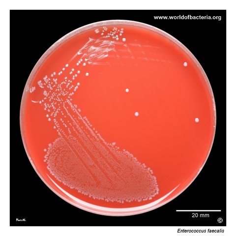
Enterococcus faecalis in pure culture on blood agar. Ten microlitres of midstream urine were inoculated and spread across the agar surface. The specimen was obtained from a 32-year-old woman with the diagnosis N30.0, acute cystitis. Culture conditions: 24 hours at 37 °C in ambient air. This image shows a typical urinary isolate of E. faecalis, a species frequently encountered in urinary tract infections and notable for its ability to grow well under routine culture conditions.
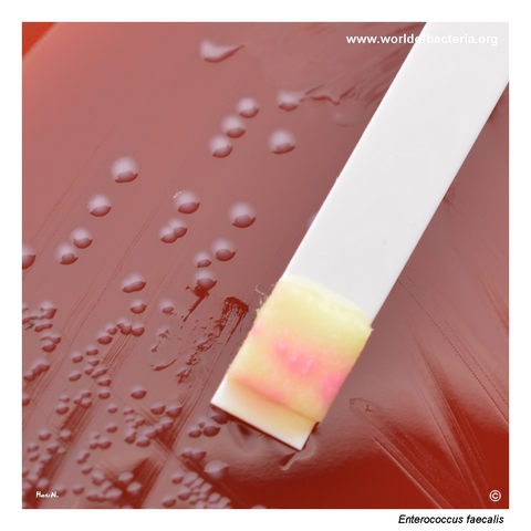
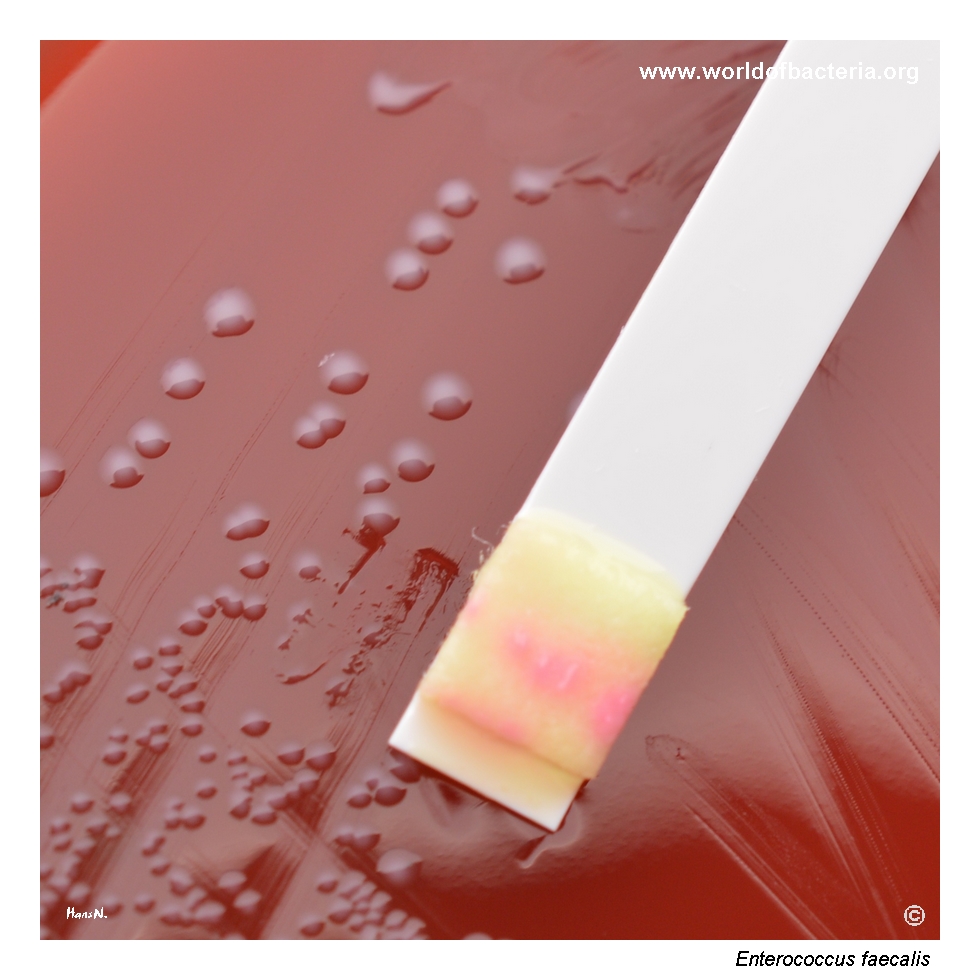
Positive PYR test. Enterococcus faecalis, like E. faecium, gives a positive PYR reaction. After the indicator reagent is added to the test strip carrying several transferred colonies, the development of a red colour confirms the presence of the enzyme pyrrolidonyl arylamidase in the tested strain. In routine microbiology, this reaction remains a useful rapid clue when differentiating enterococci from many other Gram-positive cocci.
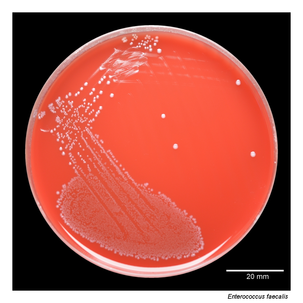
Close-up view of Enterococcus faecalis colonies on blood agar. The colonies are smooth, convex, grey-white, and round with entire margins. Most strains do not produce haemolysis on blood agar, although a smaller proportion may be alpha-haemolytic or beta-haemolytic and can then resemble beta-haemolytic streptococci. Culture conditions: 24 hours at 37 °C in ambient air. This variability is important, because haemolysis alone is not a reliable identification feature.
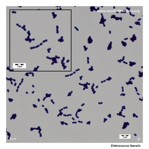
Gram-stained smear prepared from a colony of Enterococcus faecalis grown on blood agar. The organism appears as Gram-positive cocci, usually in pairs, but also arranged in short chains and small clusters. This microscopic appearance is fully compatible with enterococci, although final identification in routine practice should always be based on correlation with colony morphology, rapid biochemical tests, and, where needed, modern identification methods.
Diagnostic and Clinical Notes
This page set is useful because it combines a typical urine culture image with a positive PYR test, showing both the ordinary culture appearance and one of the classical rapid biochemical clues for enterococci.
The pure urinary growth is didactically important. Enterococcus faecalis is so common in routine material that its diagnostic significance can be underestimated, yet in the correct clinical setting it is a fully credible urinary pathogen.
The PYR image adds practical value because it links colony morphology to bench-level identification. Even in the era of MALDI-TOF MS, PYR remains a memorable and useful orienting test for enterococci.
The colony close-up also illustrates an important limitation: most E. faecalis isolates are non-haemolytic, but haemolytic variants do occur. This means that haemolysis should never be treated as a definitive exclusion criterion for enterococci.
Overall, the page reflects the core challenge of enterococcal microbiology: the organism often looks routine and familiar, but correct interpretation still depends on specimen type, simple biochemical logic, and awareness of its clinical relevance.
Laboratory Identification
Colony Morphology
On blood agar, Enterococcus faecalis typically forms smooth, convex, grey-white colonies with entire margins. Most isolates are non-haemolytic, but alpha-haemolytic or beta-haemolytic strains may occur. This means that the usual colony picture is helpful, but not sufficiently specific to identify the species by appearance alone.
Microscopy
Gram staining shows Gram-positive cocci, usually in pairs and short chains, with occasional small clusters. This pattern is fully compatible with enterococci but overlaps with other catalase-negative Gram-positive cocci, so microscopy must be interpreted together with colony morphology and rapid tests.
Key Identification Clues
Catalase-negative Gram-positive cocci in pairs and short chains
PYR-positive reaction
Typical growth on routine blood agar after 24 hours
Usually non-haemolytic, but haemolysis is variable and not decisive on its own
Need to distinguish from Enterococcus faecium and other enterococci with similar appearance
Modern Identification Methods
Modern identification is straightforward with MALDI-TOF MS and other validated laboratory systems. In routine work, however, simple biochemical logic remains valuable because a PYR-positive, catalase-negative Gram-positive coccus from urine or other typical material already strongly suggests an enterococcus while formal species identification is being completed.
Antibiotic Characteristics
Antimicrobial interpretation should never be inferred from colony appearance alone. Enterococcus faecalis often retains susceptibility patterns different from those of E. faecium, so species-level identification remains clinically relevant.
In practical diagnostics, the most important contribution of the morphology shown here is early recognition of an enterococcal isolate rather than prediction of susceptibility from haemolysis or colony size.
Note: Because enterococci have intrinsic and acquired resistance traits of major therapeutic importance, complete interpretation always depends on the individual susceptibility profile and the clinical syndrome.