Bacillus cereus is a large spore-forming bacillus of the Bacillus cereus group, typically recognized on blood agar by broad beta-haemolysis, grey rough colonies, and a distinctly coarse colonial appearance.
Basic Characteristics
Taxonomy
Domain: Bacteria
Phylum: Bacillota
Class: Bacilli
Order: Bacillales
Family: Bacillaceae
Genus:Bacillus
Species:Bacillus cereus
Microscopy & Gram Stain
Medium-sized to large Gram-positive rods, often occurring singly, in pairs, or in short chains
Endospore formation is characteristic; in the present image set the smear shows a subterminal oval spore that does not distend the cell
The Gram stain fits well with the genus Bacillus, but species identification always depends on culture features and confirmatory testing rather than microscopy alone
Compared with many coccoid skin contaminants, the large rod morphology is usually easy to appreciate once the isolate is recognized as a bacillus
Oxygen Relationship
Aerobic to facultatively anaerobic spore-forming bacillus
Grows readily on routine blood agar and develops large colonies within 24 hours under ordinary laboratory conditions
Unlike slow-growing or fastidious organisms, B. cereus usually declares itself quickly by robust growth and strong haemolysis
Rapid Identification Tests
Haemolysis on blood agar: often broad and strongly beta-haemolytic
Spore formation: characteristic of the species and genus
Modern diagnostics: MALDI-TOF MS and related methods are well suited for routine species identification, while clinical interpretation still depends on the specimen source
Ecology and Clinical Relevance
Natural Habitat
Bacillus cereus is an environmental spore-forming bacterium widely associated with soil, dust, and a broad range of external surfaces
It may also appear as a transient contaminant in clinical sampling because spores and vegetative cells are common in the environment
The species belongs to the wider Bacillus cereus group, whose members may be closely related phenotypically
Common Clinical Specimens
Depending on context, the organism may be recovered from wounds, blood cultures, catheter-associated material, ocular specimens, or food-related investigations
It may also be encountered in skin-associated or environmentally contaminated material, where the key question is whether the isolate is incidental or clinically meaningful
The image set shown here comes from a skin swab enriched in broth before subculture, which is a setting where striking colony morphology helps the isolate stand out
Clinical Significance
Bacillus cereus is clinically important both as a cause of toxin-mediated foodborne illness and as an opportunistic pathogen in selected invasive infections
At the same time, the genus contains many environmental bacilli, so interpretation of an isolate must always take specimen type and culture context into account
For atlas purposes, the main teaching value of this page is the combination of large grey colonies, rough texture, and conspicuous beta-haemolysis
Differential Considerations
Bacillus anthracis, especially because both species may form rough white-to-grey colonies, although B. anthracis is typically non-haemolytic
Other members of the Bacillus cereus group
Large aerobic Gram-positive rods recovered from mixed skin or wound material
Any strongly haemolytic, coarse colony-forming bacillus on routine blood agar
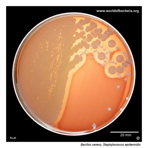
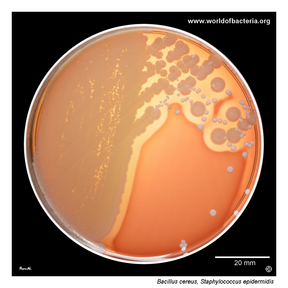
Bacillus cereus and Staphylococcus epidermidis on blood agar. The colonies of B. cereus are surrounded by a broad zone of beta-haemolysis. The specimen was a skin swab collected before blood culture sampling, enriched in broth and then subcultured onto blood agar. Culture conditions: 24 hours at 36 °C in ambient air. This image illustrates how B. cereus may stand out clearly from accompanying skin flora because of its large colonies and striking haemolysis.
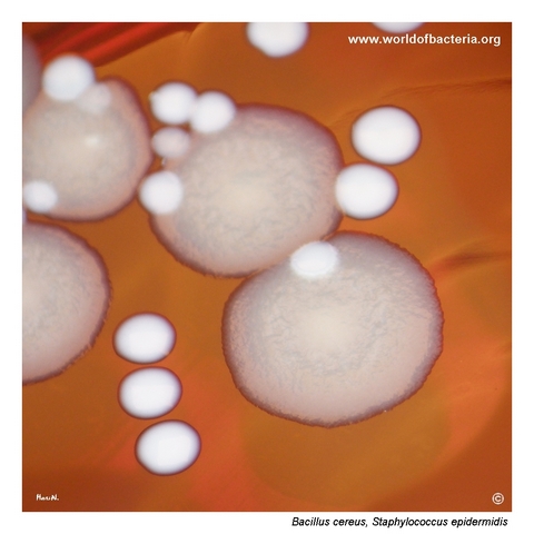
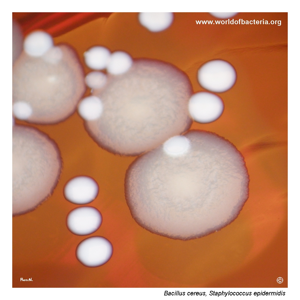
Bacillus cereus typically forms large circular colonies on blood agar with an irregular edge and a rough surface. The colony colour is greyish, which is well appreciated here in comparison with the white colonies of Staphylococcus epidermidis. Colonies of B. cereus on blood agar are often strongly beta-haemolytic. This combination of grey colour, rough surface, and marked haemolysis is one of the classic visual features of the species.
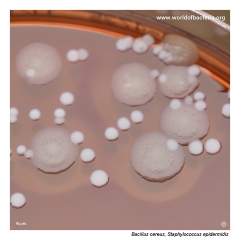
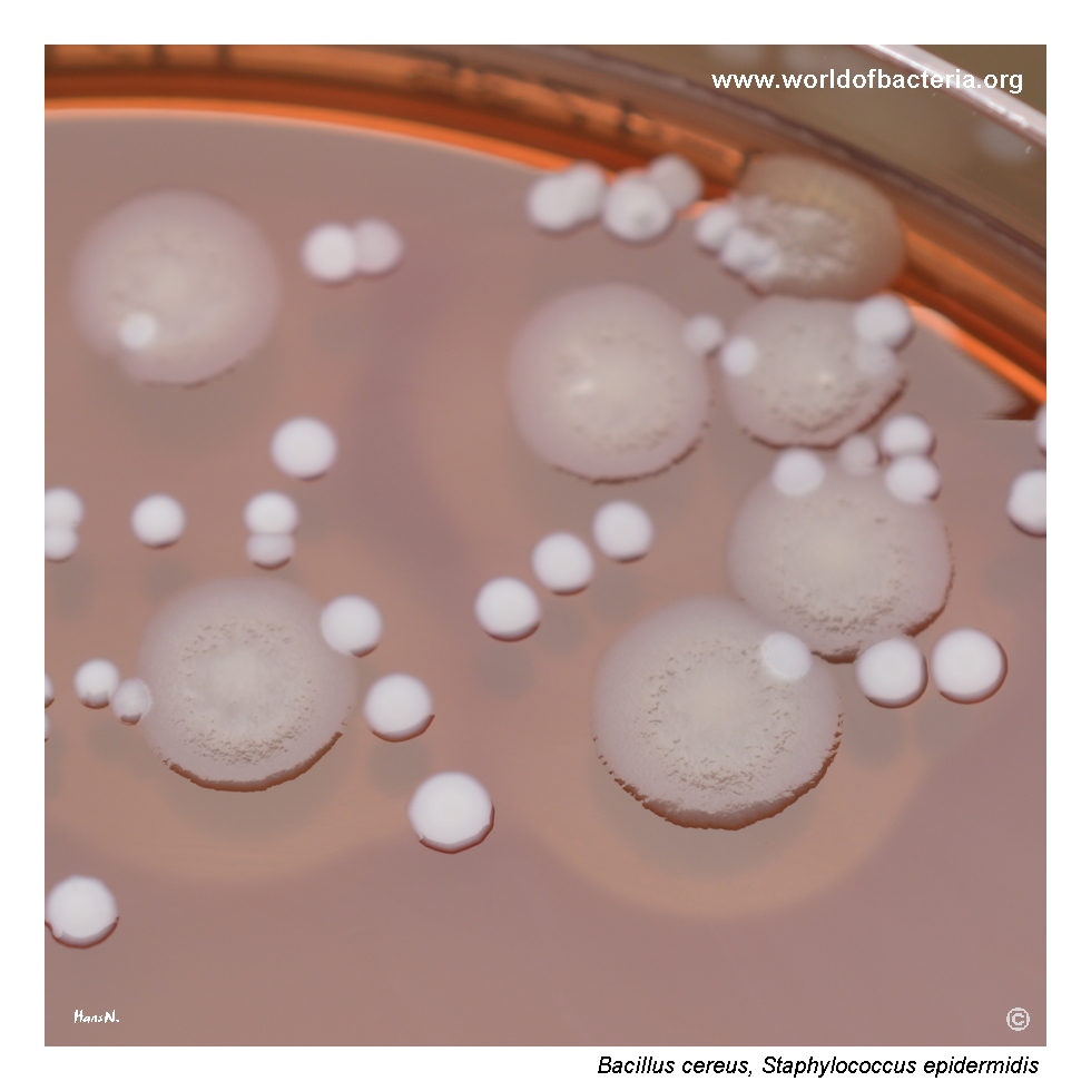
Close-up view of Bacillus cereus and Staphylococcus epidermidis colonies on blood agar after 24 hours of incubation at 36 °C in ambient air. The uneven, matt surface is typical of B. cereus. Colony shape may vary from circular to irregular, with margins ranging from entire to undulate, crenate, or fimbriate. Such variation gives the culture a distinctly coarse appearance compared with the smoother colonies of accompanying staphylococci.
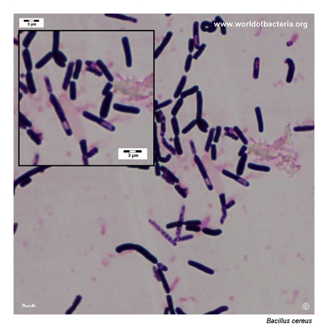
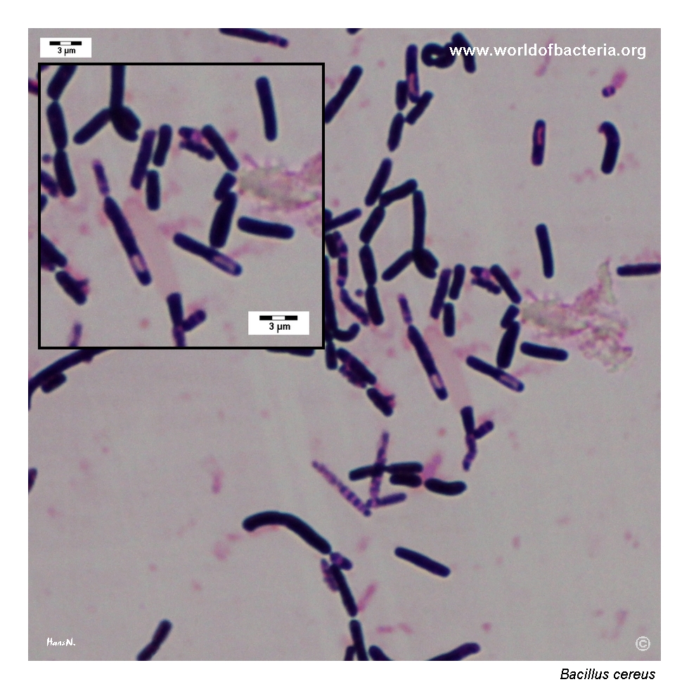
Gram-stained smear prepared from a colony of Bacillus cereus grown on blood agar. The organism appears as medium-sized to large Gram-positive rods, often with a subterminal oval spore. When present, the spore does not distend the diameter of the bacterial cell. This microscopic picture is typical of the species and fits well with its robust colonial morphology on routine solid medium.
Diagnostic and Clinical Notes
This page is didactically useful because Bacillus cereus can dominate a mixed plate visually. In the first image, broad beta-haemolysis and large colony size make the organism stand out clearly against accompanying Staphylococcus epidermidis, even though both were present in the same enriched specimen.
The colony close-ups show how much of the first impression comes from surface texture. B. cereus usually looks coarse, matt, and more irregular than the smooth white colonies of common skin staphylococci. That visual contrast is often more helpful during plate reading than colour alone.
At the same time, the page also warns against overconfidence. Large rough haemolytic colonies are suggestive, but species identification should still be confirmed, especially within the wider Bacillus cereus group and when clinically significant specimens are involved.
The comparison with Bacillus anthracis remains especially important in teaching. Both species may form large rough colonies, but B. cereus is typically haemolytic and motile, whereas B. anthracis is classically non-haemolytic and non-motile.
Overall, this image set is valuable because it shows a species that is both visually distinctive and diagnostically contextual: sometimes a meaningful pathogen, sometimes an environmental or specimen-related contaminant, but almost always recognizable once its colony morphology is appreciated.
Laboratory Identification
Colony Morphology
On blood agar, Bacillus cereus typically forms large greyish colonies with a rough matt surface and an irregular edge. Strong beta-haemolysis is common and is a major reason why the species often draws attention immediately during plate reading. Compared with smooth staphylococcal colonies, the culture usually has a more rugged and coarse appearance.
Microscopy
The Gram-stained smear shows medium-sized to large Gram-positive rods, with a subterminal oval spore visible in the illustrated preparation. This morphology is entirely compatible with the genus Bacillus, but the final identification of B. cereus still depends on the full combination of colony appearance, haemolysis, motility, and confirmatory species identification.
Key Identification Clues
Large grey rough-surfaced colonies on blood agar
Broad beta-haemolysis
Medium-sized to large Gram-positive rods
Subterminal oval spore that does not distend the cell when visible
Typical contrast with smooth white colonies of accompanying skin flora
Important differential relationship to Bacillus anthracis and other members of the Bacillus cereus group
Modern Identification Methods
Routine species identification is best supported by MALDI-TOF MS or other validated laboratory methods. This is especially useful because the broader Bacillus cereus group contains several closely related species whose colony morphology may overlap. The atlas value of the present page lies mainly in phenotype recognition, not in morphology-only identification.
Antibiotic Characteristics
No simple therapeutic assumption should be made from colony morphology alone. Clinical interpretation of Bacillus cereus depends on the specimen source, the probability of contamination versus infection, and formal susceptibility testing of the isolate.
B. cereus is often more resistant than casual plate morphology might suggest, so clinically important isolates should always be handled as full identification-and-susceptibility problems rather than as routine environmental bacilli.
Note: For this atlas page, the educational emphasis is on recognition of the large beta-haemolytic rough colony phenotype and its differential interpretation, not on treatment guidance.