Corynebacterium diphtheriae is a medically important coryneform species best known as the classical cause of diphtheria when toxigenic strains are involved, while non-toxigenic isolates may also be recovered in clinical microbiology and require careful laboratory interpretation.
Basic Characteristics
Taxonomy
Domain: Bacteria
Phylum: Actinomycetota
Class: Actinomycetes
Order: Corynebacteriales
Family: Corynebacteriaceae
Genus:Corynebacterium
Species:Corynebacterium diphtheriae
Microscopy & Gram Stain
Gram-positive coryneform rods
Cells are typically club-shaped or slightly pleomorphic
Bacteria may remain attached in pairs, V-shaped arrangements, or irregular palisades
Microscopy supports the genus-level impression but does not by itself determine toxigenicity or full species identity
Oxygen Relationship
Aerobic organism that grows under routine laboratory incubation conditions
Good growth can be obtained on blood agar and similar non-selective media
In practical diagnostics, the organism behaves as a conventional cultivable coryneform isolate rather than as a highly fastidious bacterium
Rapid Identification Tests
Catalase: positive
Morphology: coryneform Gram-positive rod
Important limitation: colony appearance on blood agar is not sufficient to distinguish toxigenic from non-toxigenic strains
Public health note: if C. diphtheriae is identified, additional testing is needed to determine whether the strain is toxigenic
Practical caveat: on non-selective media the colonies may resemble less significant Gram-positive cocci or coryneforms
Ecology and Clinical Relevance
Natural Habitat
Humans are the main reservoir, with carriage involving the respiratory tract and skin
The organism may be recovered from respiratory material, skin lesions, and occasionally from other clinical sources
The isolate shown on this page is a non-toxigenic external quality assessment strain rather than a routine patient isolate
Common Clinical Specimens
Throat and nasopharyngeal swabs when diphtheria is suspected
Cutaneous swabs or wound material from skin lesions
Occasionally other clinical samples when invasive infection is investigated
Clinical Significance
Toxigenic strains are the classical cause of diphtheria and have major public health relevance
Non-toxigenic strains are still clinically relevant because they may be recovered from cutaneous or invasive infections
Once the species is identified, interpretation must include both the clinical setting and the toxigenic status of the isolate
Differential Considerations
Coagulase-negative staphylococci and enterococci when only small non-haemolytic colonies on blood agar are considered
Other coryneform bacteria such as Corynebacterium pseudodiphtheriticum or Corynebacterium ulcerans
Low-significance diphtheroids that may appear similar on initial plate reading but differ markedly in clinical importance
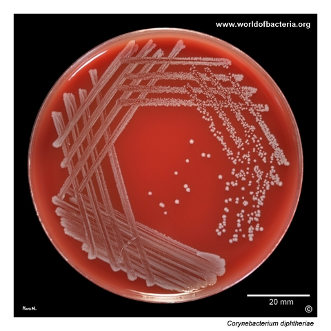
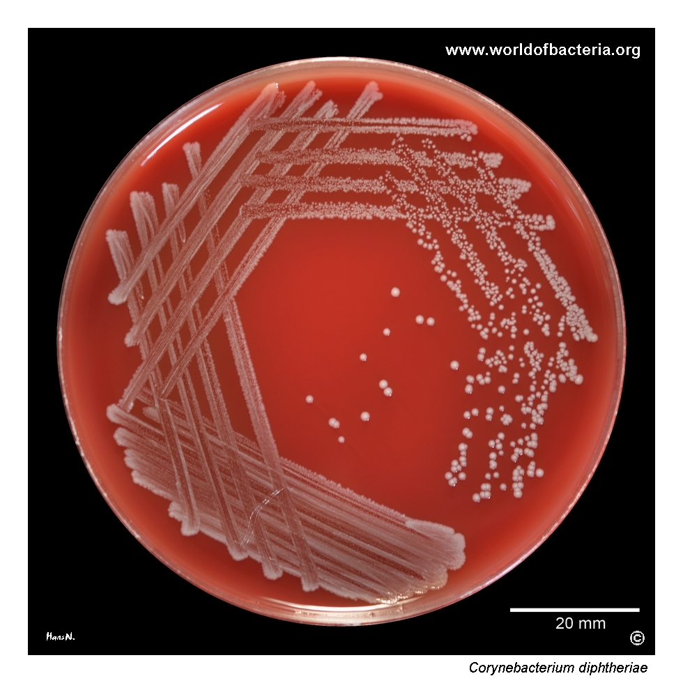
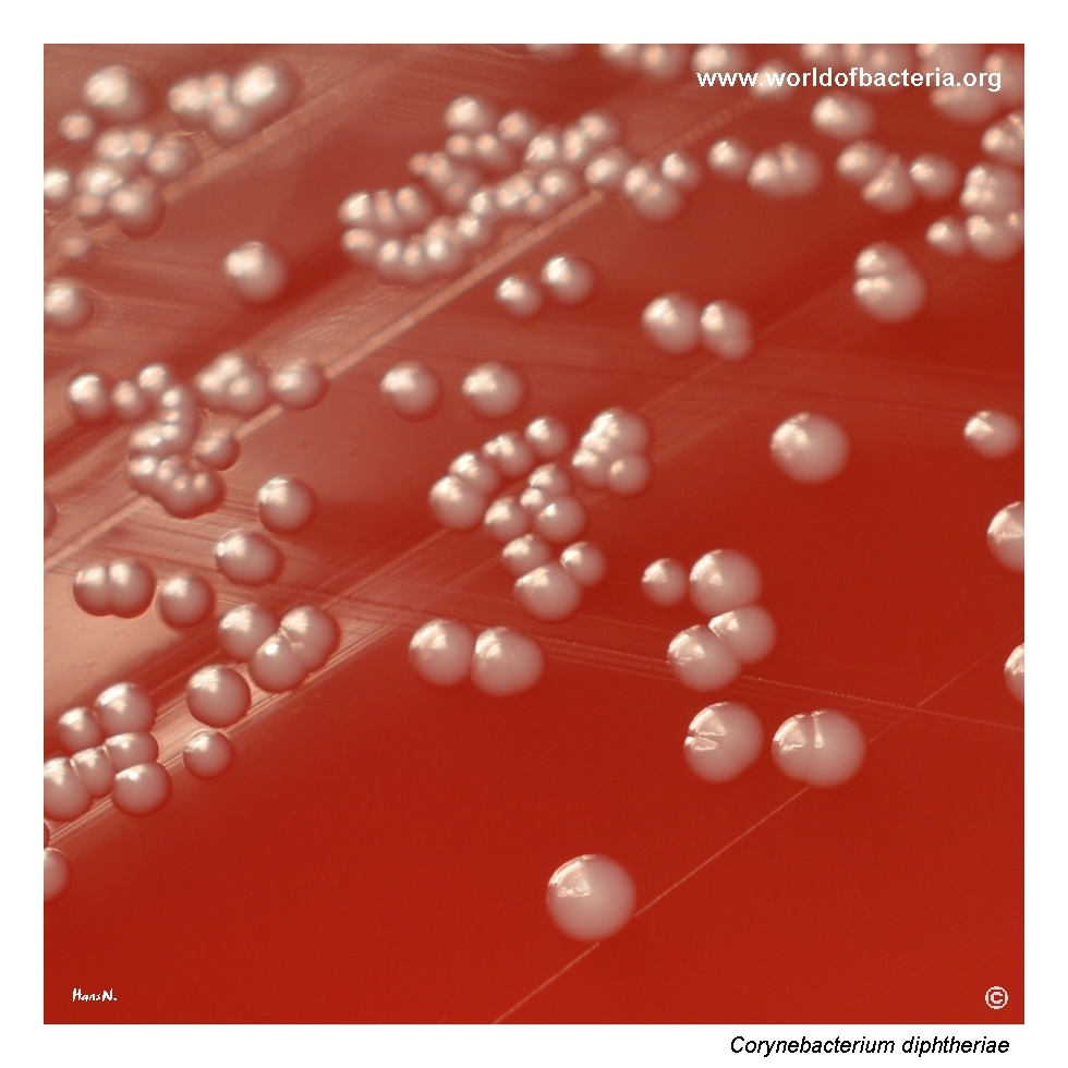
Corynebacterium diphtheriae on blood agar. Incubation: 24 hours at 36 °C in 5% CO2. This was a non-toxigenic strain provided as a control sample within an external quality assessment programme.
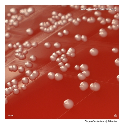
Colonies of Corynebacterium diphtheriae on blood agar. On a non-selective medium such as blood agar, the colonies may resemble non-haemolytic coagulase-negative staphylococci or enterococci. Incubation: 24 hours at 36 °C in 5% CO2.
Close-up view of Corynebacterium diphtheriae colonies. On blood agar, the organism forms smooth, convex colonies with entire margins and no haemolysis in this strain. Incubation: 24 hours at 36 °C in 5% CO2.
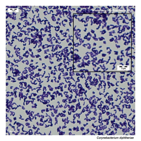
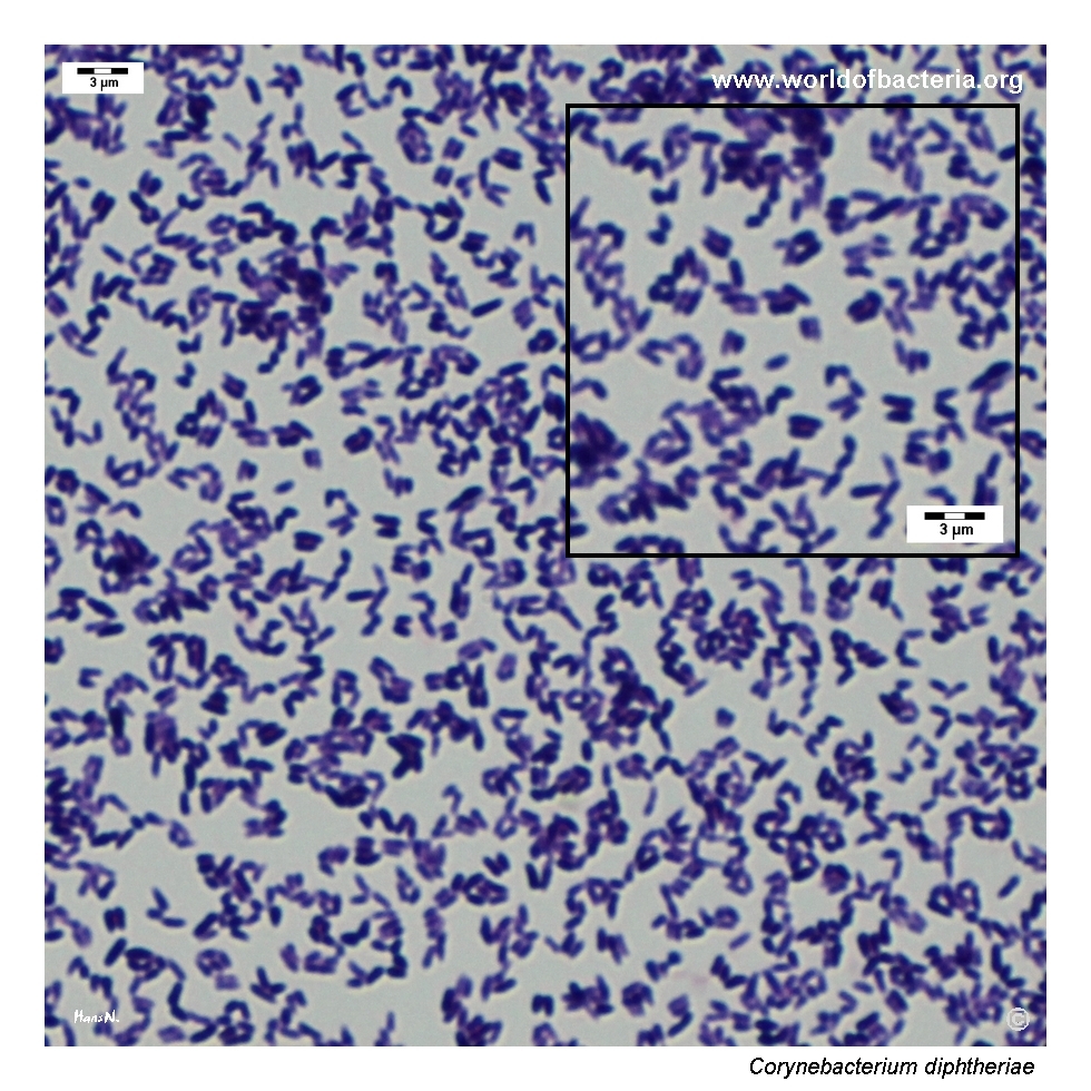
Smear prepared from a colony of Corynebacterium diphtheriae grown on blood agar and stained by the Gram method. Gram-positive coryneform rods are visible, often with club-shaped ends and arrangements in pairs or V-shaped forms.
Diagnostic and Clinical Notes
This page is intentionally instructive because it shows how inconspicuous Corynebacterium diphtheriae may appear on ordinary blood agar. Without a targeted diagnostic context, the colonies could easily be mistaken for less important organisms such as non-haemolytic coagulase-negative staphylococci, enterococci, or routine diphtheroids.
That practical point matters because the name of the species carries immediate clinical and public health significance. However, species identification alone does not answer the most important question: whether the strain is toxigenic. The isolate shown here was explicitly non-toxigenic, which is an essential part of its interpretation.
Microscopy provides a classic coryneform image, with Gram-positive rods that may broaden at one end and remain attached in characteristic angular arrangements. This morphology is highly useful for orientation but is not sufficiently specific to separate C. diphtheriae from all other corynebacteria.
For routine clinical work, the key lesson is therefore twofold: first, seemingly unremarkable colonies on blood agar should not be dismissed when the species identity is important; second, whenever C. diphtheriae is identified, toxigenicity testing and public health procedures become part of the diagnostic pathway.
Laboratory Identification
Colony Morphology
On blood agar, Corynebacterium diphtheriae may form smooth, convex, non-haemolytic colonies with entire margins. In this respect, the organism can appear deceptively ordinary on non-selective media and may resemble several less significant Gram-positive bacteria encountered in routine culture.
Microscopy
Gram staining shows Gram-positive coryneform rods with a clubbed or slightly pleomorphic appearance. Cells may be arranged in pairs, V-shaped forms, or irregular palisades. This is fully compatible with the genus Corynebacterium and is a classic teaching morphology in diagnostic microbiology.
Key Identification Clues
Gram-positive coryneform rods
Club-shaped cells with angular arrangements in pairs or V-forms
Small smooth colonies on blood agar that may mimic other organisms
Catalase positive
Major interpretive importance of toxigenicity testing
Need to distinguish the species from other clinically encountered corynebacteria
Modern Identification Methods
Modern species identification is commonly achieved by MALDI-TOF mass spectrometry when the database coverage is adequate. Once the organism is identified as Corynebacterium diphtheriae, additional reference or public health laboratory testing is needed to determine whether the strain is toxigenic, because this cannot be inferred from morphology alone.
Antibiotic Characteristics
Antimicrobial interpretation for Corynebacterium diphtheriae should never be reduced to a simple rule based on plate appearance. The first critical distinction is clinical relevance, and the second is whether the isolate is toxigenic.
For confirmed clinical infection, therapy and public health management are guided by the overall syndrome, laboratory susceptibility testing, and official recommendations rather than by presumptive morphology on culture media.
In routine laboratory work, the most urgent practical issue is often correct recognition of the species and timely communication when diphtheria is a possibility.
Note: Antimicrobial therapy and public health action should always follow formal laboratory identification, toxigenicity testing, and the clinical context.