Gardnerella vaginalis is a small Gram-variable urogenital species strongly associated with bacterial vaginosis and notable for beta-haemolysis on media containing rabbit or human erythrocytes, while remaining much less conspicuous on routine sheep-blood agar.
Basic Characteristics
Taxonomy
Domain: Bacteria
Phylum: Actinomycetota
Class: Actinomycetes
Order: Bifidobacteriales
Family: Bifidobacteriaceae
Genus:Gardnerella
Species:Gardnerella vaginalis
Microscopy & Gram Stain
Small pleomorphic rods or coccobacillary cells with characteristically poor and often Gram-variable staining
Although the cell wall places the species among Gram-positive bacteria, routine smears may look partly Gram-negative or only weakly stained
In this page set, the stained preparation shows small pleomorphic rods that can be diagnostically confusing at first glance
Microscopy is useful for recognizing the organism as a small urogenital rod, but species identification requires correlation with culture conditions and laboratory methods
Oxygen Relationship
Facultatively anaerobic organism that grows best under enriched culture conditions such as 5% CO2
In this page set, clear colony development was documented after 3 days at 36 °C in 5% CO2
Growth is often relatively delicate and slower than that of many routine urogenital bacteria, which is why adequate incubation time is important
Rapid Identification Tests
Gram stain: Gram-variable pleomorphic rods
Haemolysis: beta-haemolysis may be demonstrated on media containing rabbit or human erythrocytes
Routine sheep-blood agar: growth may occur, but haemolysis is often absent
Atmosphere: growth is favoured by 5% CO2 and prolonged incubation
Practical note: modern identification methods are often preferred because visual morphology alone may be misleading
Ecology and Clinical Relevance
Natural Habitat
Gardnerella vaginalis is strongly associated with the human urogenital tract, especially the vaginal microbiota
The species may be detected in asymptomatic colonization as well as in dysbiotic states, so microbiological interpretation always depends on the overall clinical and ecological context
It is best understood as part of a broader vaginal microbial community rather than as a solitary organism acting in isolation
Common Clinical Specimens
Vaginal and other urogenital specimens
Material examined for bacterial vaginosis or vaginal dysbiosis
Specialized culture media used for detection of gardnerellas
More rarely, unusual extra-genital or invasive clinical material
Clinical Significance
Gardnerella vaginalis is one of the best-known organisms associated with bacterial vaginosis, where it participates in a polymicrobial shift away from Lactobacillus-dominated flora
The species is clinically important not because every isolate is automatically pathogenic, but because its recovery may support the interpretation of an altered vaginal microbial ecosystem
In routine practice, the organism is especially relevant when correlated with compatible symptoms, discharge characteristics, microscopy, and other laboratory findings such as clue cells or molecular tests
This page set focuses on the classical phenotypic presentation on haemolysis-supporting medium and on the distinctive Gram-variable appearance of the cells
Differential Considerations
Other small coryneform or coccobacillary bacteria from the urogenital tract
Other Gardnerella species now recognized by genomic methods, even though G. vaginalis remains the recommended medical name in current nomenclature
Mixed vaginal flora in which the organism may be present together with anaerobes, lactobacilli, yeasts, or other accompanying bacteria
Misinterpretation as a Gram-negative organism because of the characteristic Gram-variable staining pattern
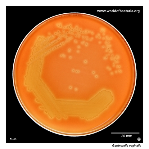
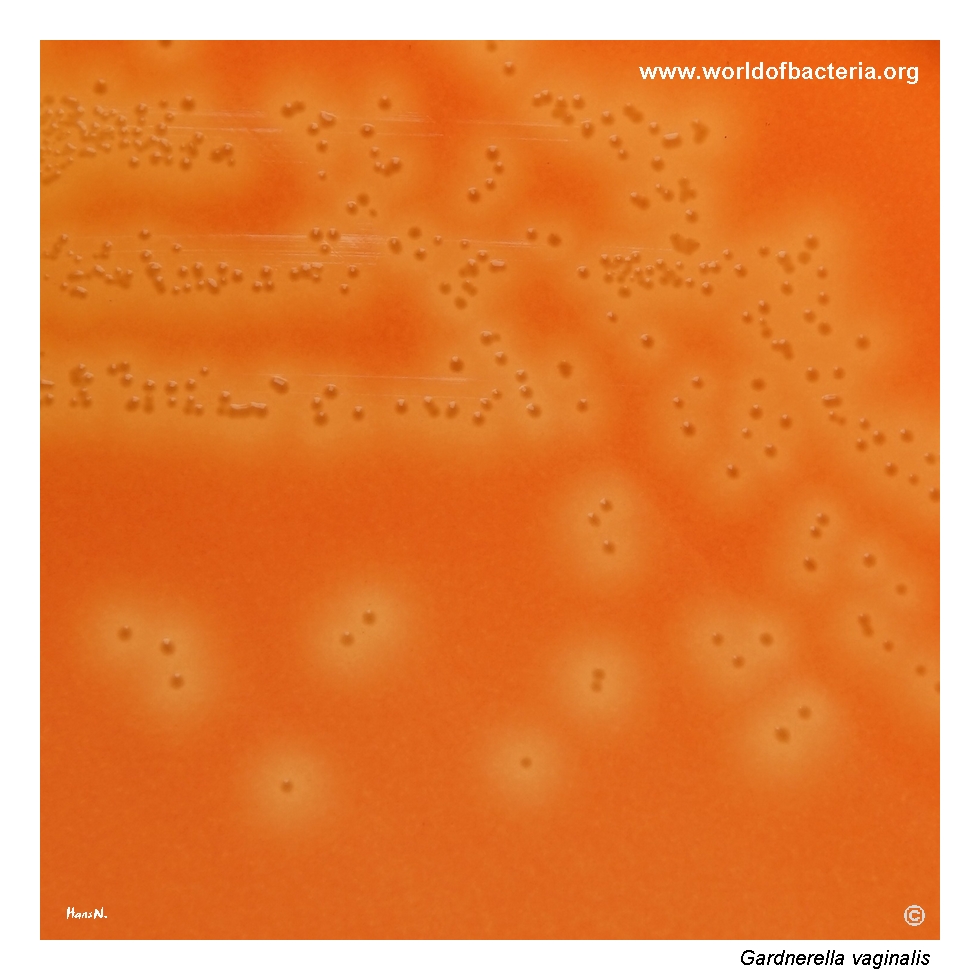
Gardnerella vaginalis, collection strain CIP 82.14, grown on agar containing rabbit erythrocytes. This is a specially designed culture medium used for the detection of gardnerellas in material from the urogenital tract. A clear zone of beta-haemolysis is visible around the grown culture. Culture conditions: 3 days at 36 °C in 5% CO2. This image illustrates one of the classic phenotypic features that may be demonstrated on suitable haemolysis-supporting media.
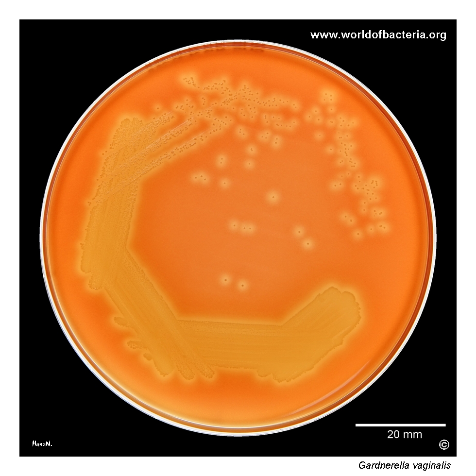
Colonies of Gardnerella vaginalis surrounded by a zone of beta-haemolysis. G. vaginalis is able to haemolyse rabbit or human erythrocytes. The strains are also capable of growth on blood agar containing sheep erythrocytes, but in that setting haemolysis is typically not produced. Culture conditions: 3 days at 36 °C in 5% CO2. This difference is diagnostically useful and helps explain why the choice of medium may strongly influence the visible phenotype.
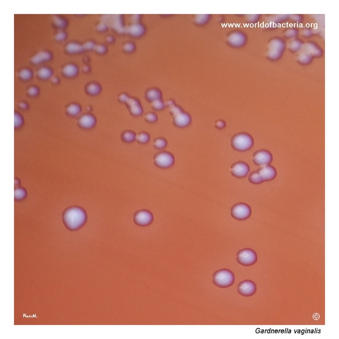
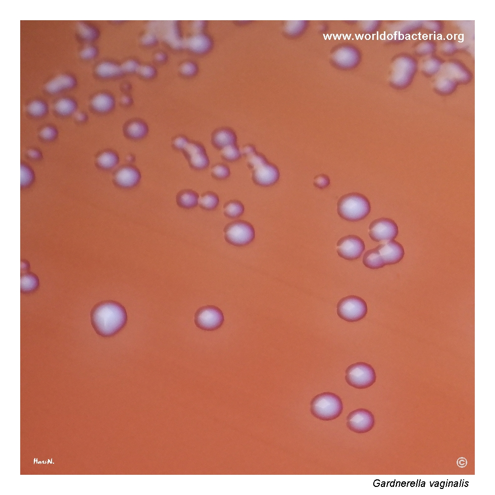
Close-up view of Gardnerella vaginalis colonies. The colonies are small, convex, and round with entire margins, measuring approximately 1 mm in diameter. Culture conditions: 3 days at 36 °C in 5% CO2. Their small size and relatively delicate appearance mean that they may be overlooked easily unless the plate is examined carefully after sufficient incubation time.
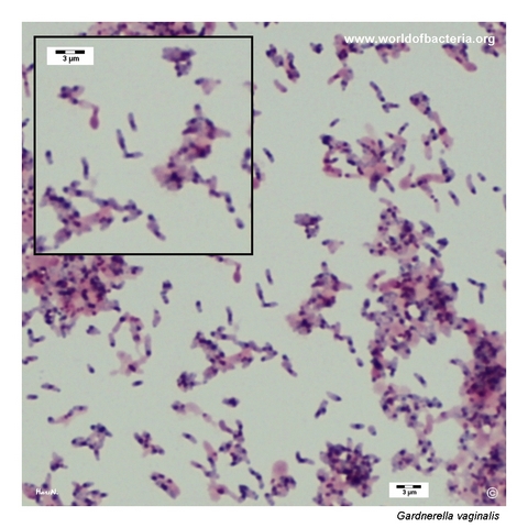
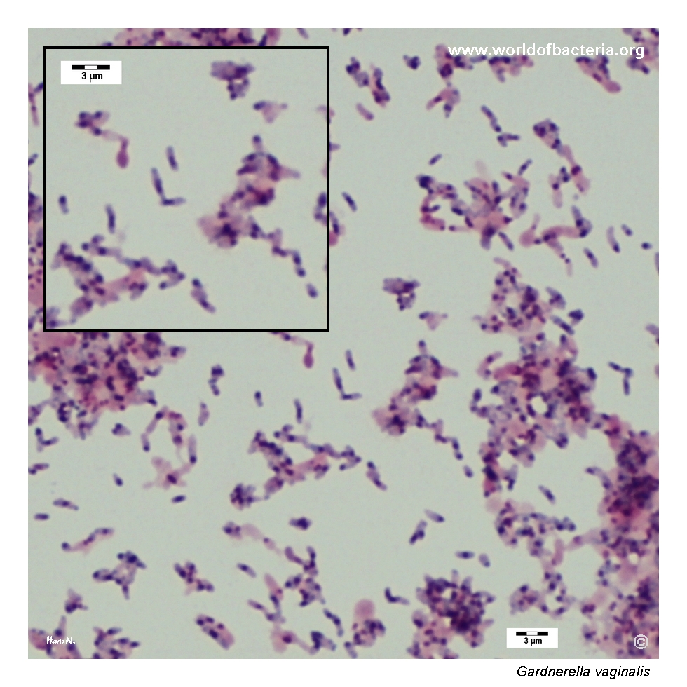
Gram-stained smear prepared from colonies of Gardnerella vaginalis, strain CIP 82.14, after 48 hours of incubation. The organism appears as small pleomorphic rods that stain poorly by the Gram method. Although the cell wall structure places the species among Gram-positive bacteria, cultures often look more like Gram-negative organisms under the microscope. This Gram-variable appearance is one of the characteristic features that can complicate routine microscopic interpretation.
Diagnostic and Clinical Notes
This page set is didactically valuable because it shows the organism on a medium that actually reveals one of its classic phenotypic traits: beta-haemolysis on rabbit-erythrocyte agar.
The contrast between rabbit- or human-blood haemolysis and the much less conspicuous behaviour on routine sheep-blood agar is important in practice. Without the appropriate medium, the phenotype may look far less distinctive.
The small colony size after prolonged incubation is another key point. Even when growth is present, the organism may be overlooked if the plate is read too early or if visual attention is drawn to more robust accompanying flora.
The Gram-stained smear reinforces a familiar laboratory pitfall: despite belonging among Gram-positive bacteria, Gardnerella vaginalis often appears Gram-variable or even deceptively Gram-negative.
Taken together, the images explain why classical phenotypic recognition of G. vaginalis depends strongly on culture conditions, incubation time, and careful microscopic interpretation.
Laboratory Identification
Colony Morphology
On suitable haemolysis-supporting media, Gardnerella vaginalis forms small, convex, round colonies with entire margins, typically around 1 mm in diameter after 3 days of incubation. Beta-haemolysis may be clear on rabbit- or human-erythrocyte media, whereas on routine sheep-blood agar the colonies may remain much less distinctive. This dependence on medium composition is one of the reasons why the species can be underestimated in routine culture.
Microscopy
Gram staining shows small pleomorphic rods or coccobacillary forms with characteristically variable staining. Some fields may suggest Gram-negative bacteria even though the organism belongs among Gram-positive taxa. This Gram-variable appearance is one of the most important routine clues and should always be interpreted together with the culture phenotype and specimen source.
Key Identification Clues
Small, delicate colonies after prolonged incubation in 5% CO2
Beta-haemolysis on rabbit- or human-erythrocyte media
Growth without the same haemolytic phenotype on sheep-blood agar
Gram-variable pleomorphic rods that may superficially resemble Gram-negative coccobacilli
Strong association with urogenital specimens and bacterial-vaginosis work-up
Modern Identification Methods
Modern identification is best achieved with validated MALDI-TOF MS databases or molecular methods. This is increasingly relevant because the genus Gardnerella now includes multiple genomically defined species. Even so, current nomenclature continues to retain Gardnerella vaginalis as the correct and medically recommended name in LPSN, which makes the species especially important as a practical reference point in clinical microbiology.
Antibiotic Characteristics
This page is primarily about phenotypic recognition rather than species-specific susceptibility prediction. In clinical practice, the significance of Gardnerella vaginalis usually arises in the context of bacterial vaginosis or other urogenital dysbiosis rather than as an isolated susceptibility-testing problem.
No simple species-level susceptibility rule should be inferred from colony morphology or haemolysis. In unusual invasive isolates, antimicrobial testing should be performed on the individual strain.
Note: The main practical lesson from this page is that correct recognition of G. vaginalis depends more on appropriate culture conditions and careful interpretation than on any single routine susceptibility pattern.