Corynebacterium aurimucosum is an uncommon nondiphtherial corynebacterial species that may appear as very small, easily overlooked colonies in urine culture, making prolonged incubation and careful plate reading especially useful.
Basic Characteristics
Taxonomy
Domain: Bacteria
Phylum: Actinomycetota
Class: Actinomycetes
Order: Corynebacteriales
Family: Corynebacteriaceae
Genus:Corynebacterium
Species:Corynebacterium aurimucosum
Microscopy & Gram Stain
Short Gram-positive coryneform rods typical of the genus Corynebacterium
The Gram stain is particularly helpful when colonies on blood agar are small and visually nondistinctive
In this page set, microscopy supports correct placement within the corynebacterial group rather than among Gram-positive cocci with superficially similar colonies
As with other nondiphtherial corynebacteria, morphology alone does not establish the species
Oxygen Relationship
Routine aerobic coryneform isolate that grows on standard blood agar under ordinary laboratory conditions
In these images, colony development is illustrated after 24 and 48 hours at 36 °C in ambient air
The main practical issue is not fastidiousness but the fact that early colonies may be so small that they are easy to overlook
Rapid Identification Tests
Morphology: Gram-positive coryneform rod
Colony phenotype: small non-haemolytic colonies that may be minute after 24 hours and more typical after prolonged incubation
Useful visual clue: some colonies develop a raised central area that becomes easier to appreciate after 48 hours
Clinical setting: interpretation in urine depends on quantitative growth, accompanying flora, and the patient's symptoms
Important limitation: reliable species-level identification generally requires MALDI-TOF MS or another modern identification method
Ecology and Clinical Relevance
Natural Habitat
Corynebacterium aurimucosum is a human-associated nondiphtherial corynebacterium, but its routine ecological role is less familiar in clinical practice than that of better-known skin corynebacteria
The species has been recovered from human clinical material and should be regarded as an occasional rather than everyday laboratory finding
For urine cultures in particular, significance must be judged cautiously because not every coryneform isolate represents true urinary tract infection
Common Clinical Specimens
Urinary specimens, including midstream urine
Other human clinical material in selected case reports or sporadic laboratory recovery
Specimens in which small coryneform colonies might easily be underestimated during primary culture reading
Clinical Significance
Corynebacterium aurimucosum is best regarded as an uncommon human clinical isolate whose significance is strongly specimen-dependent
In urinary microbiology, the organism should not automatically be dismissed, because published reports support the possibility of true urinary infection in selected cases
At the same time, interpretation still requires attention to colony count, purity of growth, accompanying flora, and the overall clinical picture
Differential Considerations
Small colonies of coagulase-negative staphylococci or enterococci when only the plate is viewed macroscopically
Other nondiphtherial corynebacteria recovered from urine or mixed clinical material
Corynebacterium amycolatum and Corynebacterium tuberculostearicum, which can also appear as visually modest coryneform isolates on blood agar
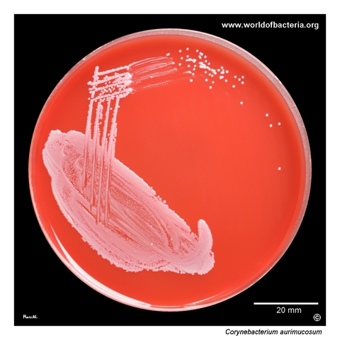
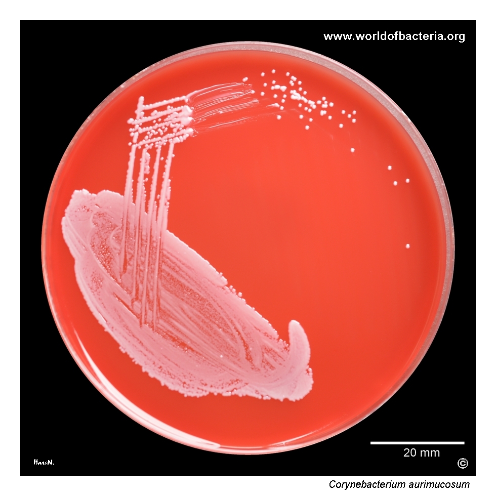
Corynebacterium aurimucosum. Midstream urine, 10 µl, inoculated and streaked onto blood agar. The specimen came from a 79-year-old woman with the diagnosis N30.0, acute cystitis. Culture conditions: 24 hours at 36 °C in ambient air. In urinary specimens, recovery of coryneform bacteria must always be interpreted in relation to colony count, accompanying flora, and the clinical picture, because some isolates may represent only incidental colonization.
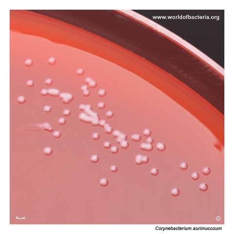
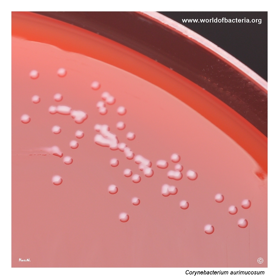
Colonies of Corynebacterium aurimucosum on blood agar after 24 hours of incubation at 36 °C. In this primary culture, the colonies are conspicuously small. Such minute colonies may be easy to overlook during early plate reading, especially in urine culture where attention is often focused on more rapidly growing and more typical uropathogens. Prolonged incubation can therefore be helpful when inconspicuous coryneform bacteria are present.
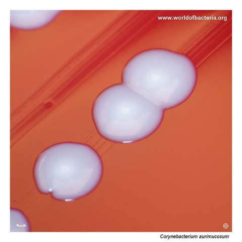
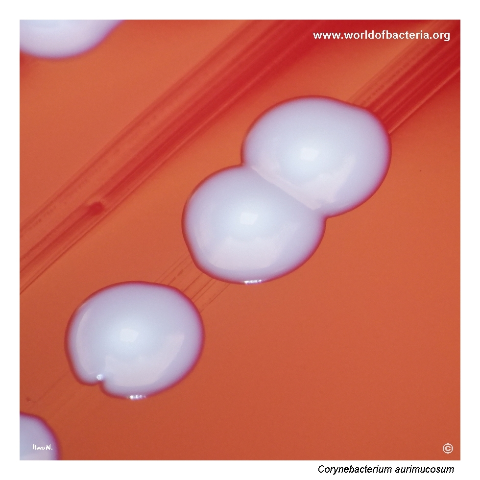
Close-up view of Corynebacterium aurimucosum colonies after 48 hours of incubation at 36 °C in ambient air. This image also shows a primary urine culture, but here the colonies have reached a more typical size of about 3 mm in diameter and display a raised central area. The comparison with the previous image illustrates how colony size and surface profile may become much more evident after prolonged incubation.
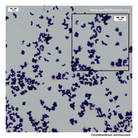
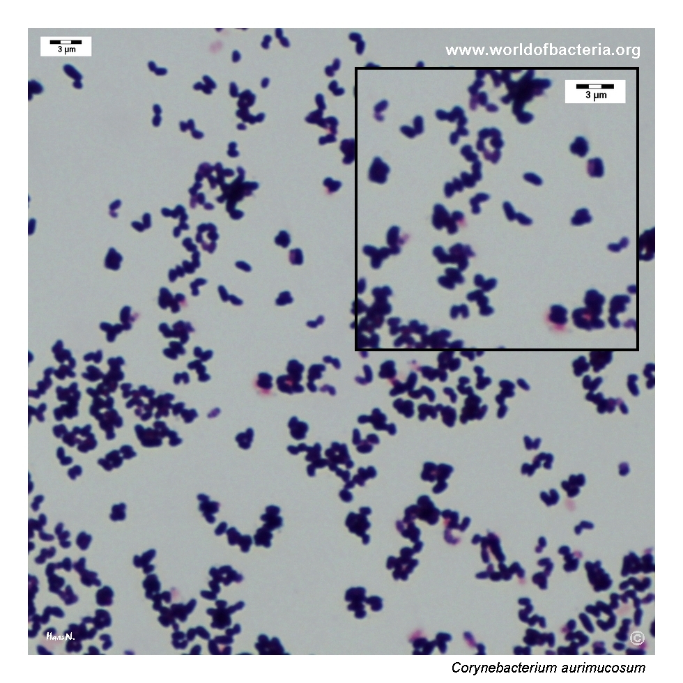
Gram-stained smear prepared from a colony of Corynebacterium aurimucosum grown on blood agar. The organism appears as short Gram-positive coryneform rods. This microscopic morphology supports placement within the corynebacterial group, although definitive species identification requires correlation with culture characteristics and, where needed, modern laboratory identification methods.
Diagnostic and Clinical Notes
This page is instructive mainly because it shows how easily an uncommon corynebacterial isolate can remain inconspicuous in urine culture. At 24 hours, the colonies are still very small, and without deliberate attention they might be overshadowed by more familiar uropathogens or dismissed as minor background growth.
The comparison between the early and prolonged incubation images is particularly useful. After 48 hours, the colonies become larger and their raised central profile becomes clearer, demonstrating how additional incubation can materially improve visual recognition of a subtle isolate.
Interpretation, however, remains specimen-driven rather than purely descriptive. The isolate came from midstream urine from a 79-year-old woman with acute cystitis, so the central microbiological question is whether the organism represents incidental colonization or a clinically relevant urinary isolate. That decision depends on quantity, purity, accompanying flora, and the clinical scenario rather than on morphology alone.
The broader teaching point is that not all coryneform bacteria in urine are automatically negligible. Corynebacterium aurimucosum is uncommon, but published case material indicates that true urinary tract infection is possible, which supports a balanced interpretation rather than reflex dismissal.
Laboratory Identification
Colony Morphology
On blood agar, Corynebacterium aurimucosum forms small non-haemolytic colonies that may initially be strikingly minute. In this page set, the early image after 24 hours shows colonies that are easy to miss, whereas the 48-hour image demonstrates more typical colonies of about 3 mm with a raised central area. This time-dependent change is one of the most useful practical observations for routine plate reading.
Microscopy
Gram staining shows short Gram-positive coryneform rods compatible with the genus Corynebacterium. This is diagnostically important because the colonies on blood agar may provide only limited visual clues and could otherwise be interpreted as small coccoid colonies of another Gram-positive group. As in other nondiphtherial corynebacteria, microscopy supports correct generic placement but does not establish the species on its own.
Key Identification Clues
Initially minute non-haemolytic colonies on blood agar in urine culture
More evident colony size and a raised central area after prolonged incubation
Short Gram-positive coryneform rods on Gram stain
Specimen context in which interpretation depends on colony count and accompanying flora
Need for MALDI-TOF MS or another reliable modern method for definitive species-level identification
Modern Identification Methods
Reliable identification is best achieved by MALDI-TOF mass spectrometry when the database is suitable for nondiphtherial corynebacteria. This is particularly relevant for C. aurimucosum, because the species is uncommon and its colonies may look too ordinary or too small to inspire confident presumptive identification from morphology alone. In selected situations, especially when clinical significance is uncertain, additional phenotypic or molecular confirmation may be useful.
Antibiotic Characteristics
For Corynebacterium aurimucosum, no simple susceptibility pattern should be inferred from colony appearance. The visual phenotype helps recognition, but not treatment prediction.
When the organism is considered clinically relevant, antimicrobial susceptibility testing should be performed on the individual isolate. This is especially important in urinary material, where the decision to report and fully work up a coryneform isolate already depends on a careful clinical-microbiological judgment.
The practical first step is therefore interpretive: deciding whether the isolate is likely to represent incidental colonization or a meaningful urinary finding. Once that threshold is crossed, therapy should be guided by isolate-specific susceptibility data rather than by assumptions about corynebacteria as a group.
Note: The didactic value of this page lies less in a distinctive resistance profile than in showing that an initially inconspicuous corynebacterial urine isolate may still deserve thoughtful evaluation.