Corynebacterium mucifaciens is an uncommon nondiphtherial corynebacterial species known for its small yellow mucoid colonies and occasional recovery from human blood and other clinical material, where interpretation often depends strongly on catheter status and sampling context.
Basic Characteristics
Taxonomy
Domain: Bacteria
Phylum: Actinomycetota
Class: Actinomycetes
Order: Corynebacteriales
Family: Corynebacteriaceae
Genus:Corynebacterium
Species:Corynebacterium mucifaciens
Microscopy & Gram Stain
Gram-positive coryneform rods typical of the genus Corynebacterium
In this page set, Gram staining helps distinguish the isolate from Gram-positive cocci that can produce superficially similar small colonies on blood agar
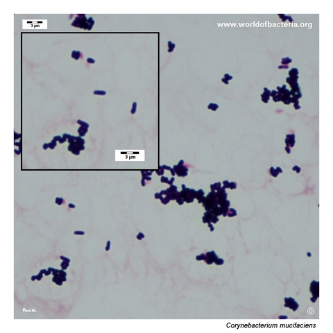
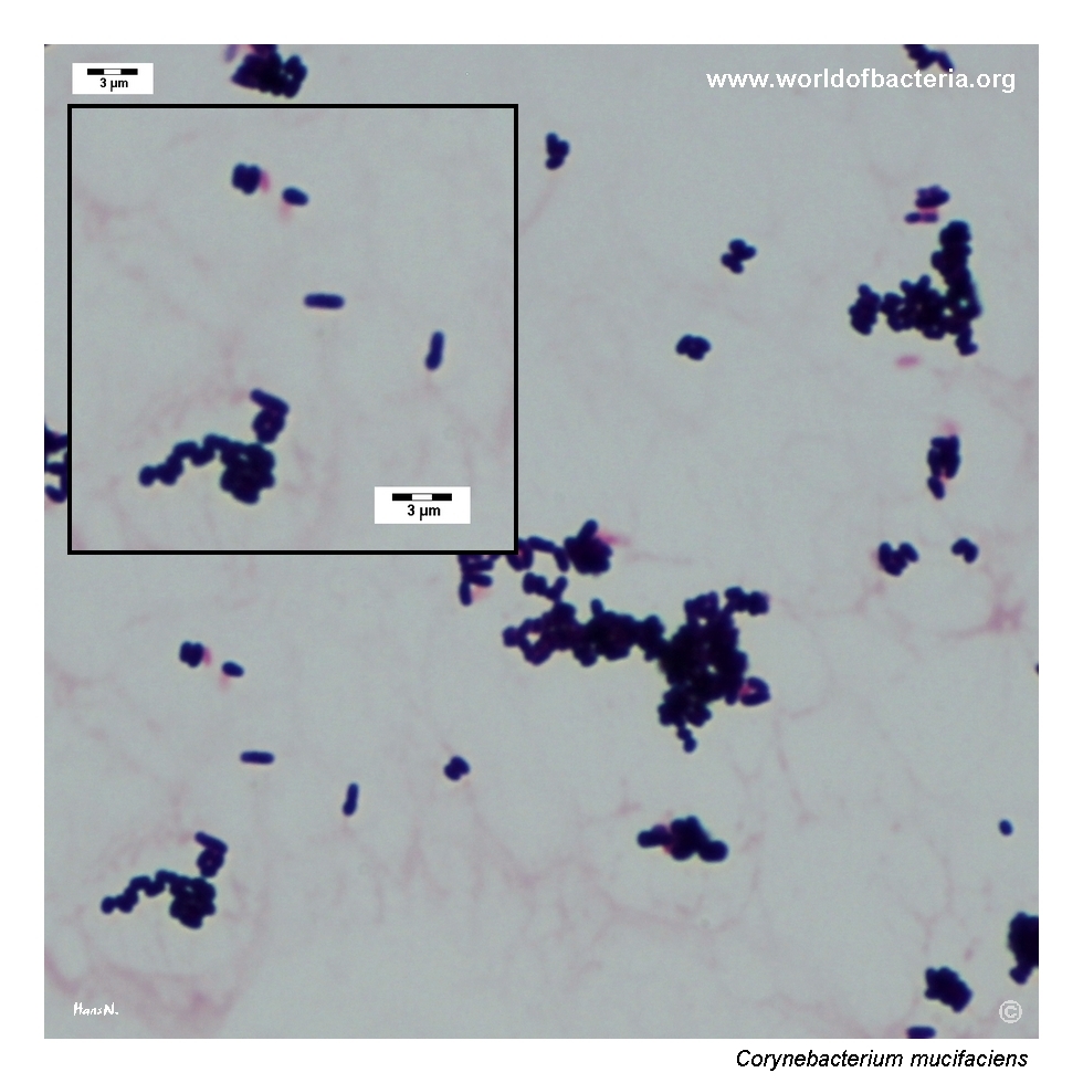
The smear from colony growth shows predominantly clustered coryneform rods, fully compatible with nondiphtherial corynebacteria
As with other members of the genus, morphology supports generic placement but does not by itself secure species identification
Oxygen Relationship
Routine aerobic coryneform isolate that grows on standard blood agar under ordinary diagnostic conditions
In these images, the organism is illustrated after incubation at 36 °C in 5% CO2, with colony changes becoming clearer after prolonged incubation
The practical challenge is not fastidiousness but the time-dependent change from more distinctly mucoid early colonies to drier, more pigmented colonies later on
Rapid Identification Tests
Morphology: Gram-positive coryneform rod
Colony phenotype: small yellow-pigmented mucoid colonies that become more conspicuous with incubation time
Useful visual clue: raised centres and a glossy surface that may partially dry with prolonged incubation
Clinical setting: blood culture drawn through an indwelling arterial catheter, where contamination, colonization, and true device-related significance all need consideration
Important limitation: definitive species-level identification generally requires MALDI-TOF MS or another reliable modern method
Ecology and Clinical Relevance
Natural Habitat
Corynebacterium mucifaciens is an uncommon human-associated nondiphtherial corynebacterium described from human clinical material rather than as a routine environmental isolate
Published experience suggests recovery from skin, blood, and other normally sterile body fluids, although the species remains infrequent in everyday laboratory work
Its significance therefore often has to be judged case by case, especially when isolated from vascular-access–related material
Common Clinical Specimens
Blood culture material, including catheter-associated or device-related sampling
Skin and other human clinical specimens reported sporadically in the literature
Selected normally sterile body fluids or uncommon invasive case material
Clinical Significance
Corynebacterium mucifaciens is best regarded as a rare human clinical isolate whose importance varies from probable contamination or colonization to genuine opportunistic infection
The species should not be dismissed automatically when recovered from clinically important material, because true disease has been reported, including bacteraemia and other invasive presentations
At the same time, single-isolate recovery from catheter-drawn blood culture must always be interpreted in relation to access site, sampling route, repeat cultures, and the overall clinical picture
Differential Considerations
Other nondiphtherial corynebacteria recovered from blood culture, especially visually modest species such as Corynebacterium amycolatum or Corynebacterium striatum
Small pigmented Gram-positive colonies that could be overinterpreted on plate appearance alone
Device-associated coryneform isolates for which the central question is contamination versus clinically relevant catheter colonization or infection
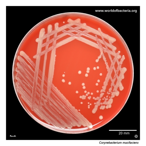
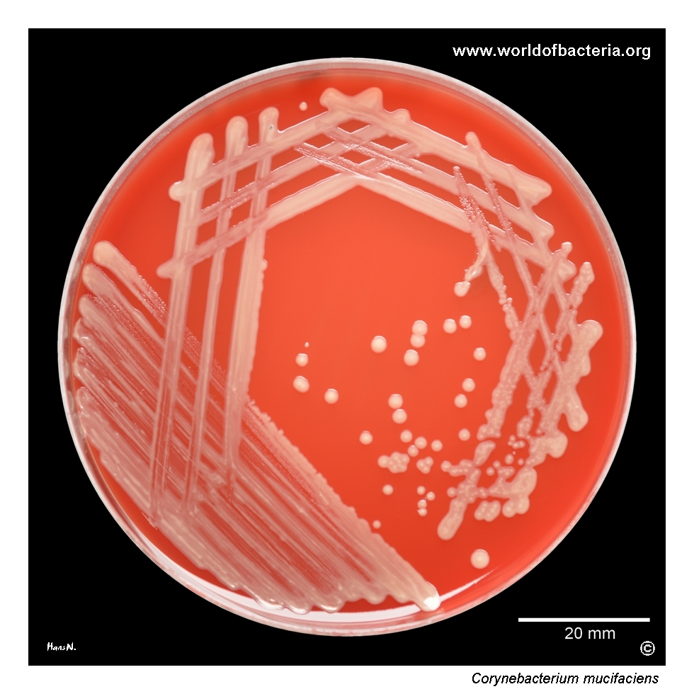
Corynebacterium mucifaciens on blood agar after 48 hours of incubation at 36 °C in 5% CO2. The isolate was recovered from a positive aerobic blood culture drawn through an indwelling arterial catheter. In this case, contamination related to the long-standing vascular access is the most likely explanation. Recovery of coryneform bacteria from catheter-drawn blood cultures must therefore always be interpreted carefully in relation to sampling technique and the overall clinical situation.
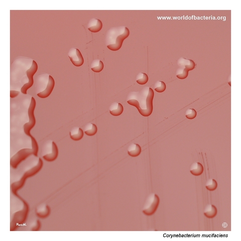
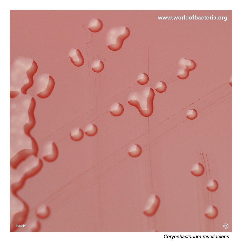
Colonies of Corynebacterium mucifaciens on blood agar after 24 hours at 36 °C in 5% CO2. The colonies are small, mucoid, and yellow-pigmented. This combination of pigmentation and surface texture makes them more visually distinctive than many other corynebacteria. Even so, species identification should not rely on appearance alone, especially when the isolate is recovered from clinically important material such as blood culture.
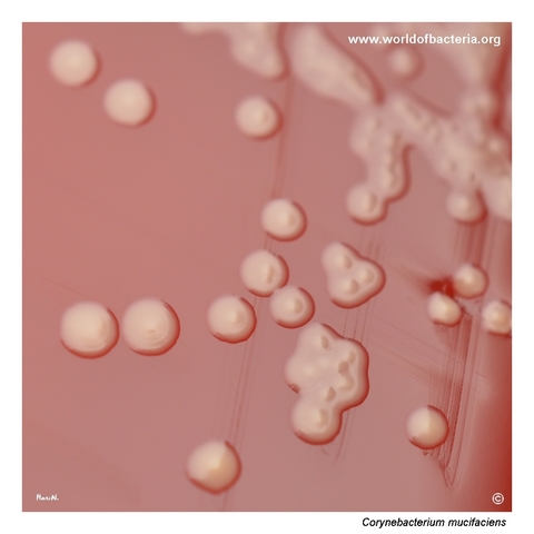
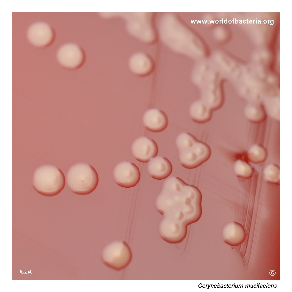
Close-up view of Corynebacterium mucifaciens colonies after 48 hours of incubation at 36 °C in 5% CO2. With prolonged incubation, the colonies begin to dry out and lose part of their original mucoid character, while the yellow pigmentation becomes more intense. The raised centres visible in most colonies are also striking. Such time-dependent changes in colony morphology are useful to appreciate when examining cultures over more than one reading interval.
Gram-stained smear prepared from a colony of Corynebacterium mucifaciens grown on blood agar. The organism appears as Gram-positive coryneform rods arranged predominantly in clusters. The strain is very likely able to form biofilm, a property that may help explain its association with colonized intravascular devices and with contamination of catheter-derived blood culture specimens.
Diagnostic and Clinical Notes
This page is particularly useful because it shows a corynebacterial isolate that is more visually distinctive than many other nondiphtherial corynebacteria. The combination of yellow pigmentation, mucoid surface texture, and later drying of the colonies provides a dynamic colony phenotype that changes noticeably between readings.
The first diagnostic question, however, is not taxonomic but interpretive. The isolate came from a positive aerobic blood culture drawn through an indwelling arterial catheter, and in that setting the key issue is whether the finding represents contamination from a long-standing vascular device, colonization of the access route, or a genuinely significant bloodstream event.
The colony images also illustrate why prolonged incubation matters. At 24 hours, the colonies already show the typical glossy yellow mucoid appearance, whereas after 48 hours they become more strongly pigmented and less mucoid, with raised centres that are easier to appreciate. This kind of time-dependent change can be diagnostically informative in routine laboratory work.
The broader teaching point is that uncommon corynebacteria recovered from blood culture should not be handled reflexively. Corynebacterium mucifaciens is rare, but it has been described from human blood and other clinical material, so interpretation should remain balanced and specimen-driven rather than automatic.
Laboratory Identification
Colony Morphology
On blood agar, Corynebacterium mucifaciens forms small colonies that are both yellow-pigmented and distinctly mucoid. In this page set, the 24-hour image shows the organism at its glistening stage, while the 48-hour image shows partial drying of the surface, more intense pigment, and conspicuous raised centres. This evolving appearance is one of the most useful practical clues for the species.
Microscopy
Gram staining shows Gram-positive coryneform rods, here arranged predominantly in clusters. The microscopic appearance is diagnostically valuable because plate morphology alone, although more distinctive than with many other corynebacteria, still does not permit secure species-level identification. As with other nondiphtherial corynebacteria, microscopy supports the correct morphological grouping but must be integrated with colony phenotype and a modern identification method.
Key Identification Clues
Small yellow-pigmented mucoid colonies on blood agar
Partial loss of mucoid character and stronger pigmentation after prolonged incubation
Raised colony centres that become easier to appreciate over time
Gram-positive coryneform rods on Gram stain rather than true cocci
Blood-culture context in which MALDI-TOF MS or another reliable method is needed for definitive species identification
Modern Identification Methods
Reliable species-level identification is best achieved by MALDI-TOF mass spectrometry when the database is suitable for nondiphtherial corynebacteria. This is especially important for C. mucifaciens, because the species is uncommon and most laboratories will not identify it confidently from morphology alone. In clinically consequential blood-culture scenarios, additional confirmatory methods may be appropriate if the result is unexpected or difficult to interpret.
Antibiotic Characteristics
For Corynebacterium mucifaciens, no treatment inference should be made from colony colour or mucoid appearance. The visual phenotype is useful for recognition, not for predicting antimicrobial susceptibility.
When the isolate is considered clinically meaningful, susceptibility testing should be performed on the individual strain. This is particularly relevant in bloodstream or device-associated contexts, where management decisions carry more weight than in clearly incidental surface contamination.
The first practical step is therefore interpretive rather than therapeutic: deciding whether the blood-culture isolate is best explained by contamination, catheter colonization, or true infection. Once the organism is judged clinically relevant, therapy should be guided by isolate-specific testing and the patient's overall situation.
Note: The main didactic value of this page lies in showing how a rare corynebacterial blood-culture isolate can look unusually distinctive on the plate while still requiring careful clinical interpretation.