Enterococcus faecium is a clinically important enterococcal species strongly associated with healthcare settings, notable for frequent antimicrobial resistance and for its role in catheter-associated and invasive infection.
Basic Characteristics
Taxonomy
Domain: Bacteria
Phylum: Bacillota
Class: Bacilli
Order: Lactobacillales
Family: Enterococcaceae
Genus:Enterococcus
Species:Enterococcus faecium
Microscopy & Gram Stain
Gram-positive cocci usually arranged in pairs and short chains, with occasional partial Gram variability in young cells
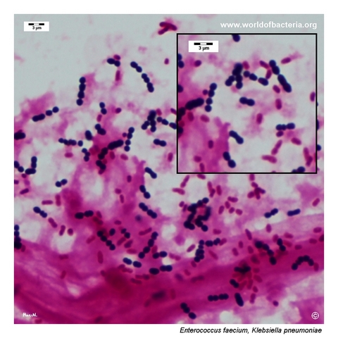
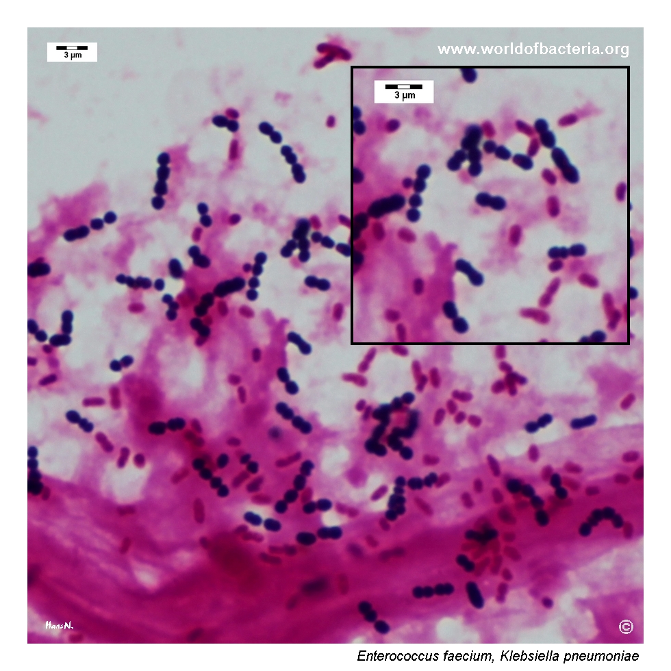
In this page set, the blood-culture Gram stain shows Gram-positive cocci in pairs and short chains together with Gram-negative rods, illustrating a polymicrobial bloodstream infection in which the enterococcal component remains morphologically recognizable
Microscopy is useful for placing the isolate among Gram-positive cocci, but it does not by itself distinguish E. faecium from E. faecalis or from some streptococci
Correlation with colony morphology, simple biochemical tests, and modern identification methods remains essential in routine practice
Oxygen Relationship
Facultatively anaerobic organism that grows readily under routine aerobic incubation conditions
In this page set, typical colony growth was documented after 24 hours at 36 °C in ambient air
Like other enterococci, the species is hardy rather than fastidious and is readily recovered on routine blood agar
Rapid Identification Tests
Catalase: negative
PYR: positive
Bile esculin: typically positive
Growth in 6.5% NaCl: typically positive
Haemolysis: many strains are non-haemolytic, but alpha-haemolytic isolates also occur and may influence plate interpretation
Ecology and Clinical Relevance
Natural Habitat
Enterococcus faecium is a human intestinal commensal, but in modern clinical microbiology it is especially notable for its persistence in the hospital environment
Because the species is adapted to healthcare settings and may colonize devices and debilitated patients, interpretation must always integrate specimen type and clinical context
Compared with E. faecalis, E. faecium is encountered less often in routine community material but more often raises concern because of resistance and healthcare association
Common Clinical Specimens
Urine, especially catheter-associated specimens
Blood cultures and other invasive specimens
Wound and device-associated material
Hospital-acquired polymicrobial samples
Clinical Significance
Enterococcus faecium is one of the most clinically important enterococci because of its strong association with healthcare-related infection and antimicrobial resistance
The species may cause urinary tract infection, bacteremia, catheter-associated infection, wound infection, and other opportunistic disease in hospitalized patients
In this page set, one image shows catheter-associated urine culture and another a positive blood culture, together illustrating both localized and invasive disease contexts
Compared with Enterococcus faecalis, E. faecium is often more difficult therapeutically because multidrug resistance is common
Differential Considerations
Enterococcus faecalis, which shares many basic enterococcal features but is often less resistant
Viridans streptococci or other catalase-negative Gram-positive cocci when only microscopy is considered
Beta-haemolytic streptococci when an enterococcal isolate shows haemolysis on blood agar
Other hospital-associated Gram-positive cocci in catheter urine or blood culture material
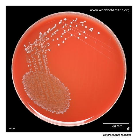
Enterococcus faecium in a urine specimen. The sample was obtained from a permanent urinary catheter that had been in place for 6 days in an 83-year-old man. Ten microlitres of urine were inoculated onto blood agar and spread across the plate. Culture conditions: 24 hours at 36 °C in ambient air. In catheter-associated urinary specimens, the recovery of E. faecium is clinically relevant because this species is well adapted to the hospital environment and is often associated with healthcare-related infection.
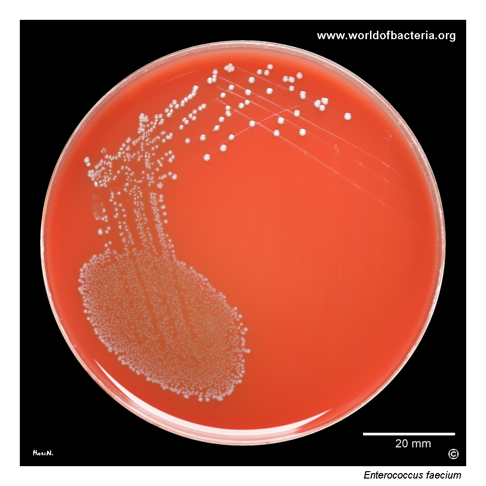
Colonies of Enterococcus faecium on blood agar. Part of the colony growth was removed from the plate, and a change in agar coloration is visible at the sites of previous growth. This reflects partial haemolysis of erythrocytes and indicates that the isolate produced alpha-haemolysis. Although many enterococci are non-haemolytic, such subtle haemolytic activity may occasionally be observed and can influence the visual interpretation of the culture plate.
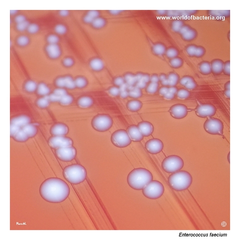
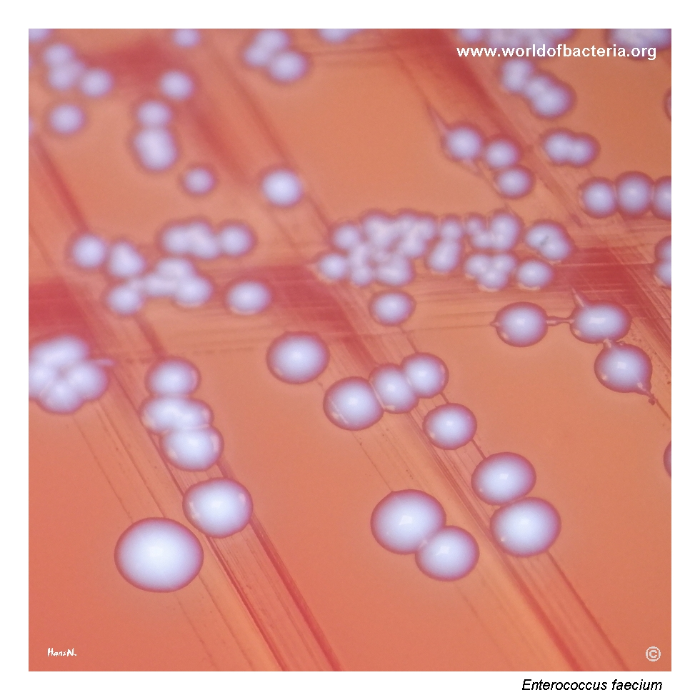
Close-up view of Enterococcus faecium colonies. The colonies are smooth, convex, and round with entire margins. Culture conditions: 24 hours at 36 °C in ambient air. Of the antibiotics tested, the isolate was susceptible only to vancomycin and linezolid. This pattern is a useful reminder that E. faecium is often considerably more resistant than E. faecalis and may present substantial therapeutic challenges in clinical practice.
Gram-stained smear from a positive anaerobic blood culture containing Enterococcus faecium and Klebsiella pneumoniae. The Gram-positive cells of E. faecium are often arranged in pairs and short chains. The specimen was obtained from a 73-year-old man hospitalized in the intensive care unit with the diagnosis J18.9, pneumonia, unspecified. This mixed microscopic picture illustrates polymicrobial bloodstream infection and highlights the importance of careful interpretation of Gram stain morphology in blood cultures.
Diagnostic and Clinical Notes
This page set is useful because it shows Enterococcus faecium in two clinically important contexts: catheter-associated urine culture and positive blood culture.
The urinary isolate is didactically relevant because catheter-associated specimens often contain hospital-adapted organisms whose significance depends on both quantity and clinical setting. In such material, E. faecium deserves particular attention.
The alpha-haemolytic plate image is also instructive. Many laboratorians expect enterococci to be non-haemolytic, yet subtle alpha-haemolysis can occur and may alter first impressions during plate reading.
The colony close-up adds an important clinical message: morphology alone cannot predict resistance, but this particular isolate showed a very restricted susceptibility profile, emphasizing why species-level identification and susceptibility testing matter.
The blood-culture smear is especially instructive because it shows E. faecium together with Klebsiella pneumoniae in an anaerobic bottle, illustrating polymicrobial bloodstream infection and the need to recognize both Gram-positive cocci and Gram-negative rods during direct smear interpretation.
Laboratory Identification
Colony Morphology
On blood agar, Enterococcus faecium typically forms smooth, convex, grey-white colonies with entire margins. Many isolates are non-haemolytic, but alpha-haemolytic growth may occasionally be seen, as in this page set. Because colony appearance overlaps widely with that of other enterococci and some streptococci, morphology alone is not sufficient for secure identification.
Microscopy
Gram staining shows Gram-positive cocci, usually in pairs and short chains. In this page set, the direct smear from an anaerobic blood culture also contains Gram-negative rods of Klebsiella pneumoniae, making the microscopic picture distinctly polymicrobial. The enterococcal component nevertheless remains recognizable, but final interpretation still requires correlation with culture findings and laboratory identification.
Key Identification Clues
Catalase-negative Gram-positive cocci in pairs and short chains
PYR-positive reaction and typical enterococcal biochemical profile
Ready growth on routine blood agar under standard incubation
Potential alpha-haemolysis that should not be overinterpreted as a streptococcal feature
Need to distinguish from Enterococcus faecalis, especially because resistance patterns often differ substantially
Modern Identification Methods
Modern identification is straightforward with MALDI-TOF MS and other validated laboratory systems. In routine diagnostics, however, recognition of the basic enterococcal phenotype remains important because species-level distinction between E. faecium and E. faecalis has immediate clinical implications for antimicrobial interpretation and infection-control awareness.
Antibiotic Characteristics
This page set highlights one of the defining clinical features of Enterococcus faecium: resistance matters. The isolate shown here was susceptible only to vancomycin and linezolid among the tested antibiotics, illustrating the therapeutic difficulty that this species may present.
No resistance pattern should be inferred from colony appearance alone, but once the organism is recognized as E. faecium, comprehensive susceptibility testing becomes especially important because hospital isolates are often markedly less susceptible than E. faecalis.
Note: In routine clinical microbiology, the main value of the morphology shown here is early recognition of an enterococcal isolate and prompt awareness that E. faecium may be a multidrug-resistant healthcare-associated pathogen.