Corynebacterium ulcerans is a zoonotic coryneform species that may produce striking beta-haemolytic colonies on blood agar and, when toxigenic, can cause diphtheria-like illness requiring careful laboratory evaluation.
Basic Characteristics
Taxonomy
Domain: Bacteria
Phylum: Actinomycetota
Class: Actinomycetes
Order: Corynebacteriales
Family: Corynebacteriaceae
Genus:Corynebacterium
Species:Corynebacterium ulcerans
Microscopy & Gram Stain
Gram-positive coryneform rods, often club-shaped
Cells may remain attached in V-shaped pairs or irregular palisades
The smear supports placement within the corynebacterial group but does not by itself determine species identity or toxigenicity
In practical diagnostics, microscopy is especially helpful when the blood agar colonies might otherwise be mistaken for Gram-positive cocci
Oxygen Relationship
Aerobic coryneform organism that grows under routine blood agar culture conditions
In this page set, the isolate grew well after 48 hours at 36 °C in 5% CO2, with further visual enhancement after prolonged incubation
The organism is not especially important because of fastidious growth, but because its recognition may trigger a diphtheria-related diagnostic pathway
Rapid Identification Tests
Catalase: positive
Morphology: Gram-positive coryneform rod
Practical clue: beta-haemolytic colonies on blood agar, sometimes with slight yellowish discoloration after prolonged incubation
Important limitation: colony appearance does not determine whether the strain is toxigenic
Public health note: when C. ulcerans is recovered in a diphtheria-like clinical setting, correlation with toxin testing is essential
Ecology and Clinical Relevance
Natural Habitat
Corynebacterium ulcerans is primarily an animal-associated species and is generally regarded as zoonotic in human infection
Human isolates may be linked with contact with domestic animals or other animal reservoirs, although the epidemiological source is not always obvious
In throat swabs and skin-associated material, the clinical context is therefore important for deciding whether the isolate is merely present or truly significant
Common Clinical Specimens
Throat swabs in patients with suspected diphtheria-like illness
Swabs from skin lesions or wound material
Occasionally other clinical specimens when invasive infection is investigated
Clinical Significance
Corynebacterium ulcerans is clinically important because toxigenic strains may cause disease that resembles classical diphtheria
Even before toxigenicity is known, species identification can alter the urgency of interpretation and communication
The organism should therefore not be dismissed as an ordinary diphtheroid when recovered from a compatible specimen such as a throat swab from a patient with suspected diphtheria
Differential Considerations
Corynebacterium diphtheriae, especially in respiratory specimens and other diphtheria-like presentations
Corynebacterium pseudotuberculosis, another zoonotic corynebacterial species with potential clinical relevance
Non-haemolytic or weakly haemolytic nondiphtherial corynebacteria that may be less clinically urgent but superficially similar on initial review
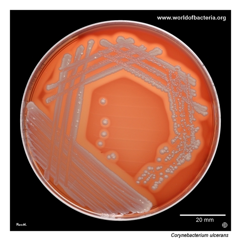
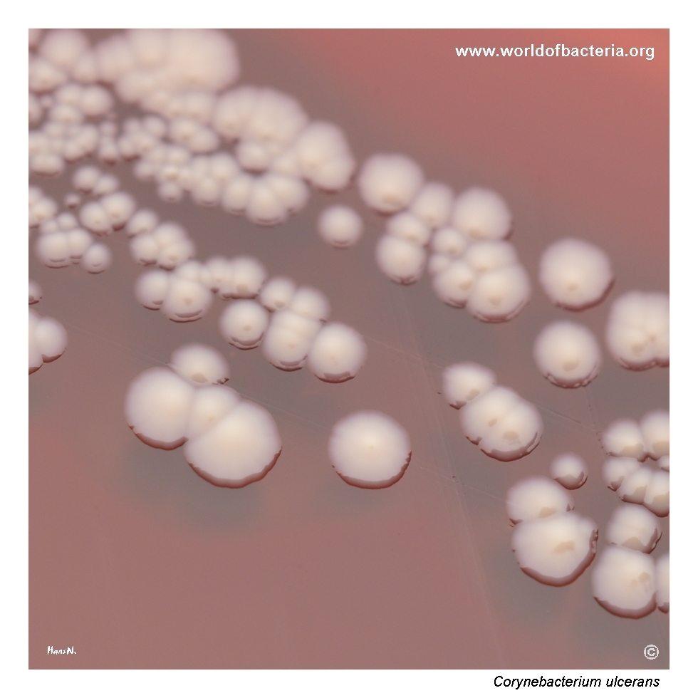
Corynebacterium ulcerans on blood agar after 48 hours of incubation at 36 °C in 5% CO2. The isolate was obtained from a throat swab from a six-year-old child with suspected diphtheria. In this clinical setting, accurate recognition of the isolate is especially important, because C. ulcerans may be associated with diphtheria-like illness and therefore requires careful laboratory evaluation and correlation with toxin testing where appropriate.
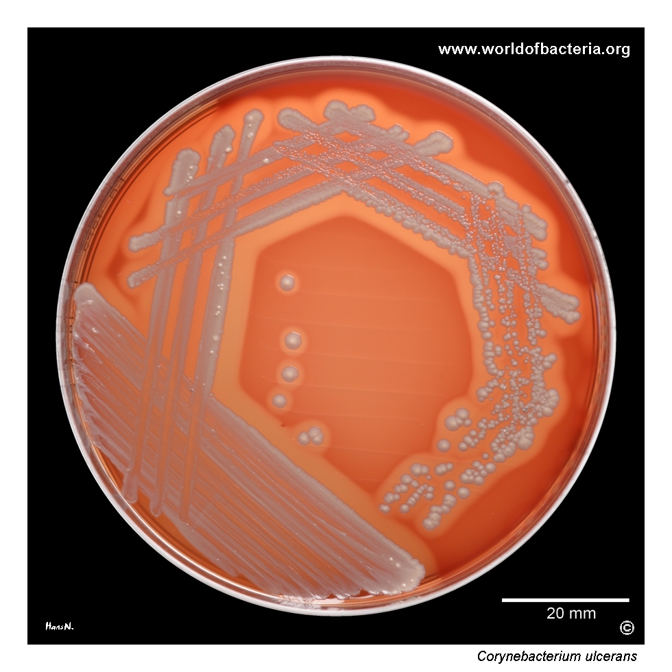
Colonies of Corynebacterium ulcerans on blood agar after 48 hours at 36 °C in 5% CO2, followed by 24 hours at room temperature. Slight yellowish discoloration of the colonies may become visible during prolonged incubation. Such subtle pigment development can influence the visual appearance of the culture plate, although the most striking feature of this species on blood agar is often the haemolytic activity around the colonies rather than colour alone.
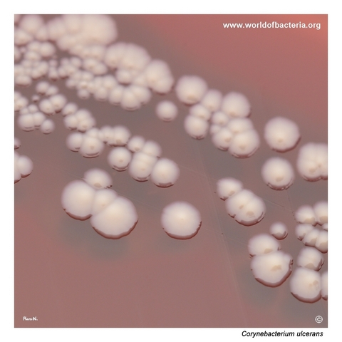
Close-up view of Corynebacterium ulcerans colonies on blood agar after 48 hours of incubation at 36 °C in 5% CO2. A conspicuous zone of beta-haemolysis surrounds the colonies. This finding is diagnostically useful because it makes the isolate more visually prominent than many other corynebacteria, which often remain non-haemolytic and much less distinctive on routine blood agar plates.
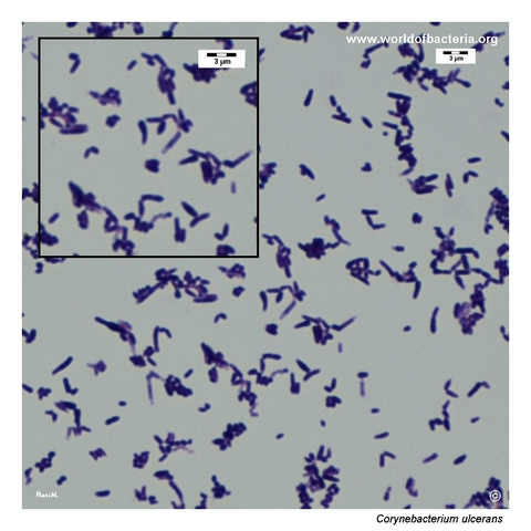
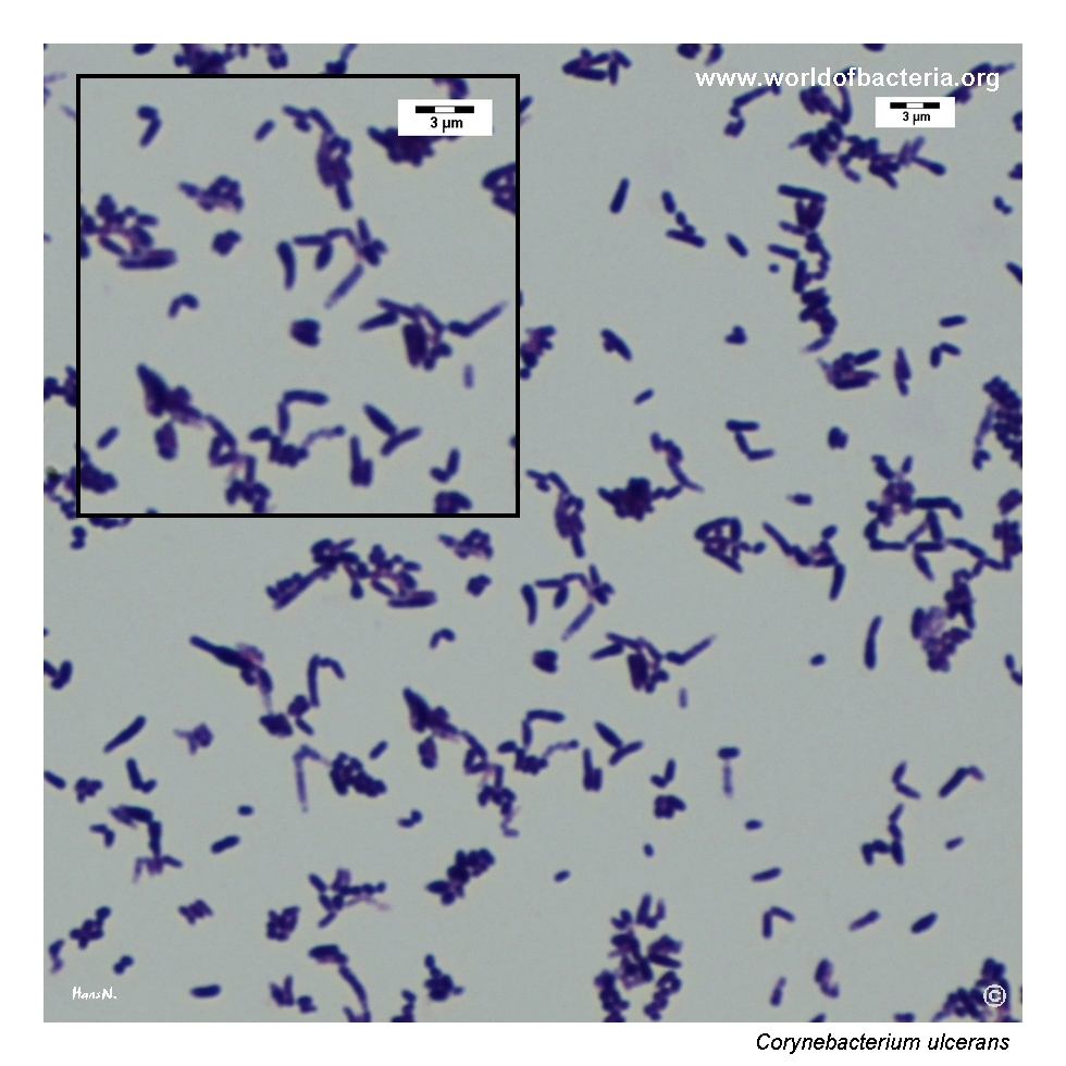
Gram-stained smear prepared from a colony of Corynebacterium ulcerans grown on blood agar. The organism appears as Gram-positive coryneform rods, often club-shaped. The bacterial cells frequently remain attached in V-shaped pairs, and in some areas palisade arrangements are visible. This morphology is typical of corynebacteria and is an important clue when distinguishing the isolate from Gram-positive cocci with superficially similar colony morphology.
Diagnostic and Clinical Notes
This page is especially useful for teaching because it shows a corynebacterial isolate recovered from a throat swab in a patient with suspected diphtheria. In such a clinical setting, even the preliminary recognition of the organism matters, because the diagnostic implications are very different from those of ordinary low-significance diphtheroids.
The colony appearance is more striking than that of many routine corynebacteria. Beta-haemolysis makes the isolate conspicuous on blood agar, and prolonged incubation may add a faint yellowish discoloration that further changes the visual impression. Even so, pigment should be considered secondary to the haemolytic phenotype and to the clinical context.
Microscopy contributes a classic coryneform pattern, with club-shaped Gram-positive rods, V-shaped pairs, and palisade-like groupings. This is highly useful for orientation and helps separate the organism from Gram-positive cocci that may look superficially similar at the colony level.
The main diagnostic lesson is that species identification alone is not the end point. Once Corynebacterium ulcerans is recognized, the crucial next question is whether the strain is toxigenic. The isolate therefore deserves careful laboratory follow-up and correlation with the clinical picture rather than routine dismissal as a minor contaminant.
Laboratory Identification
Colony Morphology
On blood agar, Corynebacterium ulcerans forms colonies that are made diagnostically notable by a conspicuous zone of beta-haemolysis. In this image set, slight yellowish discoloration becomes visible after prolonged incubation, but the haemolytic effect remains the more important visual clue. The overall appearance is therefore more prominent than that of many other nondiphtherial corynebacteria.
Microscopy
Gram staining shows Gram-positive coryneform rods, often club-shaped, with frequent V-shaped pairs and palisade arrangements. This is a classical corynebacterial morphology and is particularly helpful when the culture plate might otherwise suggest Gram-positive cocci. As usual, however, microscopy alone does not determine species identity or toxigenicity.
Key Identification Clues
Throat swab or another clinically compatible specimen in a diphtheria-like setting
Beta-haemolytic colonies on blood agar
Possible slight yellowish discoloration after prolonged incubation
Gram-positive coryneform rods with club-shaped cells, V-shaped pairs, and palisades
Need for toxin testing once the species is identified or strongly suspected
Modern Identification Methods
Reliable species identification is best achieved by MALDI-TOF mass spectrometry when the database is sufficiently current. In diagnostically sensitive cases, additional reference laboratory work may be needed to assess tox gene presence and, most importantly, actual toxin production. This distinction is crucial because morphology alone cannot establish whether an isolate is capable of causing diphtheria-like disease.
Antibiotic Characteristics
For Corynebacterium ulcerans, antimicrobial interpretation should not be reduced to a simple rule based on colony appearance. The first critical issue is recognition of the species, and the second is assessment of toxigenicity in clinically compatible disease.
When a strain is recovered from a patient with suspected diphtheria-like illness, laboratory management extends beyond routine susceptibility thinking. Correct species identification may influence public health communication, further toxin testing, and the urgency of clinical interpretation.
Therapeutic decisions should therefore follow the clinical syndrome, isolate-specific susceptibility testing, and the formal diagnostic pathway used for possible diphtheria-associated organisms.
Note: Morphology on blood agar is diagnostically helpful, but therapy and any public health response must be guided by toxigenicity assessment, susceptibility results, and the full clinical context.