Corynebacterium pseudotuberculosis is an animal-associated coryneform species and occasional zoonotic human pathogen that may form striking beta-haemolytic colonies on blood agar, especially in suppurative lymph node infections.
Basic Characteristics
Taxonomy
Domain: Bacteria
Phylum: Actinomycetota
Class: Actinomycetes
Order: Corynebacteriales
Family: Corynebacteriaceae
Genus:Corynebacterium
Species:Corynebacterium pseudotuberculosis
Microscopy & Gram Stain
Very short Gram-positive rods with a coryneform appearance
Cells may appear somewhat pleomorphic and in some fields almost coccoid at first glance
Microscopy is useful for recognizing the isolate as a coryneform bacterium, but the species cannot be determined from smear morphology alone
The combination of coryneform rods on Gram stain and haemolytic colonies on blood agar makes the organism more conspicuous than many other nondiphtherial corynebacteria
Oxygen Relationship
Aerobic to facultatively anaerobic coryneform organism that grows under routine blood agar culture conditions
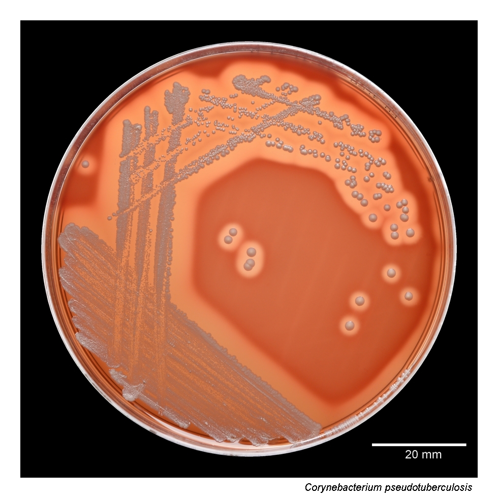
In this page set, growth is documented after 48 hours at 36 °C in 5% CO2, with colonies remaining easy to appreciate because of the marked haemolysis
In practical diagnostics, the species is not important because of fastidious growth, but because epidemiological context and haemolytic colony morphology may provide an early clue
Rapid Identification Tests
Catalase: positive
Microscopy: Gram-positive coryneform rod
Practical clue: beta-haemolytic coryneform colonies in purulent material, especially with possible animal exposure
Clinical clue: suppurative lymphadenitis or other abscess-forming infection in a patient with zoonotic contact history
Important caveat: reliable species assignment requires MALDI-TOF MS or another up-to-date identification method
Ecology and Clinical Relevance
Natural Habitat
Corynebacterium pseudotuberculosis is primarily an animal-associated species, especially linked with small ruminants and other farm animals
Human infection is uncommon and usually raises the possibility of zoonotic transmission
The epidemiological setting can therefore be highly informative, particularly when purulent lymph node disease follows contact with livestock or other farm animals
Common Clinical Specimens
Purulent lymph node material
Abscess aspirates
Wound material from suppurative lesions
Occasionally other invasive human specimens in a zoonotic context
Clinical Significance
Corynebacterium pseudotuberculosis is a well-known veterinary pathogen and only an occasional human pathogen, but human cases are clinically meaningful because the organism is not part of routine skin flora
Suppurative lymphadenitis is an especially suggestive presentation, and the clinical history may be decisive for interpreting the isolate correctly
When recovered from human infection, the bacterium deserves careful attention because it points beyond ordinary opportunistic corynebacterial colonization and toward a true zoonotic process
Differential Considerations
Corynebacterium diphtheriae and Corynebacterium ulcerans, especially when haemolytic corynebacterial colonies are encountered
Other coryneform rods recovered from purulent material that might appear less haemolytic or less epidemiologically distinctive
Short Gram-positive rods that may be visually underestimated if microscopy is interpreted without the accompanying culture phenotype
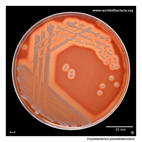
Isolate of Corynebacterium pseudotuberculosis from a patient with suppurative lymphadenitis. The patient reported recent contact with farm animals. On blood agar after 48 hours of incubation at 36 °C in 5% CO2, the colonies are surrounded by a prominent zone of beta-haemolysis. This species is of particular interest because it is associated with animals and only occasionally recovered from human infection, where epidemiological context may provide an important diagnostic clue.
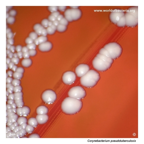
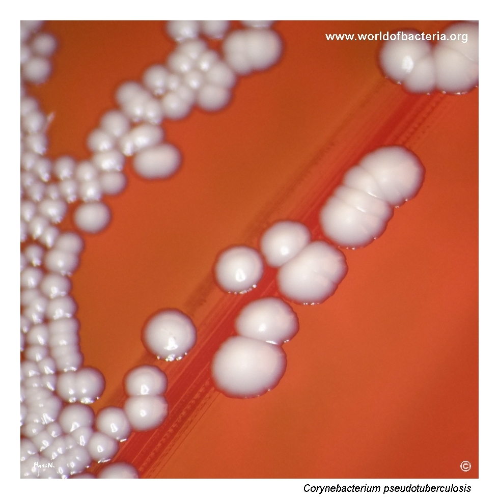
Colonies of Corynebacterium pseudotuberculosis on blood agar after 48 hours at 36 °C in 5% CO2. In contrast to the more inconspicuous appearance of many other corynebacteria, this species may attract attention because of the surrounding haemolysis. Such a finding can be diagnostically useful when evaluating purulent material, especially if the clinical history suggests possible zoonotic exposure.
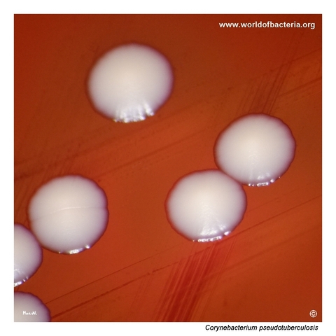
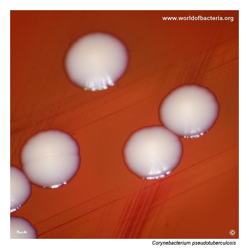
Close-up view of Corynebacterium pseudotuberculosis colonies on blood agar after 48 hours at 36 °C in 5% CO2. The colony morphology is shown in greater detail together with the pronounced haemolytic effect. In routine culture reading, the combination of coryneform rods on microscopy and beta-haemolytic colonies on blood agar makes this isolate more striking than many other members of the genus.
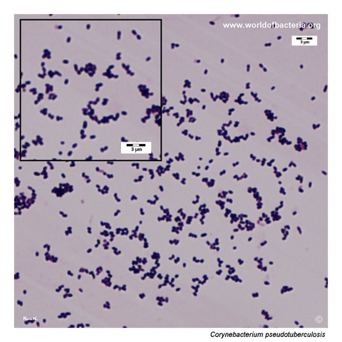
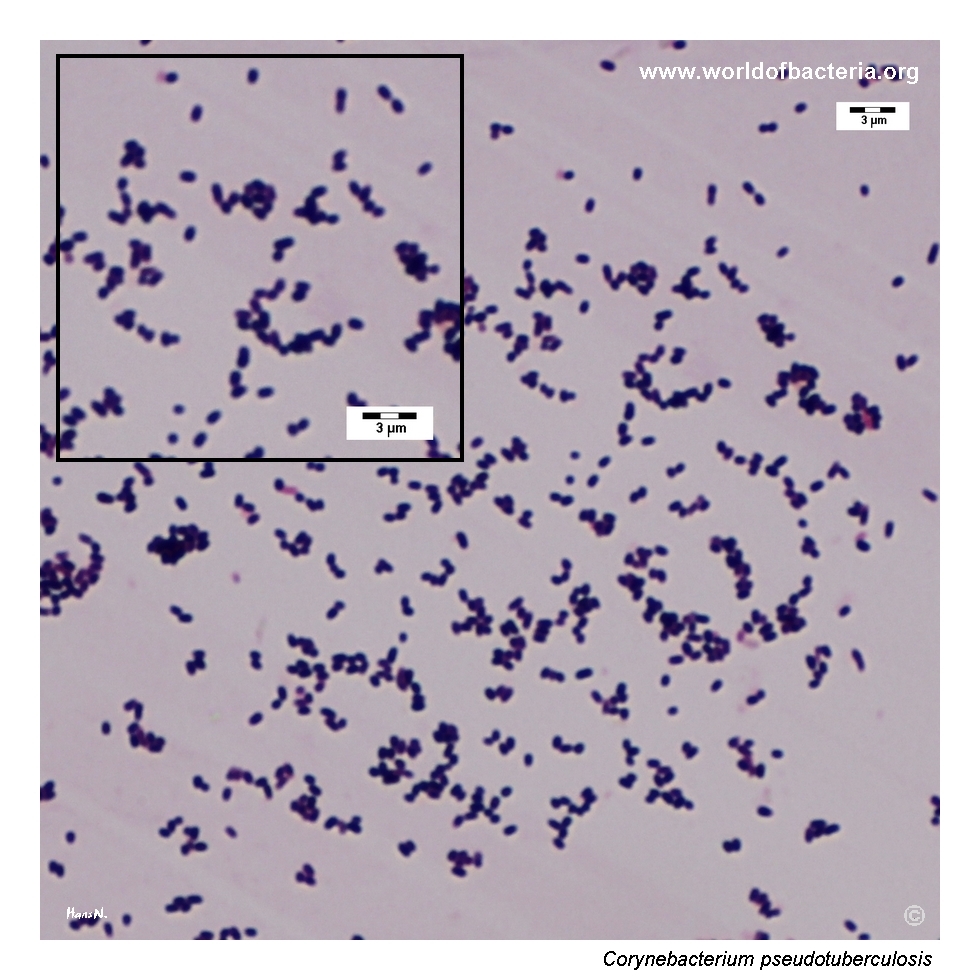
Gram-stained smear prepared from a colony of Corynebacterium pseudotuberculosis grown on blood agar. The organism appears as very short Gram-positive rods, in some places almost resembling cocci. This can make the microscopic picture slightly misleading at first glance, but the overall morphology remains compatible with coryneform bacteria when interpreted together with the colony appearance on solid medium.
Diagnostic and Clinical Notes
This page is diagnostically valuable because it shows a coryneform isolate that is more visually assertive than many routine nondiphtherial corynebacteria. The pronounced beta-haemolysis makes the colonies stand out on blood agar, which is unusual enough to attract attention during plate reading and to encourage a broader differential diagnosis.
The epidemiological context is just as important as the colony morphology. In this case, suppurative lymphadenitis together with recent contact with farm animals immediately raises the possibility of zoonotic infection, which is a key practical clue for interpreting the isolate correctly.
Microscopy contributes another useful teaching point. The cells are very short Gram-positive rods and in some fields may look almost coccoid, which can be slightly misleading at first glance. The diagnosis becomes much more secure when the smear is interpreted together with the haemolytic growth pattern on blood agar.
Overall, the image set illustrates a species that should not be treated as an ordinary diphtheroid contaminant. In the right clinical setting, a haemolytic corynebacterial isolate from purulent lymph node material may represent a genuine zoonotic pathogen and deserves species-level identification.
Laboratory Identification
Colony Morphology
On blood agar, Corynebacterium pseudotuberculosis forms colonies that are made especially notable by the surrounding beta-haemolysis. In this page set, the haemolytic effect is already prominent after 48 hours of incubation, making the isolate visually more striking than many other corynebacteria. The colony morphology itself is best interpreted together with the clinical picture and the coryneform appearance on microscopy.
Microscopy
Gram staining shows very short Gram-positive rods with a coryneform appearance. In some microscopic fields the cells may appear almost coccoid, which can be misleading if the smear is read in isolation. Species-level identification therefore depends on correlating the microscopy with colony morphology and with modern identification methods.
Key Identification Clues
Purulent lymph node or abscess material rather than routine skin flora context
Recent contact with farm animals or another suggestive zoonotic exposure history
Beta-haemolytic colonies on blood agar
Very short Gram-positive coryneform rods that may look almost coccoid in places
Need for reliable species-level identification because the clinical implications differ from those of ordinary nondiphtherial corynebacteria
Modern Identification Methods
Reliable identification is best achieved by MALDI-TOF mass spectrometry when the reference database is sufficiently current. In difficult cases, sequencing-based methods may be used for confirmation. This is particularly relevant for C. pseudotuberculosis because the species is uncommon in human microbiology and the decisive interpretation often depends on integrating the species name with the epidemiological history.
Antibiotic Characteristics
Corynebacterium pseudotuberculosis should not be assigned an expected susceptibility pattern solely from morphology or from its veterinary reputation. Human infection is uncommon, and antimicrobial management should be guided by isolate-specific testing together with the clinical syndrome.
The more important teaching point on this page is the diagnostic value of recognizing the isolate at all. When a haemolytic coryneform rod is recovered from suppurative lymphadenitis in a patient with animal exposure, laboratory identification may influence not only therapy but also epidemiological interpretation.
Because this is primarily an animal-associated pathogen, the microbiological finding carries broader significance than that of many routine corynebacterial isolates. The laboratory report should therefore be integrated carefully with the clinical history and the possibility of zoonotic acquisition.
Note: Therapeutic decisions should always follow the clinical picture, any need for drainage of suppurative lesions, and actual susceptibility results.