Corynebacterium tuberculostearicum is a lipophilic coryneform species that forms small non-haemolytic colonies on blood agar, may resemble coagulase-negative staphylococci, and requires careful interpretation in catheter-associated clinical material.
Basic Characteristics
Taxonomy
Domain: Bacteria
Phylum: Actinomycetota
Class: Actinomycetes
Order: Corynebacteriales
Family: Corynebacteriaceae
Genus:Corynebacterium
Species:Corynebacterium tuberculostearicum
Microscopy & Gram Stain
Gram-positive coryneform rods, sometimes slightly curved
V-shaped pairs may be present, helping to separate the organism from Gram-positive cocci with superficially similar colonies
The smear supports placement within the corynebacterial group but does not by itself determine species identity
In practical diagnostics, microscopy is particularly useful because the colonies on blood agar may resemble coagulase-negative staphylococci
Oxygen Relationship
Aerobic coryneform organism that grows under routine blood agar culture conditions
In this page set, the isolate formed visible colonies after 48 hours at 36 °C in 5% CO2
The organism is not visually striking on solid medium, so recognition depends more on careful plate reading and microscopy than on any dramatic growth phenotype
Rapid Identification Tests
Morphology: Gram-positive coryneform rod
Colony phenotype: small non-haemolytic colonies on blood agar that may resemble coagulase-negative staphylococci
Physiology: lipophilic species
Practical clue: growth from catheter-associated or other device-related material should not be dismissed without correlation with the clinical context
Important limitation: routine visual inspection alone may not distinguish this species reliably from other nondiphtherial corynebacteria or from staphylococci
Ecology and Clinical Relevance
Natural Habitat
Corynebacterium tuberculostearicum is regarded as part of the human skin-associated and mucosal microbiota
Because of this background, recovery from catheter-associated material or blood-culture-related pathways always requires careful clinical interpretation
The organism may therefore act either as a colonizer or as a clinically relevant opportunist, depending on the full microbiological picture
Common Clinical Specimens
Catheter-associated material and other device-related specimens
Wound or surgical-site specimens
Occasionally blood culture or other hospital-associated material where interpretation can be difficult
Clinical Significance
Corynebacterium tuberculostearicum is generally an uncommon clinical isolate, but published work suggests that it may be underrecognized rather than entirely insignificant
Its importance is greatest when isolated from patients with indwelling devices, prolonged hospitalization, or complicated underlying disease
In dialysis-catheter material, the key question is whether the isolate represents colonization or a clinically meaningful catheter-associated infection
Differential Considerations
Coagulase-negative staphylococci, because the colonies may look deceptively similar on blood agar
Corynebacterium jeikeium and other nondiphtherial corynebacteria recovered from hospital-associated material
Other small non-haemolytic Gram-positive organisms from catheter-associated or mixed specimens
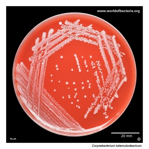
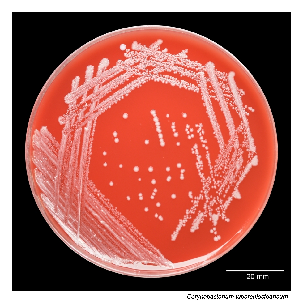
Corynebacterium tuberculostearicum on blood agar after 48 hours of incubation at 36 °C in 5% CO2. The isolate originated from a dialysis catheter from a 38-year-old man with chronic kidney disease. In catheter-associated material, organisms of this type must be interpreted carefully, because corynebacteria may represent either colonization or clinically relevant infection depending on the overall microbiological and clinical context.
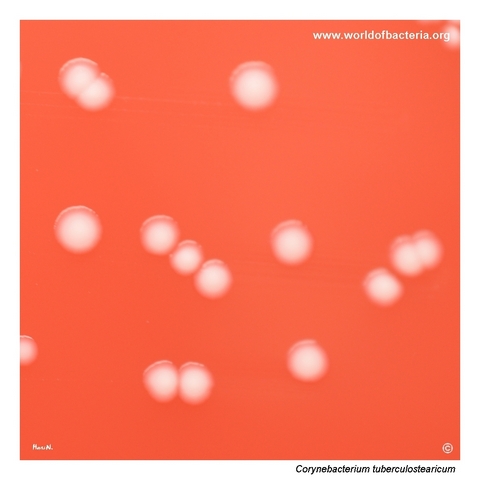
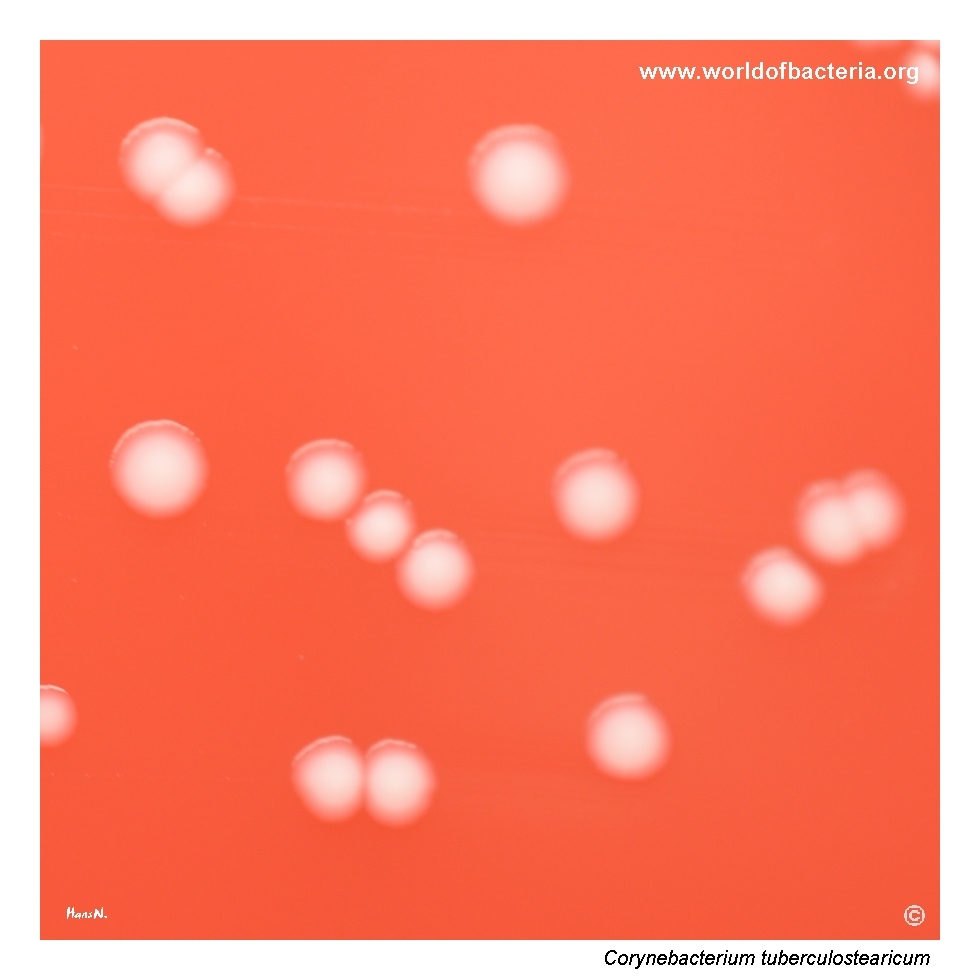
Colonies of Corynebacterium tuberculostearicum on blood agar after 48 hours at 36 °C in 5% CO2. The colonies are non-haemolytic and resemble coagulase-negative staphylococci in appearance. This similarity is diagnostically relevant because small corynebacterial colonies may easily be misread as staphylococci unless Gram staining or a modern identification method is performed.
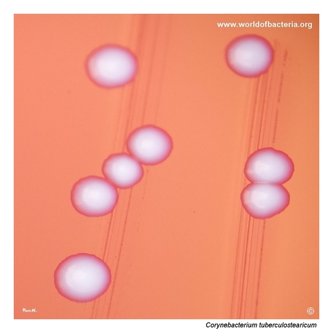
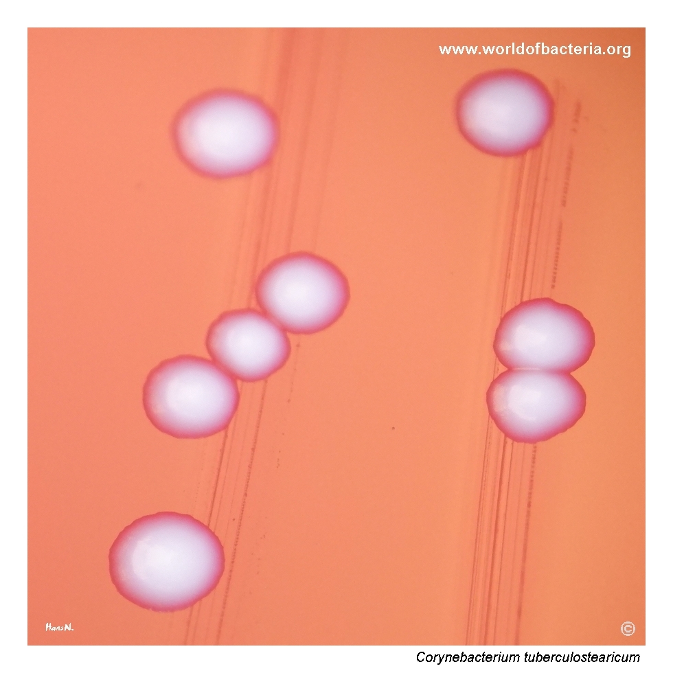
Close-up view of Corynebacterium tuberculostearicum colonies after 48 hours of incubation at 36 °C in 5% CO2. The colonies are smooth, convex, and visually similar to staphylococcal colonies on blood agar. Their rather inconspicuous morphology provides only limited clues on solid medium, so correct recognition depends heavily on microscopic examination and species identification beyond colony appearance alone.
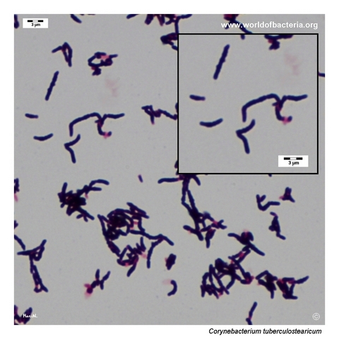
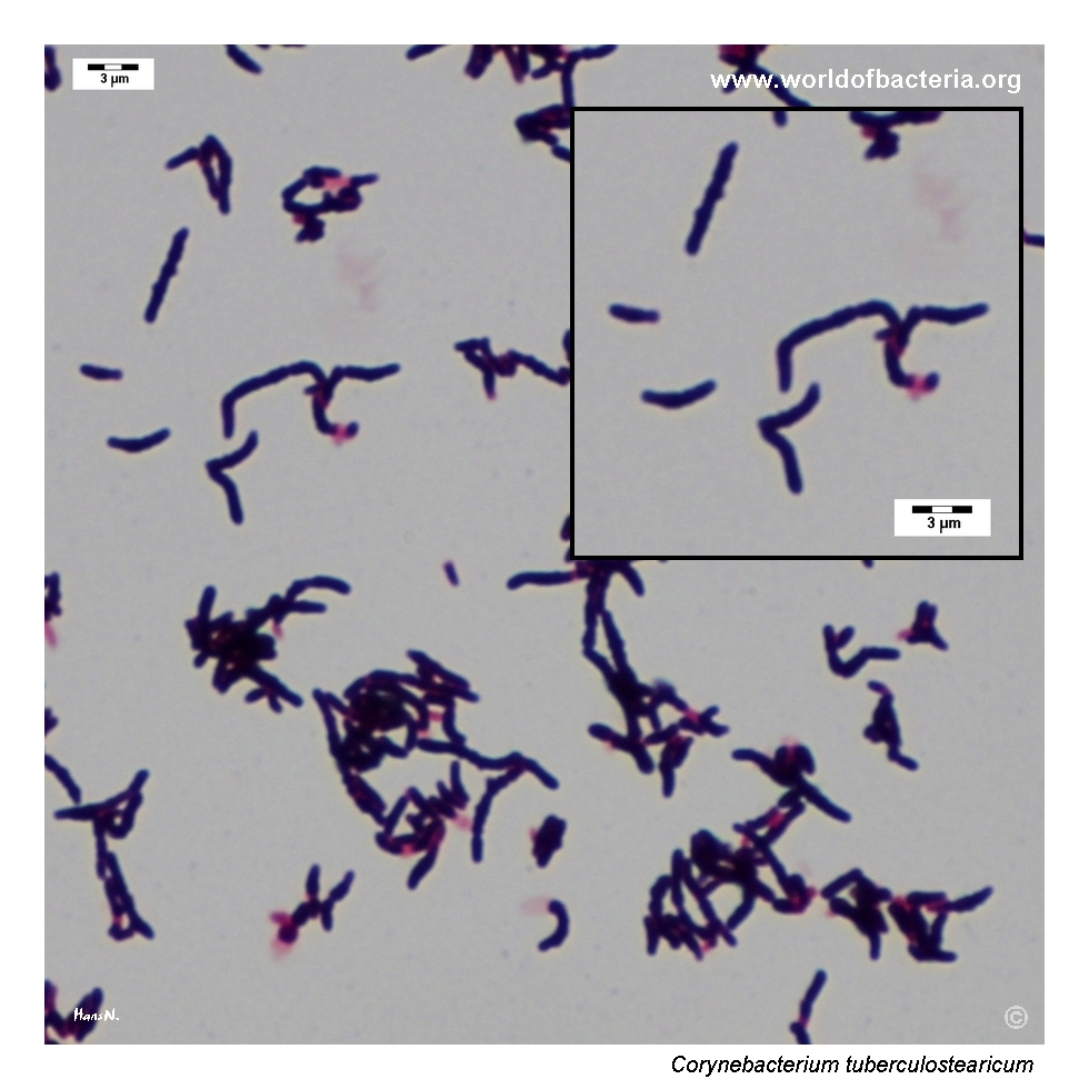
Gram-stained smear prepared from a colony of Corynebacterium tuberculostearicum grown on blood agar. The organism appears as Gram-positive rods, sometimes slightly curved, with occasional V-shaped pairs. This coryneform microscopic pattern is helpful in distinguishing the isolate from Gram-positive cocci that may produce superficially similar colonies on blood agar.
Diagnostic and Clinical Notes
This page is especially useful because it shows a corynebacterial species that can be visually misleading on routine blood agar. The colonies are small, smooth, convex, and non-haemolytic, closely resembling coagulase-negative staphylococci unless microscopy or instrumental identification is added.
The dialysis-catheter origin of the isolate makes the interpretive problem very practical. In device-related material, corynebacteria may reflect simple colonization, but they may also be clinically relevant opportunists. The laboratory therefore has to combine morphology, specimen type, quantity, and clinical context rather than relying on appearance alone.
Microscopy provides the decisive first correction to the plate impression. Instead of Gram-positive cocci, the smear shows Gram-positive coryneform rods, occasionally slightly curved and sometimes arranged in V-shaped pairs. This shifts the diagnostic pathway away from staphylococci and toward nondiphtherial corynebacteria.
The main diagnostic lesson is that an inconspicuous colony phenotype does not imply insignificance. Species such as Corynebacterium tuberculostearicum may be underrecognized precisely because they look ordinary on solid medium. In hospital-associated material, especially catheter-related specimens, reliable identification is therefore more important than the plate appearance might suggest.
Laboratory Identification
Colony Morphology
On blood agar, Corynebacterium tuberculostearicum forms small, smooth, convex, non-haemolytic colonies. In this image set, the colonies are visually very similar to coagulase-negative staphylococci, which is one of the most important practical teaching points of the page. Their morphology is therefore informative mainly because it demonstrates how easily the species can be overlooked or misclassified during routine culture reading.
Microscopy
Gram staining shows Gram-positive coryneform rods, sometimes slightly curved, with occasional V-shaped pairs. This microscopic pattern is much more helpful than colony appearance for separating the isolate from staphylococci. As with other nondiphtherial corynebacteria, microscopy supports the generic placement of the organism but does not by itself establish species identity.
Key Identification Clues
Small non-haemolytic colonies on blood agar that may resemble coagulase-negative staphylococci
Gram-positive coryneform rods rather than Gram-positive cocci
Recovery from catheter-associated or other hospital-related specimens
Need for careful interpretation in the context of device colonization versus infection
Importance of MALDI-TOF MS or another modern identification method for final species assignment
Modern Identification Methods
Reliable identification is best achieved by MALDI-TOF mass spectrometry when an up-to-date database is available. This is especially relevant for C. tuberculostearicum because visually it offers few distinctive clues and historically has been liable to underrecognition or misidentification. In diagnostically difficult cases, additional phenotypic or molecular work may be required, particularly when the isolate is recovered from clinically significant device-associated material.
Antibiotic Characteristics
For Corynebacterium tuberculostearicum, no simple therapeutic assumption should be made from colony appearance alone. The visually bland phenotype on blood agar gives little information about pathogenic potential or resistance pattern.
Published clinical work has associated this species with hospital-associated material and with patients who have substantial underlying disease or prior antimicrobial exposure. In such settings, isolate-specific susceptibility testing is more informative than any species stereotype inferred from morphology alone.
Because catheter-associated specimens may reflect either colonization or true infection, antimicrobial decisions should be integrated with the overall device-related clinical picture and with the strength of microbiological evidence for significance.
Note: The key diagnostic task is first to recognize that the isolate is a corynebacterium rather than a staphylococcus and then to interpret the result in the light of specimen type, clinical context, and isolate-specific susceptibility testing.