Corynebacterium macginleyi is a lipophilic ocular-associated coryneform species that typically forms very small non-pigmented colonies on blood agar and is most often recovered from conjunctival material.
Basic Characteristics
Taxonomy
Domain: Bacteria
Phylum: Actinomycetota
Class: Actinomycetes
Order: Corynebacteriales
Family: Corynebacteriaceae
Genus:Corynebacterium
Species:Corynebacterium macginleyi
Microscopy & Gram Stain
Gram-positive coryneform rods
Small pleomorphic bacilli with the typical diphtheroid appearance
Cells may occur singly, in pairs, or in short angular arrangements
Microscopy supports the coryneform nature of the isolate but does not by itself determine the species
Oxygen Relationship
Facultatively anaerobic coryneform organism
Grows under routine aerobic culture conditions and often grows well in 5% CO2
In practice the species is slow and forms very small colonies rather than being truly fastidious in the classical sense
Rapid Identification Tests
Catalase: positive
Oxidase: negative
Microscopy: Gram-positive coryneform rod
Practical clue: very small grey non-haemolytic colonies after 48 hours, especially in conjunctival material
Important caveat: final identification usually requires MALDI-TOF MS or another reliable species-level method
Ecology and Clinical Relevance
Natural Habitat
The species has a strong association with the ocular surface and conjunctival material
Its lipophilic character may help explain its affinity for the lipid-rich environment of the eye
Rare extra-ocular infections have been reported, but ocular specimens remain the most characteristic source
Common Clinical Specimens
Conjunctival swabs
Corneal material in keratitis
Other ocular surface samples in ophthalmic microbiology
Only rarely non-ocular clinical specimens
Clinical Significance
Usually regarded as a low-virulence but genuine ocular opportunist rather than a routine major invasive pathogen
Can be associated with conjunctivitis and keratitis, especially when recovered in a compatible clinical setting
Interpretation depends on specimen type, quantity of growth, and the presence of inflammatory findings
Differential Considerations
Corynebacterium pseudodiphtheriticum and other small coryneform rods from ocular specimens
Non-haemolytic coagulase-negative staphylococci when only tiny colonies on blood agar are considered
Other lipophilic ocular corynebacteria that may require instrumental identification for reliable differentiation
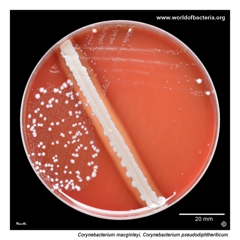
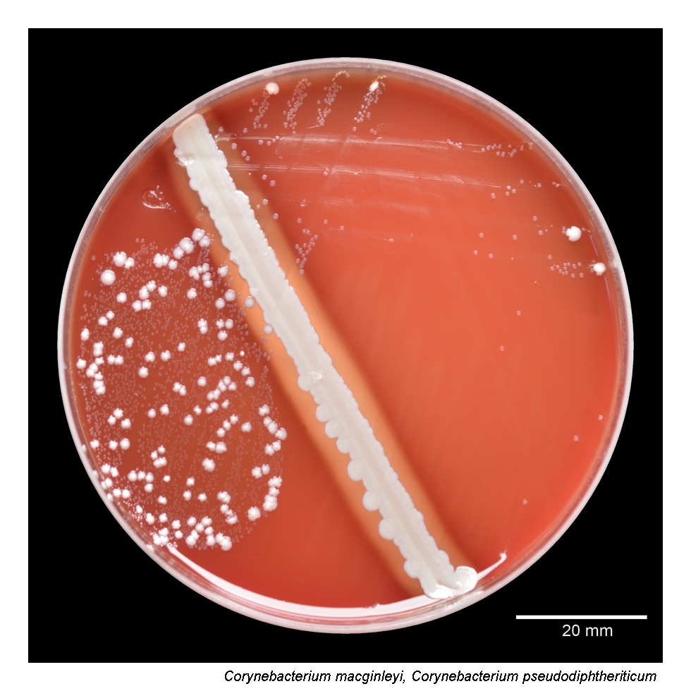
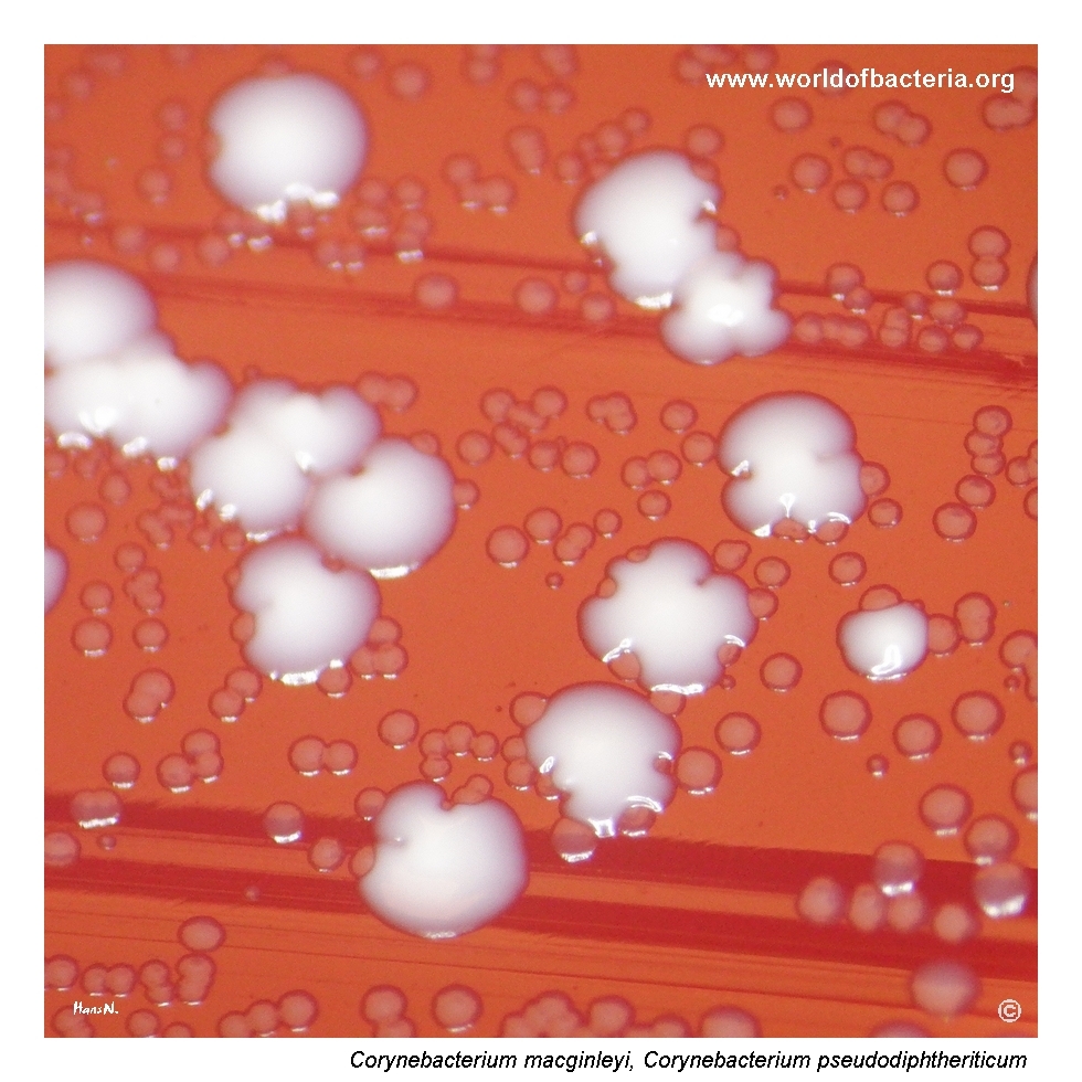
Conjunctival swab after subculture from enrichment broth. Tiny colonies of Corynebacterium macginleyi are visible together with larger white colonies of C. pseudodiphtheriticum. Incubation: 48 hours at 36 °C in 5% CO2. Patient: 97-year-old woman, diagnosis H100, mucopurulent conjunctivitis.
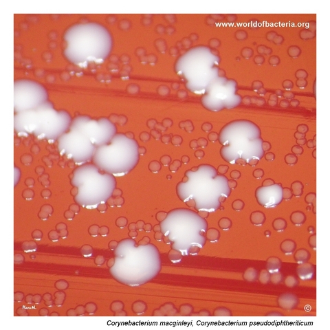
Colonies of C. macginleyi and C. pseudodiphtheriticum on blood agar. Even after 48 hours of incubation, the colonies of C. macginleyi remain very small, greyish, and non-haemolytic. Incubation: 48 hours at 36 °C in 5% CO2.
Close-up view of C. macginleyi colonies. The colonies are tiny, raised, non-pigmented, and measure approximately up to 1 mm in diameter. Incubation: 48 hours at 36 °C in 5% CO2.
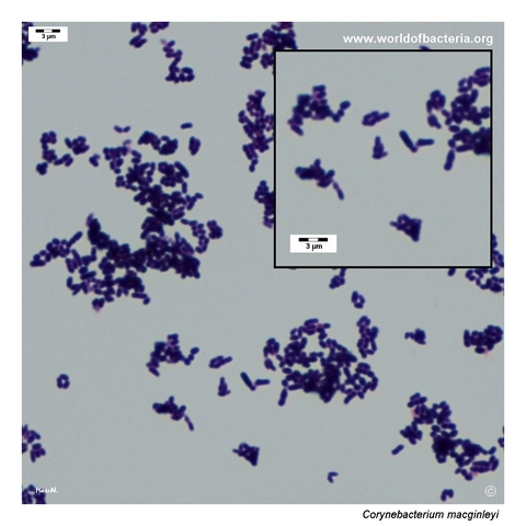
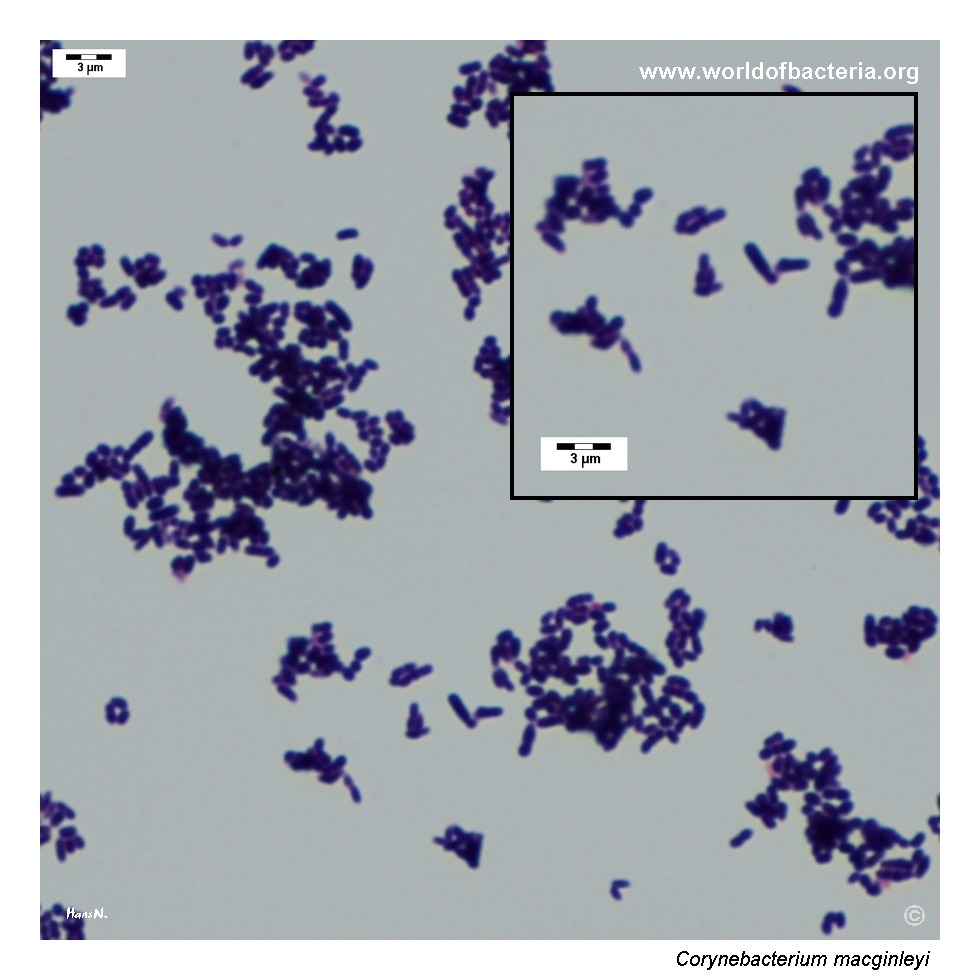
Smear prepared from colonies of Corynebacterium macginleyi grown on blood agar and stained by the Gram method. Gram-positive coryneform rods are visible.
Diagnostic and Clinical Notes
This page is didactically useful because Corynebacterium macginleyi shows how an organism of real ocular relevance can remain visually very inconspicuous on ordinary blood agar. Even after 48 hours, the colonies stay tiny, greyish, and non-haemolytic, so they can easily be overlooked in mixed cultures or dismissed as insignificant diphtheroid growth.
The comparison with Corynebacterium pseudodiphtheriticum is especially instructive. On the same plate, C. macginleyi remains clearly smaller and less prominent, which is exactly the kind of subtle difference that matters during careful reading of ocular cultures.
Microscopy confirms a classic coryneform pattern rather than a staphylococcal one, but that still does not solve the species-level question. In practice, the main value of microscopy here is orientation, while the decisive step is reliable instrumental identification.
Clinically, this species should be interpreted in context. In conjunctival or corneal material, repeated or quantitatively relevant recovery may support a pathogenic role, whereas the same visual phenotype would be much easier to dismiss if the ocular association of the species were not appreciated.
Laboratory Identification
Colony Morphology
On blood agar, Corynebacterium macginleyi forms very small, smooth to slightly raised, non-pigmented colonies with entire margins. The colonies are typically greyish and non-haemolytic, and even after 48 hours they may remain around 1 mm in diameter. This combination of tiny colony size and slow visual development is one of the most useful practical clues on primary culture.
Microscopy
Gram staining shows Gram-positive coryneform rods with the usual diphtheroid appearance. The cells may occur singly, in pairs, or in short angular arrangements. This morphology fits well with the genus Corynebacterium but is not sufficiently specific for final species assignment.
Key Identification Clues
Very small grey non-haemolytic colonies on blood agar
Raised colony profile with limited enlargement even after 48 hours
Gram-positive coryneform rods
Strong association with conjunctival and other ocular specimens
Potential confusion with other small corynebacteria or insignificant diphtheroid flora
Need for reliable species-level identification in ophthalmic material
Modern Identification Methods
Modern identification is best achieved by MALDI-TOF mass spectrometry when the database includes this species adequately. In difficult or unusual cases, sequencing-based methods may still be required. This is particularly relevant because the colony phenotype is subtle and can easily be grouped generically as “diphtheroids” unless instrumental identification is actively pursued.
Antibiotic Characteristics
No simple species-level susceptibility rule should be assumed from appearance alone for Corynebacterium macginleyi. In ocular microbiology, treatment decisions should be guided by the clinical syndrome, specimen type, and actual susceptibility testing of the isolate.
Older literature often regarded the organism as susceptible to commonly used topical ophthalmic agents, but more recent studies have shown that ocular isolates may display clinically relevant fluoroquinolone resistance. This makes routine assumptions less reliable than they once seemed.
For this reason, the practical laboratory lesson is straightforward: do not dismiss the species as a harmless coryneform without context, and do not infer susceptibility solely from historical expectations.
Note: Antimicrobial therapy should always follow the clinical setting and, whenever possible, isolate-specific susceptibility testing.