Corynebacterium striatum is a nondiphtherial corynebacterial species of increasing clinical importance that may still appear deceptively ordinary on blood agar, where its small smooth colonies can resemble coagulase-negative staphylococci.
Basic Characteristics
Taxonomy
Domain: Bacteria
Phylum: Actinomycetota
Class: Actinomycetes
Order: Corynebacteriales
Family: Corynebacteriaceae
Genus:Corynebacterium
Species:Corynebacterium striatum
Microscopy & Gram Stain
Short Gram-positive coryneform rods typical of the genus Corynebacterium
The Gram stain is especially important because the colonies may resemble coagulase-negative staphylococci on blood agar
Microscopy is therefore the key step that redirects interpretation from Gram-positive cocci toward a nondiphtherial corynebacterium
As in other corynebacteria, morphology supports generic placement but does not by itself establish the species
Oxygen Relationship
Aerobic or aerotolerant coryneform organism that grows well under routine laboratory conditions on blood agar
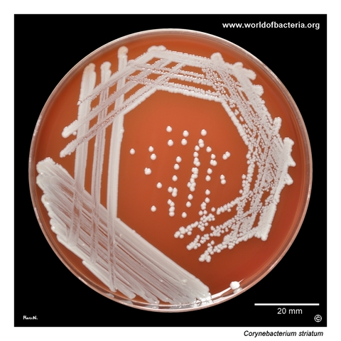
In this page set, visible colonies are shown after 48 hours of incubation at 36 °C in 5% CO2
The species is not visually demanding in culture, but careful plate reading is needed because the colonies are relatively inconspicuous
Rapid Identification Tests
Morphology: Gram-positive coryneform rod
Colony phenotype: small, smooth, convex, non-haemolytic colonies that may resemble coagulase-negative staphylococci
Practical clue: ordinary-looking colonies on blood agar should still be checked by Gram stain when isolated from clinically important wound material
Clinical context: interpretation depends heavily on specimen type, quantity of growth, and accompanying flora
Important limitation: reliable species-level identification generally requires MALDI-TOF MS or another modern identification method
Ecology and Clinical Relevance
Natural Habitat
Corynebacterium striatum is associated mainly with human skin and mucosal surfaces and may be recovered as part of the accompanying flora of superficial specimens
Because of this ecological background, recovery from wound material does not automatically prove pathogenic significance
At the same time, the species is increasingly recognized as an opportunistic hospital-associated organism and should not always be dismissed as a harmless bystander
Common Clinical Specimens
Wound swabs and chronic wound material
Respiratory and device-associated specimens
Blood culture or hospital-associated material in selected clinically significant cases
Clinical Significance
Corynebacterium striatum occupies an intermediate position between accompanying flora and true opportunistic pathogen, which makes interpretation strongly context-dependent
The species has gained attention because it can cause clinically relevant infections, especially in hospitalized or otherwise predisposed patients
In wound microbiology, significance is best judged from specimen quality, abundance, associated organisms, and the broader clinical picture rather than from species identity alone
Differential Considerations
Coagulase-negative staphylococci, because the colonies may appear deceptively coccoid in culture
Other nondiphtherial corynebacteria with small non-haemolytic colonies on blood agar
Corynebacterium amycolatum and Corynebacterium tuberculostearicum, which may also appear in wound or device-associated material
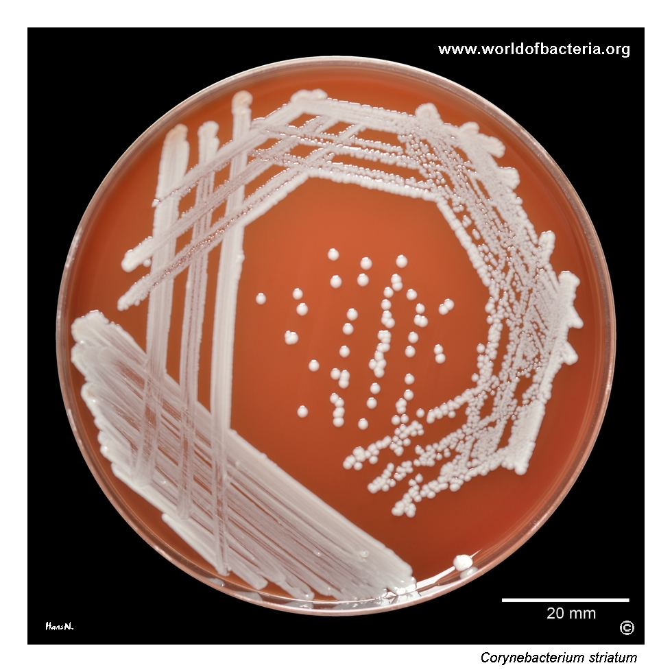
Corynebacterium striatum on blood agar after 48 hours of incubation at 36 °C in 5% CO2. The isolate was recovered from a lower-limb wound swab from a 74-year-old man. In wound microbiology, this species may occur either as part of accompanying flora or as a more significant opportunistic organism, so its role must always be assessed in relation to the type of specimen, quantity of growth, and the presence of other bacteria.
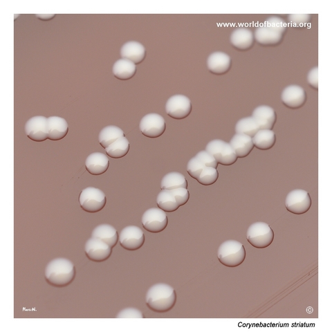
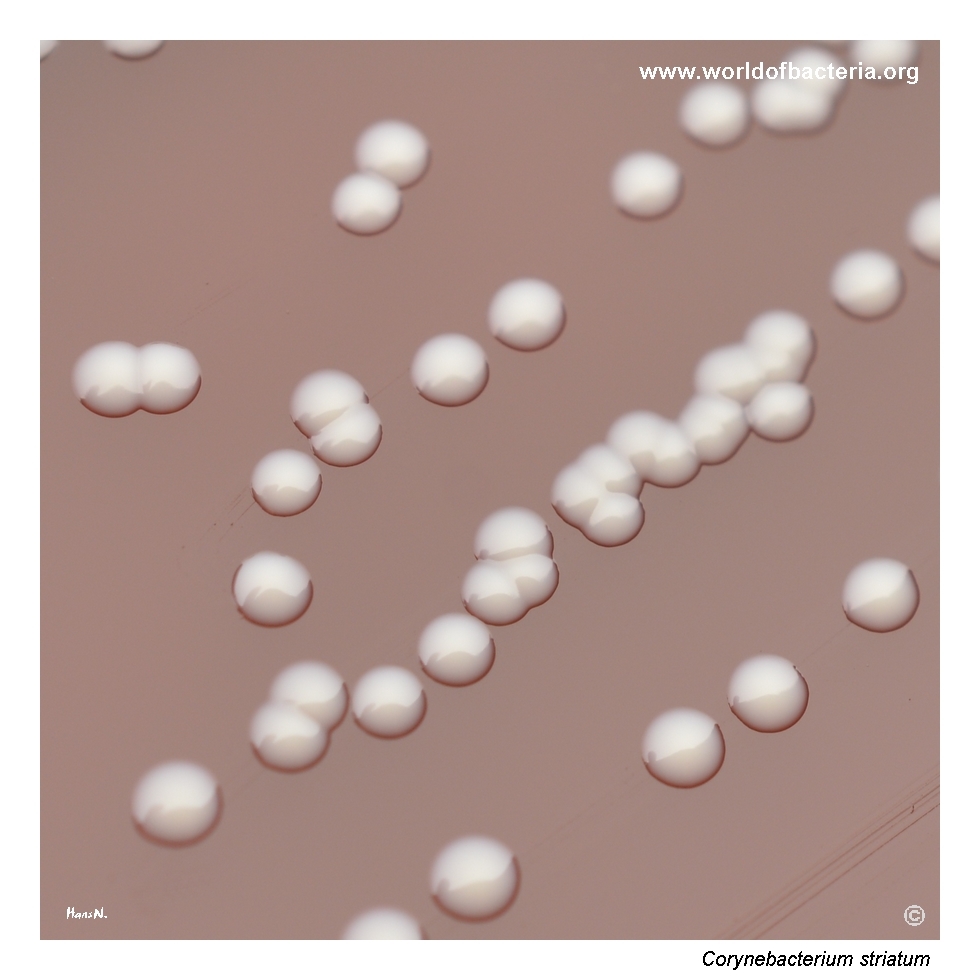
Colonies of Corynebacterium striatum on blood agar after 48 hours at 36 °C in 5% CO2. The colony morphology is relatively inconspicuous on routine medium, which is one reason why this organism may be underestimated during primary culture reading. Careful interpretation is especially important in wound specimens, where mixed growth often complicates assessment of microbiological significance.
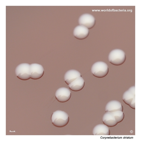
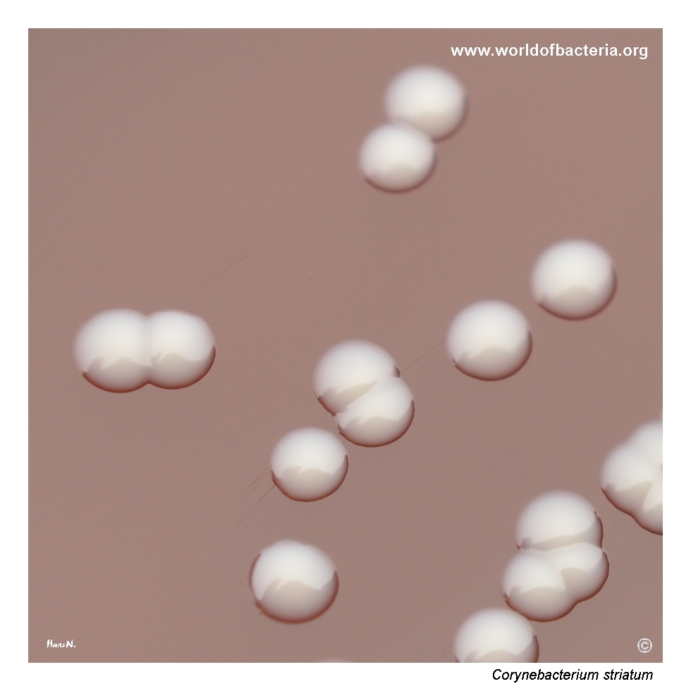
Close-up view of Corynebacterium striatum colonies on blood agar after 48 hours of incubation at 36 °C in 5% CO2. The colonies are smooth, convex, and round with entire margins, resembling coagulase-negative staphylococci in appearance. This similarity is a useful reminder that seemingly ordinary small colonies on blood agar may represent corynebacteria rather than true cocci.
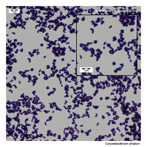
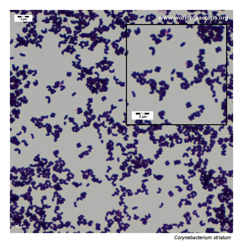
Gram-stained smear prepared from a colony of Corynebacterium striatum grown on blood agar. The organism appears as short Gram-positive coryneform rods. Although the microscopic morphology is typical for the genus, species-level identification still depends on correlation with the culture pattern and further laboratory testing, particularly when the isolate is recovered from clinically significant wound material.
Diagnostic and Clinical Notes
This page is intentionally didactic because Corynebacterium striatum shows how an organism of increasing clinical importance can still look surprisingly ordinary on a routine blood agar plate. The small smooth colonies shown here do not immediately demand attention, which is precisely why the species may be underestimated during primary culture reading.
In wound microbiology, interpretation must remain cautious. The isolate came from a lower-limb wound swab, and in such material the central question is rarely species identification alone. What matters most is whether the organism is present merely as accompanying flora or whether its abundance, persistence, and clinical context support a more direct role in infection.
Microscopy is the decisive corrective step. The Gram-stained smear shows short Gram-positive coryneform rods and immediately redirects the observer away from coagulase-negative staphylococci, which the colonies may resemble macroscopically. This is one of the most useful teaching points of the image set.
The broader clinical lesson is that nondiphtherial corynebacteria should not automatically be ignored when isolated from clinically meaningful specimens. Corynebacterium striatum is one of the species that most clearly illustrates this shift from “background flora” to potentially significant opportunistic pathogen in modern clinical microbiology.
Laboratory Identification
Colony Morphology
On blood agar, Corynebacterium striatum forms relatively inconspicuous non-haemolytic colonies. In this isolate the colonies are smooth, convex, and round with entire margins, a morphology that can resemble coagulase-negative staphylococci more than a striking corynebacterial phenotype. This limited distinctiveness is the main reason why the organism may be underestimated in routine culture work.
Microscopy
Gram staining shows short Gram-positive coryneform rods typical of the genus Corynebacterium. Even though this morphology does not determine the species, it is diagnostically crucial because it distinguishes the isolate from Gram-positive cocci that may appear similar on the basis of colony morphology alone. In mixed wound cultures, this shift in interpretation can substantially change the diagnostic workup.
Key Identification Clues
Small smooth non-haemolytic colonies on blood agar
Colony morphology that may mimic coagulase-negative staphylococci
Short Gram-positive coryneform rods on Gram stain
Recovery from clinically important wound material, where interpretation must be specimen-driven
Need for MALDI-TOF MS or another reliable modern method for definitive species-level identification
Modern Identification Methods
Reliable identification is best achieved by MALDI-TOF mass spectrometry when the database is current and well curated for nondiphtherial corynebacteria. This is especially relevant for C. striatum, because the species often looks unremarkable in culture while carrying greater clinical relevance than might be assumed from the plate alone. In selected cases, additional phenotypic or molecular methods may be useful, particularly when distinguishing clinically significant isolates from accompanying flora influences therapeutic decisions.
Antibiotic Characteristics
For Corynebacterium striatum, antimicrobial expectations should not be inferred from the visually bland appearance of the colonies. The plate phenotype is helpful descriptively but gives little direct information about susceptibility.
This species has become important in part because clinically significant isolates may show broad antimicrobial resistance. For that reason, species-level identification should ideally be followed by isolate-specific susceptibility testing whenever the organism is considered potentially relevant to infection.
In wound specimens, the first practical challenge is interpretive rather than purely technical: deciding whether the isolate deserves full workup at all. Once that threshold is crossed, treatment decisions should be based on the specific isolate and the overall wound microbiology rather than on general assumptions about corynebacteria.
Note:C. striatum is one of the nondiphtherial corynebacteria that most clearly demonstrates how apparently inconspicuous colonies can still belong to a clinically meaningful and sometimes multidrug-resistant opportunistic pathogen.