Corynebacterium amycolatum is a non-haemolytic nondiphtherial corynebacterial species that may occur in polymicrobial wound cultures, where its irregular lobate colonies can provide a useful visual clue among smoother accompanying bacteria.
Basic Characteristics
Taxonomy
Domain: Bacteria
Phylum: Actinomycetota
Class: Actinomycetes
Order: Corynebacteriales
Family: Corynebacteriaceae
Genus:Corynebacterium
Species:Corynebacterium amycolatum
Microscopy & Gram Stain
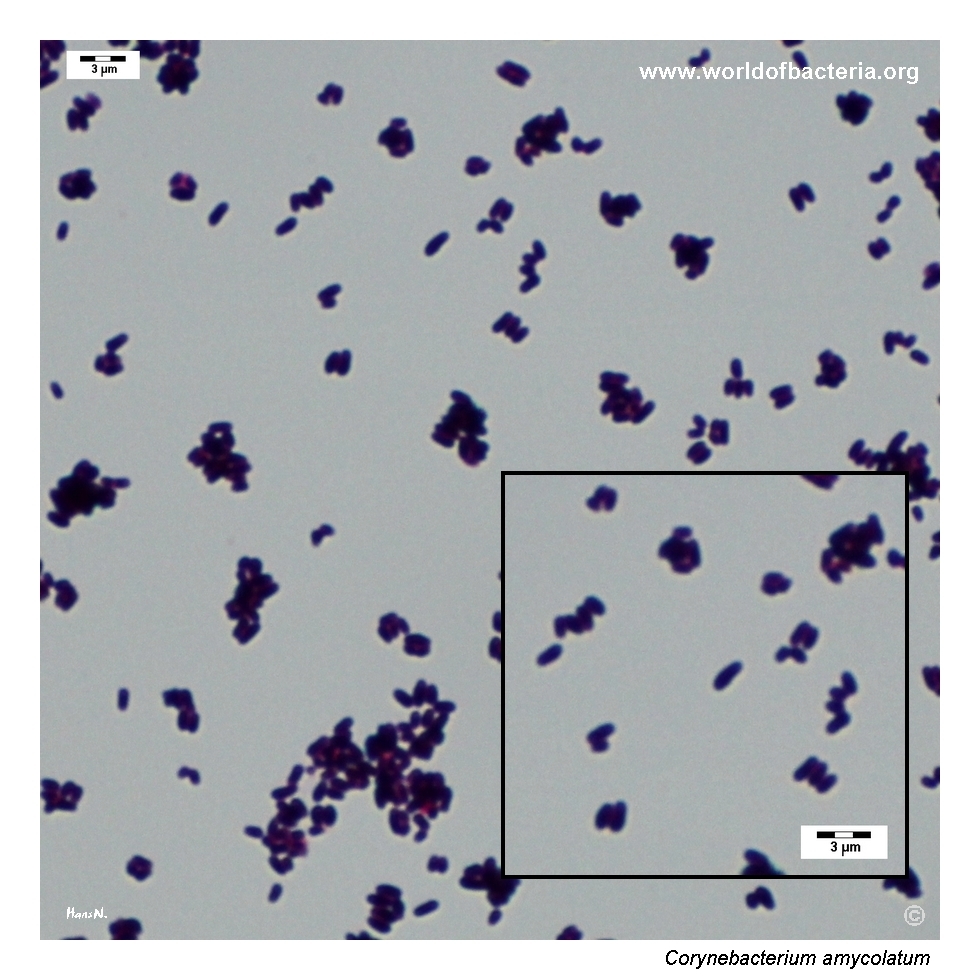
Short Gram-positive coryneform rods, sometimes arranged in palisades
Microscopy is particularly useful in mixed wound cultures because it helps distinguish the organism from Gram-positive cocci with superficially similar colonies
The smear supports placement within the corynebacterial group but does not by itself determine species identity
In practical diagnostics, the Gram stain is often the key step that prevents irregular non-haemolytic colonies from being misread as a different Gram-positive organism
Oxygen Relationship
Aerobic coryneform organism that grows under routine laboratory conditions on blood agar
In this page set, visible colonies are shown after 48 hours of incubation at 36 °C in 5% CO2
The species behaves as a conventional nondiphtherial corynebacterial isolate rather than as a fastidious organism, but careful reading is still needed in polymicrobial cultures
Rapid Identification Tests
Morphology: Gram-positive coryneform rod
Colony phenotype: non-haemolytic colonies with a lobate or serrated outline on blood agar
Practical clue: the irregular colony contour may help the species stand out among smoother colonies of accompanying wound flora
Ecological setting: commonly encountered in skin-associated or wound material and therefore subject to the usual problem of colonization versus significance
Important limitation: definitive species-level identification generally requires MALDI-TOF MS or another modern method rather than colony appearance alone
Ecology and Clinical Relevance
Natural Habitat
Corynebacterium amycolatum is best regarded as part of the human skin-associated microbiota and is often encountered in superficial or chronic wound material
Because of this ecological background, recovery from polymicrobial specimens must always be interpreted in relation to specimen type, quantity, and the rest of the culture
The organism may therefore act either as a colonizer or as an opportunistic participant in infection, depending on the clinical setting
Common Clinical Specimens
Wound swabs and tissue specimens, especially from chronic or polymicrobial wounds
Skin-associated material and superficial specimens
Occasionally blood culture or hospital-associated specimens when the species becomes clinically relevant
Clinical Significance
Corynebacterium amycolatum is usually interpreted as a low-virulence opportunist, but it should not automatically be dismissed when recovered from clinically important sites
Its significance increases when the isolate is recovered from chronic wounds, diabetic foot material, device-associated settings, or from patients with substantial underlying disease
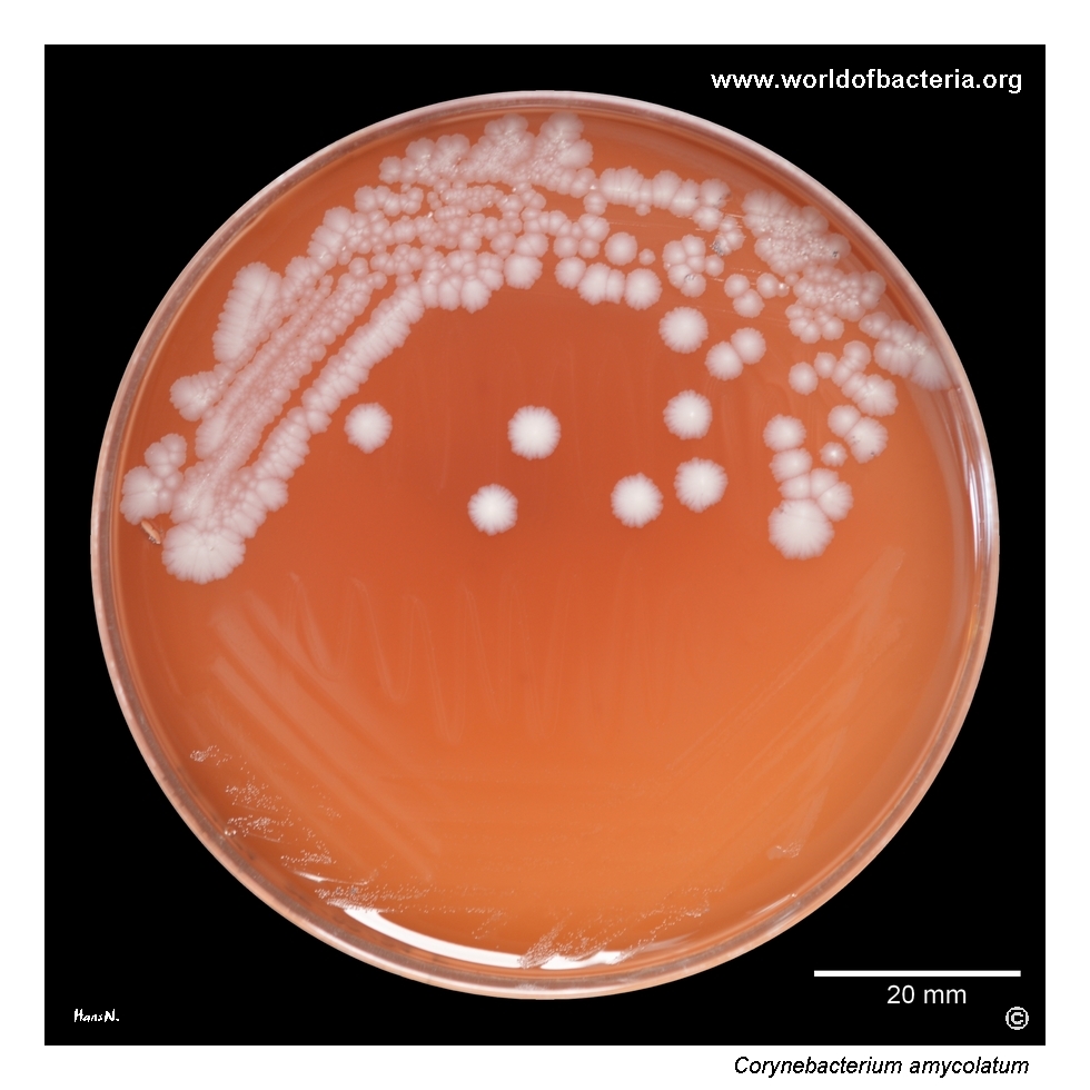
In this image set, the species formed part of a polymicrobial wound community together with Staphylococcus aureus and Enterobacter cloacae, illustrating the interpretive difficulty typical of chronic wound microbiology
Differential Considerations
Other nondiphtherial corynebacteria recovered from wound material, especially species that form small non-haemolytic colonies
Coagulase-negative staphylococci, because corynebacterial colonies in mixed culture may initially be underestimated
Corynebacterium tuberculostearicum and Corynebacterium jeikeium, which are also clinically relevant corynebacteria encountered in hospital-associated material
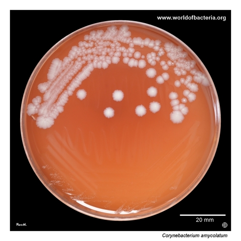
Corynebacterium amycolatum on blood agar after 48 hours of incubation at 36 °C in 5% CO2. The isolate came from a lower-limb wound in a patient with diabetes. In this specimen, C. amycolatum formed part of a polymicrobial community together with Staphylococcus aureus and Enterobacter cloacae. In chronic wounds, such mixed growth is common and requires careful interpretation to distinguish colonizing flora from organisms contributing more directly to tissue infection.
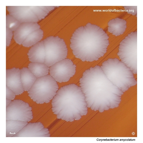
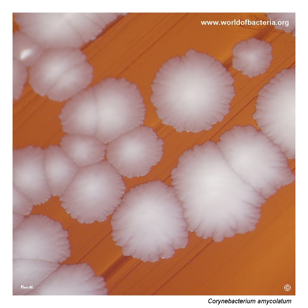
Colonies of Corynebacterium amycolatum on blood agar after 48 hours at 36 °C in 5% CO2. The colonies are lobate and show a serrated margin, giving them a more irregular outline than many other corynebacterial colonies. This somewhat distinctive morphology may provide a useful visual clue during routine plate reading, especially when the organism is present among smoother colonies of accompanying bacteria.
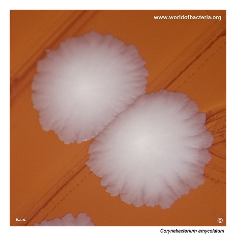
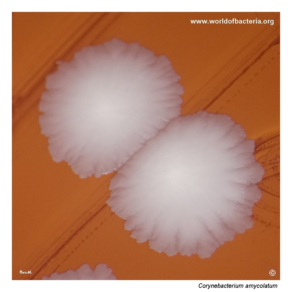
Close-up view of Corynebacterium amycolatum colonies on blood agar after 48 hours of incubation at 36 °C in 5% CO2. The image highlights the irregular colony contour and overall surface appearance of the isolate. Compared with more uniform, smooth corynebacterial colonies, this morphology may stand out on the plate and can help direct attention to the organism during evaluation of polymicrobial wound cultures.
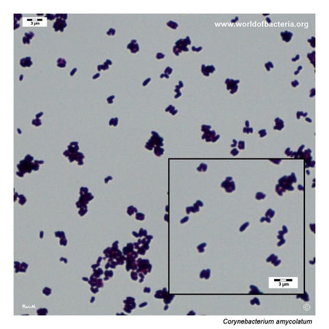
Gram-stained smear prepared from a colony of Corynebacterium amycolatum grown on blood agar. The organism appears as short Gram-positive coryneform rods, in some areas arranged in palisades. This microscopic pattern is typical of corynebacteria and provides an important clue when distinguishing the isolate from coccoid Gram-positive organisms in mixed clinical material.
Diagnostic and Clinical Notes
This page is useful because it shows a corynebacterial species that does not hide only by being small and bland. Here the colonies are still non-haemolytic, but their outline is more irregular than that of many other corynebacteria, with a lobate, serrated contour that can serve as a practical visual clue during plate reading.
The wound origin of the isolate adds a realistic interpretive problem. Chronic diabetic wounds commonly contain polymicrobial growth, and the recovery of Corynebacterium amycolatum alongside Staphylococcus aureus and Enterobacter cloacae illustrates the familiar challenge of deciding which organisms are acting mainly as colonizers and which may be contributing more directly to tissue infection.
Microscopy remains essential. The Gram-stained smear shows short Gram-positive coryneform rods, sometimes in palisades, which immediately separates the organism from Gram-positive cocci that might otherwise be suspected when reading a mixed blood agar plate. This is the decisive corrective step in the diagnostic workflow.
The main teaching point is that even when colony morphology offers a useful hint, definitive interpretation still depends on the whole picture: wound type, host factors, accompanying flora, microscopy, and reliable species identification. The page therefore demonstrates both the value and the limits of colony appearance in polymicrobial wound microbiology.
Laboratory Identification
Colony Morphology
On blood agar, Corynebacterium amycolatum forms non-haemolytic colonies that in this isolate are notable for their irregular, lobate outline and serrated margin. Compared with the smoother colonies of many other corynebacteria, this morphology is more visually distinctive and may help the organism stand out in a polymicrobial wound culture. Even so, the colony phenotype remains only a guide and does not by itself establish the species.
Microscopy
Gram staining shows short Gram-positive coryneform rods, in some areas arranged in palisades. This pattern is highly useful for placing the isolate in the corynebacterial group and for distinguishing it from coccoid Gram-positive organisms in mixed material. As with other nondiphtherial corynebacteria, however, microscopy supports generic recognition rather than definitive species assignment.
Key Identification Clues
Non-haemolytic colonies with a lobate or serrated contour on blood agar
Short Gram-positive coryneform rods, sometimes arranged in palisades
Recovery from a chronic lower-limb wound in a patient with diabetes
Presence in a polymicrobial culture together with smoother, more visually dominant accompanying species
Need for MALDI-TOF MS or another modern identification method for reliable species-level assignment
Modern Identification Methods
Reliable identification is best achieved by MALDI-TOF mass spectrometry when a current database is available. This is especially useful for C. amycolatum, because wound-associated nondiphtherial corynebacteria can overlap morphologically and may otherwise be underestimated in mixed culture. In difficult or clinically important cases, additional phenotypic or molecular methods may be helpful, particularly when therapeutic decisions depend on clarifying the significance of individual members of a polymicrobial flora.
Antibiotic Characteristics
For Corynebacterium amycolatum, therapeutic interpretation should not be based on colony appearance alone. The irregular non-haemolytic phenotype on blood agar is useful descriptively, but it gives little direct information about clinical relevance or susceptibility profile.
In chronic wound microbiology, the first practical task is to decide whether the organism is likely to be a bystander, a colonizer, or one of several bacteria contributing to the inflammatory process. Once full workup is justified, isolate-specific susceptibility testing is more informative than assumptions based only on taxonomic identity or plate appearance.
Because the species often appears in polymicrobial material, antimicrobial decisions should be integrated with the broader wound context rather than inferred from the presence of the organism in isolation.
Note: The main diagnostic challenge is not merely to identify C. amycolatum, but to decide what weight it should carry within a chronic mixed wound culture that also contains more obviously pathogenic bacteria.