Corynebacterium simulans is an uncommon nondiphtherial corynebacterial species whose non-haemolytic colonies may closely mimic coagulase-negative staphylococci on blood agar, making Gram stain and modern species identification especially important.
Basic Characteristics
Taxonomy
Domain: Bacteria
Phylum: Actinomycetota
Class: Actinomycetes
Order: Corynebacteriales
Family: Corynebacteriaceae
Genus:Corynebacterium
Species:Corynebacterium simulans
Microscopy & Gram Stain
Very short Gram-positive coryneform rods typical of the genus Corynebacterium
In this page set, the cells can appear deceptively coccoid in some areas, which helps explain why the isolate may initially be mistaken for Gram-positive cocci
The Gram stain is therefore central to correct interpretation when apparently ordinary non-haemolytic colonies are recovered on blood agar
As with other nondiphtherial corynebacteria, morphology alone does not establish the species
Oxygen Relationship
Routine aerobic coryneform isolate that grows on standard blood agar under ordinary laboratory conditions
In these images, colony development is illustrated after 48 hours of incubation in ambient air
The main diagnostic challenge is not fastidiousness but the striking resemblance of the colonies to coagulase-negative staphylococci
Rapid Identification Tests
Morphology: very short Gram-positive coryneform rods
Practical warning: plate morphology alone may be misleading and can result in misclassification as true cocci
Useful next step: Gram staining followed by MALDI-TOF MS or another reliable modern identification method
Differential issue: distinction from Corynebacterium striatum, other nondiphtherial corynebacteria, and visually similar coagulase-negative staphylococci
Ecology and Clinical Relevance
Natural Habitat
Corynebacterium simulans is a human-associated nondiphtherial corynebacterium and published clinical series suggest that it is recovered mainly from skin-associated material
Compared with more familiar corynebacteria, it appears to be an uncommon laboratory finding and is likely underrecognized when identification is based too heavily on colony appearance alone
Its name is diagnostically apt: the species can visually simulate other Gram-positive organisms on routine media
Common Clinical Specimens
Skin- and soft-tissue-associated material
Wound-related specimens and other clinically relevant material in which small non-haemolytic colonies may be overlooked
Occasional human clinical isolates that require modern identification for confident recognition
Clinical Significance
Corynebacterium simulans should be regarded as an uncommon but potentially relevant human clinical isolate rather than a species known only from taxonomy
Published clinical observations suggest that it is encountered much less often than Corynebacterium striatum, but that when recovered it is often associated with skin-related material
Its main practical significance in routine microbiology is that it may be hidden among colonies that appear at first glance to be ordinary coagulase-negative staphylococci
Differential Considerations
Coagulase-negative staphylococci when only the blood agar plate is inspected macroscopically
Corynebacterium striatum and other small nondiphtherial corynebacteria with similarly modest colony morphology
Other Gram-positive organisms producing smooth, non-haemolytic colonies that provide limited clues on solid media
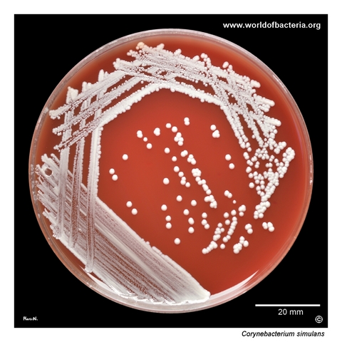
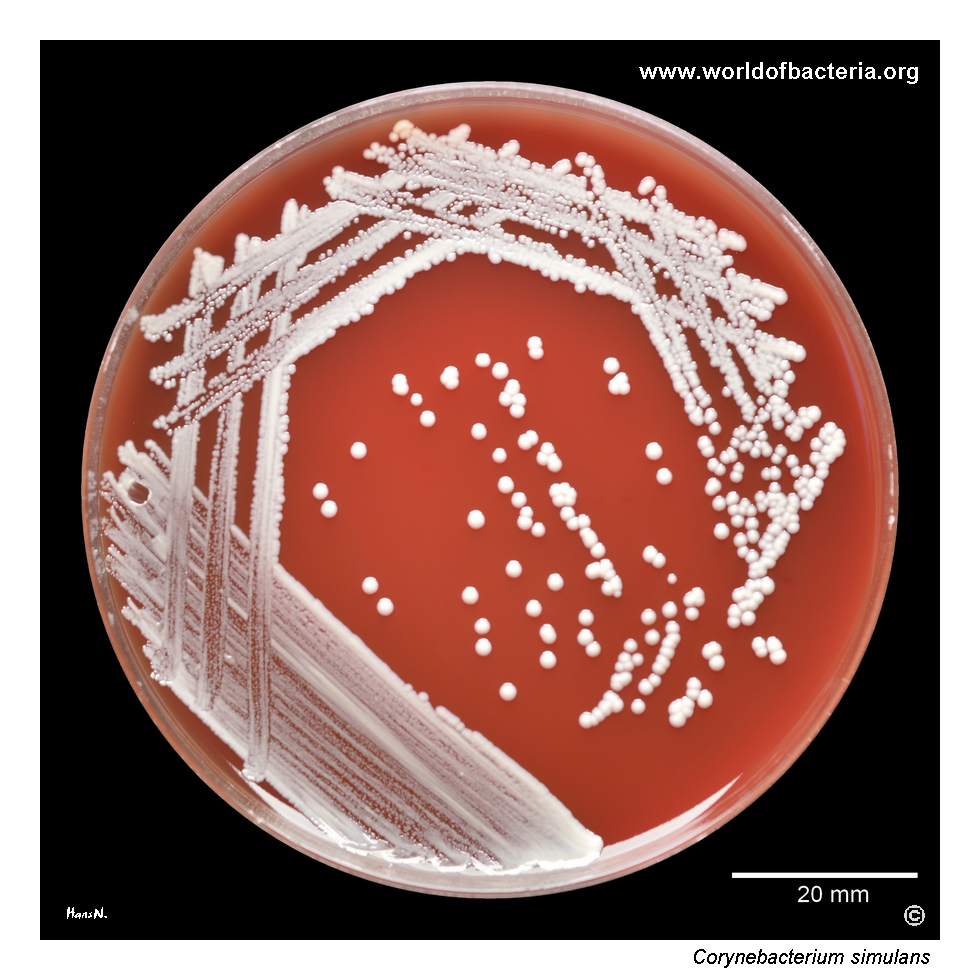
Corynebacterium simulans cultured on blood agar after 48 hours of incubation at 36 °C in ambient air. The isolated colonies closely resemble those of coagulase-negative staphylococci in routine culture. This inconspicuous appearance is diagnostically important, because corynebacteria of this type may easily be overlooked or misclassified unless Gram staining is performed and the colony morphology is interpreted with sufficient caution.
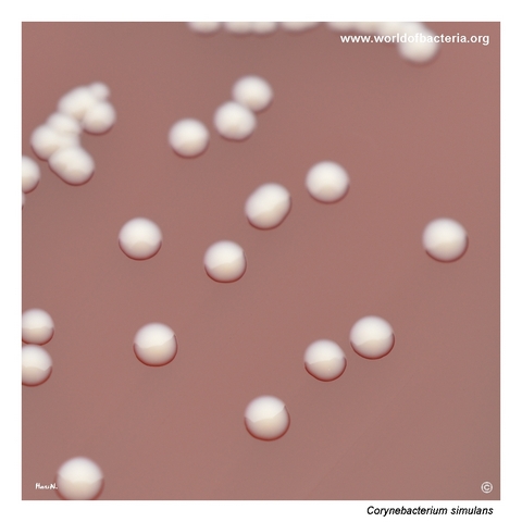
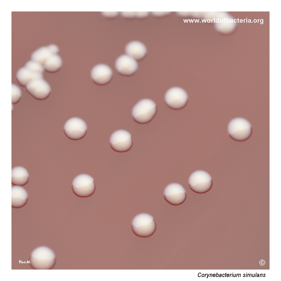
Colonies of Corynebacterium simulans on blood agar. The colonies are smooth, convex, and round with entire margins, again resembling coagulase-negative staphylococci. On routine media, such isolates provide only limited visual clues to their true identity, which makes microscopic examination particularly valuable when apparently ordinary, non-haemolytic colonies are recovered from clinically relevant material.
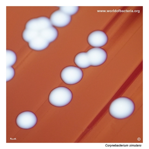
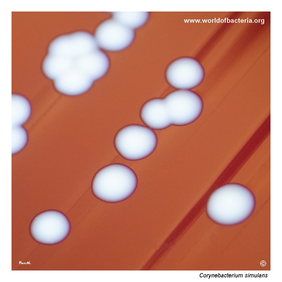
Close-up view of Corynebacterium simulans colonies after 48 hours of incubation. At this stage, the colonies are visually indistinguishable from those of other non-haemolytic coagulase-negative staphylococci growing on blood agar. This is a useful reminder that plate morphology alone may be misleading and that even very typical-looking small colonies may represent coryneform bacteria rather than true cocci.
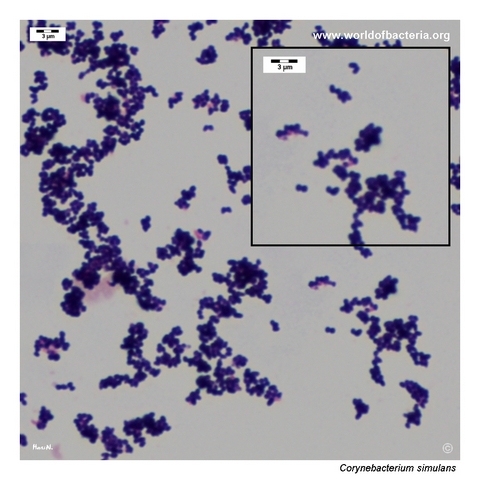
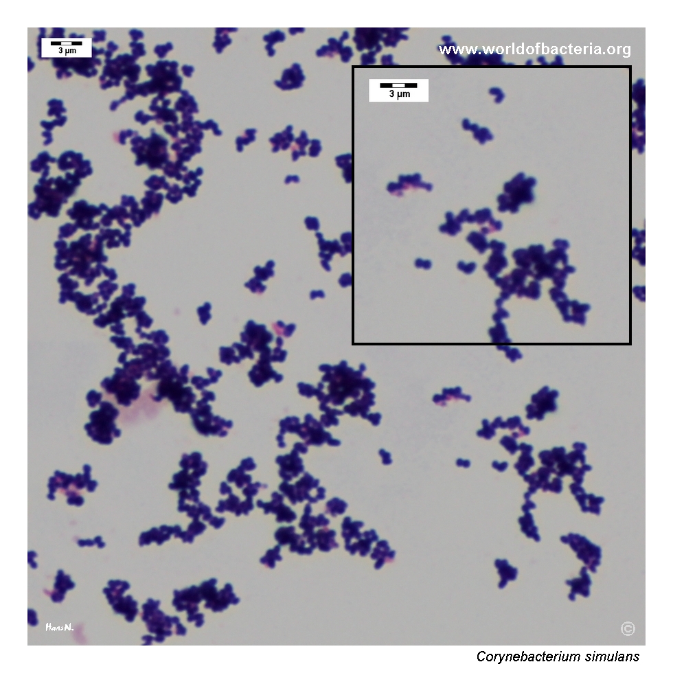
Gram-stained smear prepared from a colony of Corynebacterium simulans grown on blood agar. The organism appears as very short Gram-positive coryneform rods, in some areas resembling clusters of cocci at first glance. This microscopic appearance explains why the isolate may be confusing in routine practice and highlights the importance of careful interpretation of both colony morphology and Gram stain findings.
Diagnostic and Clinical Notes
This page is didactically useful because it shows a nondiphtherial corynebacterium that can be almost impossible to recognize correctly from plate morphology alone. All three culture images illustrate an isolate that visually simulates coagulase-negative staphylococci so closely that an unwary reading could stop at the wrong organism group altogether.
The Gram stain therefore becomes the pivotal corrective step. Instead of Gram-positive cocci, the smear reveals very short coryneform rods, some of which are so compactly arranged that they can give a coccoid impression at first glance. That tension between macroscopic and microscopic appearance is the central teaching value of this page.
The species is also microbiologically interesting because published clinical work suggests that Corynebacterium simulans is recovered mainly from skin-associated material and is much less common than Corynebacterium striatum. In routine diagnostics, that makes it both uncommon and easy to underrecognize.
The broader message is clear: when a culture plate seems to show an ordinary field of small, smooth, non-haemolytic staphylococcus-like colonies, a careful Gram stain may completely change the interpretation. For this reason, C. simulans is an excellent atlas example of why apparently simple colony morphology should not be trusted without microscopy.
Laboratory Identification
Colony Morphology
On blood agar, Corynebacterium simulans forms smooth, convex, round, non-haemolytic colonies with entire margins. In the present image set, the colonies are so similar to coagulase-negative staphylococci that they provide almost no reliable macroscopic clue to their true identity. This makes the species a useful example of how a routine blood agar plate can be visually misleading.
Microscopy
Gram staining shows very short Gram-positive coryneform rods. In some microscopic fields, the cells may superficially resemble clusters of cocci, but careful inspection supports placement within the corynebacterial group rather than among true staphylococci. This page therefore demonstrates how microscopy can overturn an initial plate-based assumption.
Key Identification Clues
Smooth, convex, round non-haemolytic colonies that mimic coagulase-negative staphylococci
Very short Gram-positive coryneform rods on Gram stain, sometimes deceptively coccoid in impression
Poor discriminatory value of plate morphology alone
Need to consider uncommon nondiphtherial corynebacteria when apparently ordinary colonies do not fit the full microscopic picture
Definitive species-level identification best achieved by MALDI-TOF MS or another reliable modern method
Modern Identification Methods
Reliable identification is best achieved by MALDI-TOF mass spectrometry when the reference database is suitable for uncommon nondiphtherial corynebacteria. This is especially relevant for C. simulans, because the species is uncommon and morphologically deceptive on blood agar. Where uncertainty remains, additional phenotypic or molecular methods may be needed, particularly when the isolate is recovered from clinically significant material.
Antibiotic Characteristics
No therapeutic conclusion should be drawn from the visual resemblance of Corynebacterium simulans to coagulase-negative staphylococci. Apparent colony type does not predict antimicrobial susceptibility.
Published clinical data suggest that C. simulans may be more susceptible overall than Corynebacterium striatum, including to several orally available agents, but this should be treated as a population-level observation rather than a rule for any individual isolate.
When the organism is considered clinically relevant, antimicrobial susceptibility testing should be performed on the individual isolate and interpreted in the full clinical context.
Note: The main didactic value of this page is diagnostic rather than therapeutic: it shows how an uncommon corynebacterium can first imitate a staphylococcal colony pattern and only later reveal its true identity through microscopy and modern identification methods.