Corynebacterium kroppenstedtii is a small-colony lipophilic coryneform species strongly associated with recurrent mastitis and other inflammatory breast lesions, where it may be easy to overlook among accompanying flora.
Basic Characteristics
Taxonomy
Domain: Bacteria
Phylum: Actinomycetota
Class: Actinomycetes
Order: Corynebacteriales
Family: Corynebacteriaceae
Genus:Corynebacterium
Species:Corynebacterium kroppenstedtii
Microscopy & Gram Stain
Gram-positive rods with a coryneform appearance
Pleomorphic diphtheroid bacilli that may look short, slightly clubbed, or irregularly arranged
Microscopy is useful for recognizing the isolate as a corynebacterium, but it does not by itself establish the species
In material from recurrent mastitis, the microscopic pattern should always be interpreted together with the small-colony growth on culture
Oxygen Relationship
Aerobic to facultatively anaerobic coryneform organism that grows under routine culture conditions
On ordinary blood agar the species may remain visually subtle, with very small colonies that become easier to appreciate after prolonged incubation
Growth in this page set is documented both in 5% CO2 and in ambient air, showing that the practical diagnostic problem is not fastidiousness but easy visual oversight
Rapid Identification Tests
Catalase: positive
Microscopy: Gram-positive coryneform rod
Practical clue: minute non-haemolytic colonies in breast-associated material, often mixed with more conspicuous flora
Biological note: an unusual lipophilic corynebacterium that lacks mycolic acids
Important caveat: reliable identification usually requires MALDI-TOF MS or another up-to-date species-level method
Ecology and Clinical Relevance
Natural Habitat
The species is regarded as a human-associated lipophilic corynebacterium and has been recovered mainly from breast-related clinical material
Its biological adaptation to lipid-rich environments is consistent with the strong association between this species and inflammatory disease of the breast
Although corynebacteria may also occur as colonizers, C. kroppenstedtii has become increasingly recognized as a genuine human pathogen in the appropriate clinical setting
Common Clinical Specimens
Nipple discharge
Breast abscess aspirates
Tissue from inflammatory breast lesions
Specimens from recurrent or granulomatous mastitis
Clinical Significance
Corynebacterium kroppenstedtii is now strongly linked with recurrent mastitis and granulomatous mastitis rather than being viewed as a routine insignificant diphtheroid
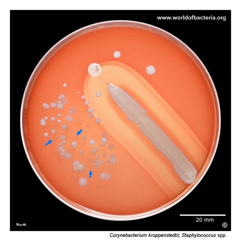
The species may be underrecognized because its colonies are tiny and can be hidden among accompanying skin flora or coagulase-negative staphylococci
Interpretation depends on specimen type, repeated recovery, and the overall inflammatory context, but breast-associated isolates deserve careful attention
Differential Considerations
Other nondiphtherial corynebacteria recovered from breast material, including lipophilic species
Coagulase-negative staphylococci when mixed cultures on blood agar are judged mainly by colony prominence
Corynebacterium jeikeium, Corynebacterium macginleyi, and other small-colony coryneform rods that may require instrumental identification for reliable differentiation
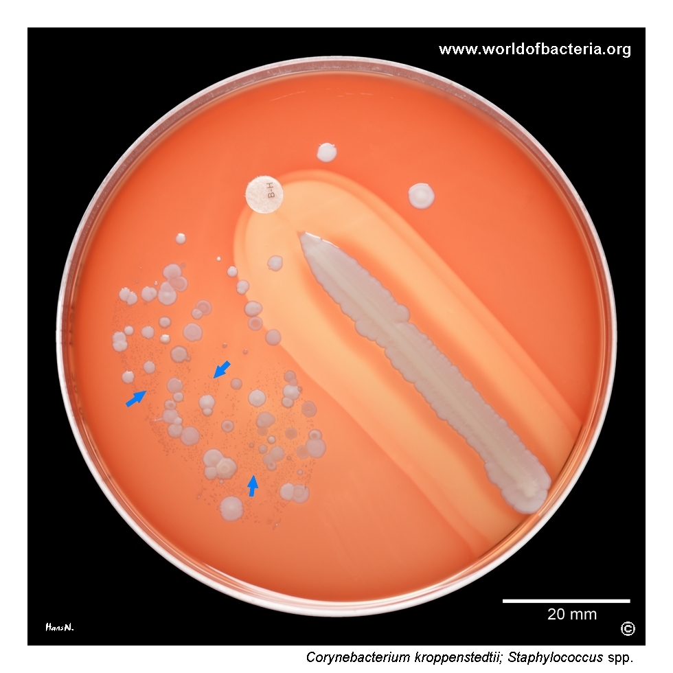
Culture of nipple discharge from a 26-year-old woman with the diagnosis L03.3, cellulitis of the trunk, with a clinical note of recurrent mastitis. Colonies of Corynebacterium kroppenstedtii, marked by blue arrows, are growing on blood agar among colonies of coagulase-negative staphylococci. A streak of Staphylococcus aureus ATCC 25923 and a bacitracin disk were also placed on the plate. The culture was incubated for 48 hours at 36 °C in 5% CO2.
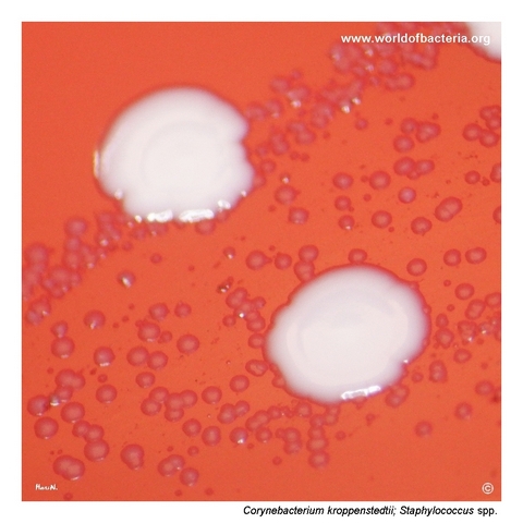
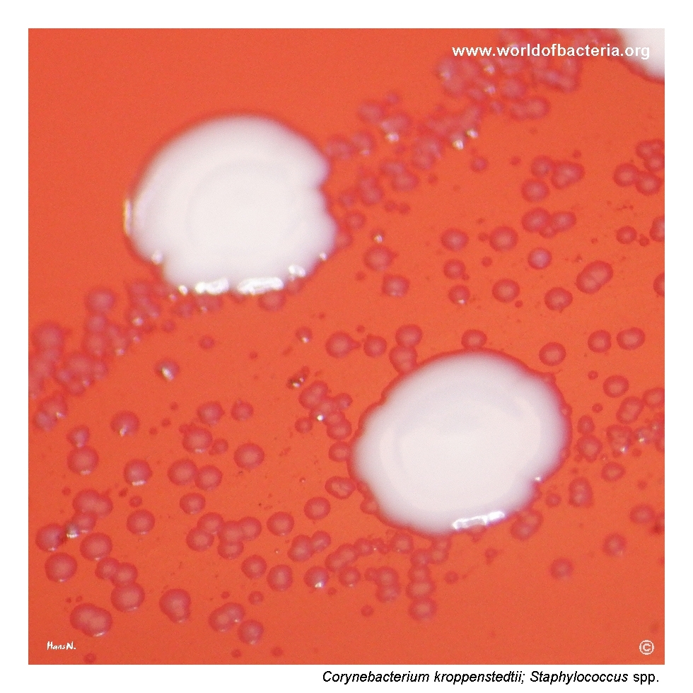
Very small non-haemolytic colonies of Corynebacterium kroppenstedtii growing on blood agar near white colonies of Staphylococcus species. The colonies are easy to miss because of their minute size and their position among more conspicuous accompanying flora. This is one reason why careful plate reading is especially important in specimens from chronic or recurrent breast inflammation. Culture conditions: 48 hours at 36 °C in 5% CO2.
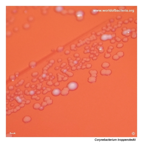
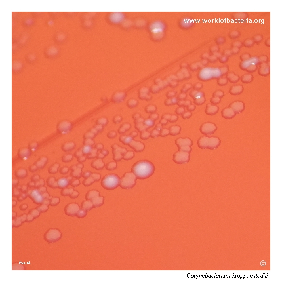
Close-up view of Corynebacterium kroppenstedtii colonies. Two colony types are visible in this image. Predominant are flattened, non-pigmented colonies with irregular margins, accompanied by white-pigmented, smooth, convex colonies. These represent two subpopulations of C. kroppenstedtii. Culture conditions: 48 hours at 36 °C in ambient air. Such variability within a single isolate may complicate visual interpretation of the primary culture.
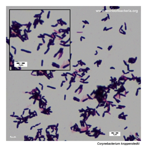
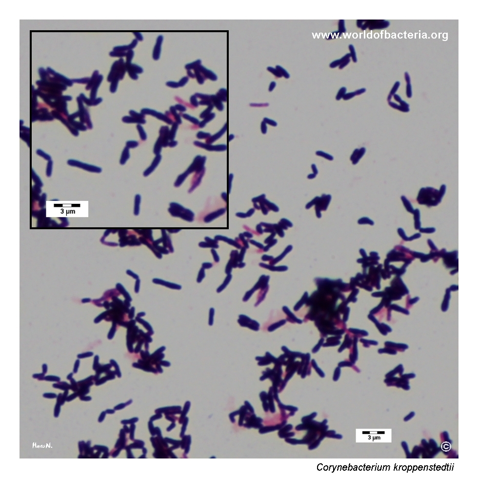
Smear prepared from colonies of Corynebacterium kroppenstedtii grown on blood agar and stained by the Gram method. The organism appears as Gram-positive rods, in some areas showing a coryneform appearance. Although the microscopic pattern is helpful for placing the isolate within the corynebacterial group, definitive identification requires correlation with the culture features and the clinical context, particularly in specimens associated with recurrent mastitis.
Diagnostic and Clinical Notes
This page is particularly useful because it demonstrates how an organism of real clinical importance can remain almost hidden on an ordinary culture plate. The colonies of Corynebacterium kroppenstedtii are minute, non-haemolytic, and easily overshadowed by accompanying coagulase-negative staphylococci or other routine flora, yet the clinical context points to a species that deserves deliberate attention.
The breast-associated setting is the main diagnostic clue. In recurrent mastitis or granulomatous inflammation, recovery of tiny coryneform colonies should not be dismissed automatically as irrelevant diphtheroid flora, especially if the same pattern recurs or the microbiological findings fit the clinical picture.
The image set also shows another practical complication: colony heterogeneity. Within one isolate, flattened irregular colonies and white-pigmented smooth convex colonies may coexist. Such variation can complicate visual interpretation and makes instrumental identification more important than morphology alone.
Microscopy confirms the corynebacterial nature of the isolate but does not resolve the clinically decisive question. The real challenge is to notice the organism at all, to relate it to the history of recurrent mastitis, and to avoid overlooking a species now well recognized in chronic inflammatory breast disease.
Laboratory Identification
Colony Morphology
On blood agar, Corynebacterium kroppenstedtii forms very small, non-haemolytic colonies that may remain inconspicuous even after 48 hours of incubation. In this page set, the isolate also shows colony heterogeneity, with flattened non-pigmented colonies with irregular margins alongside white-pigmented smooth convex colonies. This variability within a single isolate is visually striking and can complicate routine plate reading.
Microscopy
Gram staining shows Gram-positive rods with a coryneform appearance. The microscopic pattern supports placement within the corynebacterial group, but species-level identification cannot be made from microscopy alone.
Key Identification Clues
Minute non-haemolytic colonies on blood agar, often hidden among accompanying flora
Breast-associated specimen, especially recurrent mastitis or granulomatous inflammation
Gram-positive coryneform rods
Possible colony heterogeneity within a single isolate
Lipophilic biology and lack of mycolic acids
Need for MALDI-TOF MS or another up-to-date identification method for confident species assignment
Modern Identification Methods
Reliable identification is best achieved by MALDI-TOF mass spectrometry when the reference database is sufficiently current and comprehensive. For difficult or ambiguous isolates, sequencing-based methods may still be needed. This is particularly relevant for C. kroppenstedtii because the species may be underrecognized on morphology alone, and because recent taxonomic work has described closely related mastitis-associated taxa that can challenge older identification systems.
Antibiotic Characteristics
Corynebacterium kroppenstedtii should not be assigned a therapeutic expectation on the basis of colony appearance alone. Breast-associated infections caused by this species can be persistent or recurrent, and treatment decisions should be guided by the clinical syndrome together with actual susceptibility testing whenever possible.
Because the organism is lipophilic and strongly associated with chronic inflammatory breast disease, the microbiological result may have more significance than its tiny non-haemolytic colonies would suggest. The main laboratory lesson is therefore not to underestimate the isolate simply because it looks modest on blood agar.
Published clinical experience suggests that management of recurrent mastitis associated with C. kroppenstedtii can be challenging, and microbiological recognition of the species may influence diagnostic thinking even before final therapeutic decisions are made.
Note: Antimicrobial therapy should always follow the clinical setting, breast-specific pathology, and isolate-specific susceptibility data.