Clostridium perfringens is a large anaerobic spore-forming rod notable for rapid growth, robust grey colonies, and the classic double zone of haemolysis on blood agar.
Basic Characteristics
Taxonomy
Domain: Bacteria
Phylum: Bacillota
Class: Clostridia
Order: Clostridiales
Family: Clostridiaceae
Genus:Clostridium
Species:Clostridium perfringens
Microscopy & Gram Stain
Large Gram-positive anaerobic rods, often relatively short and broad, classically described as boxcar-shaped
Cells may occur singly, in pairs, or in short chains depending on culture conditions and specimen type
Spore formation is taxonomically characteristic, but spores are often sparse or not easily seen in routine clinical cultures
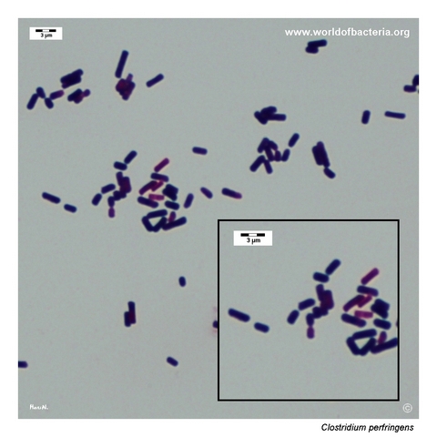
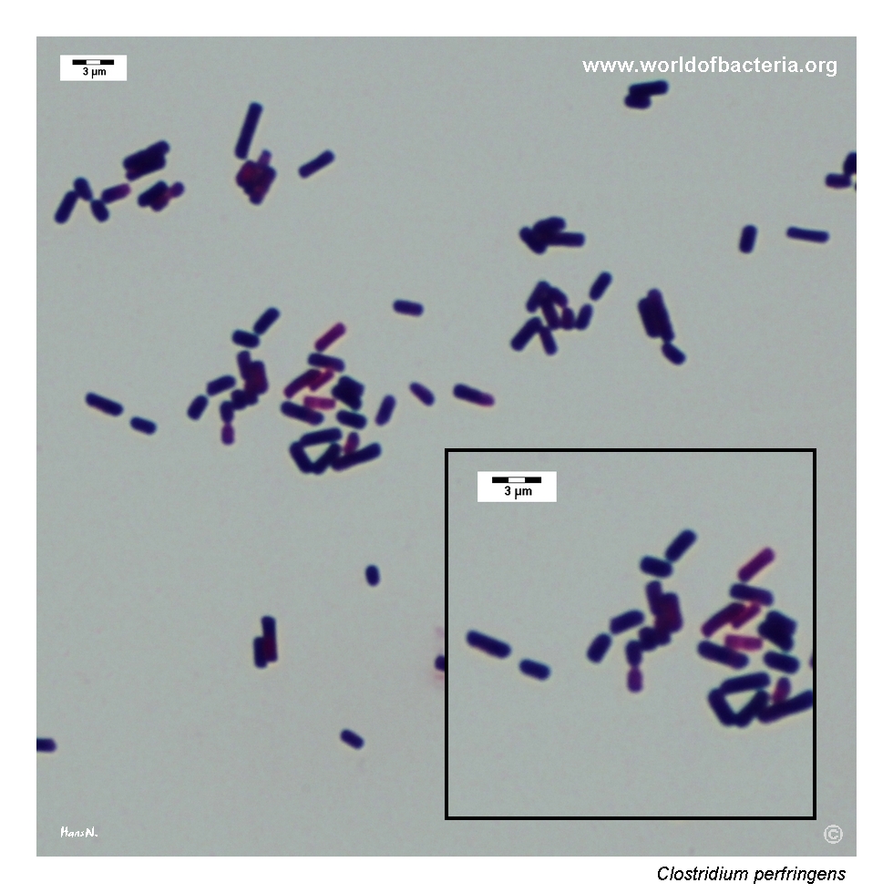
The illustrated smear shows the robust rod morphology that fits well with the classic laboratory appearance of C. perfringens
Oxygen Relationship
Obligate anaerobe
Grows best under strict anaerobic conditions and forms robust colonies on anaerobic blood agar within 18–24 hours
In routine bacteriology, the speed and strength of growth help distinguish the species from many more fastidious anaerobes
The image set shown here was cultured under an anaerobic atmosphere of 80% N2, 10% H2, and 10% CO2
Rapid Identification Tests
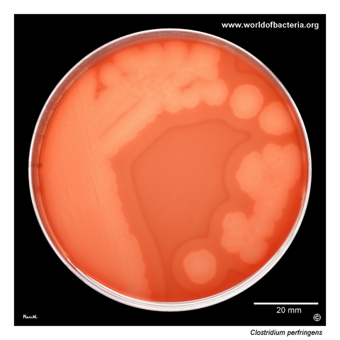
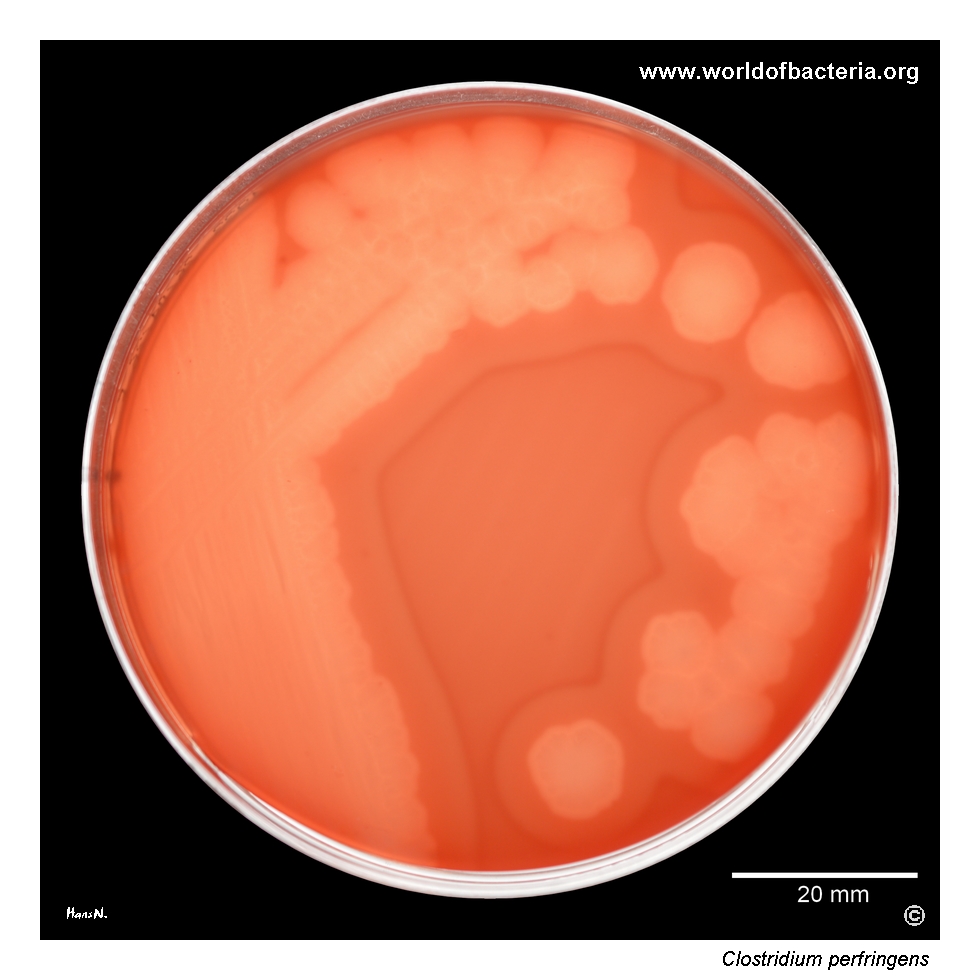
Double zone of haemolysis: classic blood agar finding with an inner zone of complete beta-haemolysis and a wider outer zone of partial haemolysis
Nagler reaction: historically important demonstration of alpha-toxin activity
Lecithinase production: characteristic feature on suitable egg-yolk-containing media
Stormy fermentation in milk: classic teaching feature of the species
Modern diagnostics: MALDI-TOF MS and validated anaerobe identification systems are well suited for routine species confirmation
Ecology and Clinical Relevance
Natural Habitat
Clostridium perfringens is widely distributed in soil, dust, decaying organic material, and the intestinal tract of humans and animals
Because of this broad environmental and enteric distribution, the organism may be recovered from a variety of contaminated or polymicrobial specimens
Its clinical significance therefore depends strongly on specimen type, quantity of growth, and the overall microbiological picture
Common Clinical Specimens
Deep wound material, necrotic tissue, abscesses, postoperative specimens, and traumatic soft-tissue samples
Blood culture in severe invasive infection
Food and stool in the setting of toxin-mediated gastrointestinal disease
Anaerobic culture material in which the species stands out by rapid, heavy growth and classic haemolysis
Clinical Significance
Clostridium perfringens is one of the most important clinically significant anaerobic pathogens
It is strongly associated with gas-producing soft-tissue infection, myonecrosis, wound infection, and toxin-mediated gastrointestinal disease
For atlas purposes, the page is especially useful because the species is one of the most visually distinctive anaerobes on blood agar
Differential Considerations
Other large anaerobic Gram-positive rods
Species of Clostridium and related anaerobic spore-formers recovered from deep tissue or wound material
Clostridium septicum and other haemolytic clostridia
Large grey colony-forming anaerobes where haemolytic pattern and colonial texture provide key first clues
Clostridium perfringens, collection strain ATCC 13124, on blood agar after 24 hours of incubation at 36 °C under anaerobic conditions (80% N2, 10% H2, 10% CO2). The image was taken in transmitted light. Particularly striking is the characteristic double zone of haemolysis: an inner zone of complete beta-haemolysis at the site of colony growth and a wider outer zone of partial haemolysis in the surrounding agar.
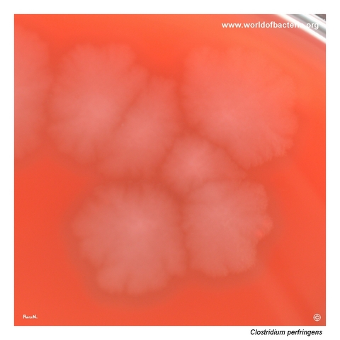
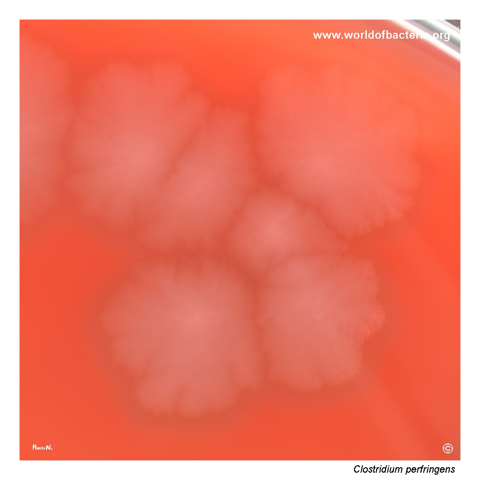
Colonies of Clostridium perfringens ATCC 13124. After 24 hours of anaerobic incubation at 36 °C, the organism forms large, flat, grey colonies with a raised centre, irregular outline, and uneven margins. This robust colonial appearance, together with the characteristic haemolytic pattern on blood agar, makes C. perfringens one of the most visually distinctive anaerobic pathogens encountered in routine culture.
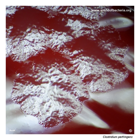
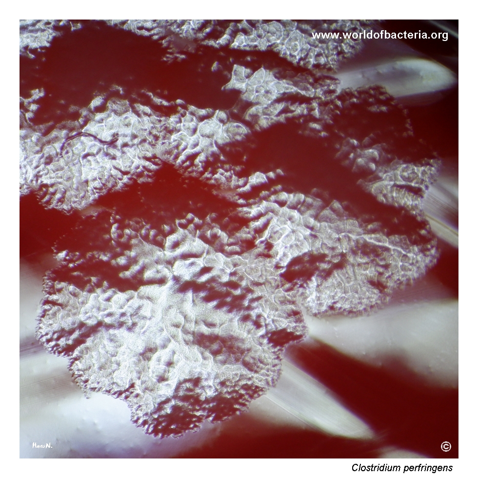
Close-up view of Clostridium perfringens colonies photographed in reflected light. At higher magnification, the colony surface often shows a characteristic texture resembling a sheet of beaten metal, commonly described as a “beaten-copper” surface. This subtle but classic morphological feature is well known in anaerobic bacteriology and adds another useful visual clue when evaluating mature colonies of this species.
Gram-stained smear prepared from a colony of Clostridium perfringens ATCC 13124 grown on blood agar. The organism appears as Gram-positive rod-shaped bacteria, in this case relatively short and often described as boxcar-shaped rods. Spore formation is usually infrequent in clinical cultures, so spores may be absent or only rarely visible despite the organism’s taxonomic placement among spore-forming anaerobes.
Diagnostic and Clinical Notes
This page is especially instructive because it shows several of the classic visual hallmarks that make Clostridium perfringens one of the most memorable anaerobes in clinical microbiology. The most important is the double zone of haemolysis, which remains a standard teaching feature of the species.
The second major teaching point is the combination of rapid anaerobic growth and robust colony size. Compared with many other anaerobes, C. perfringens often looks heavy, confident, and easy to recognize once the plate is read under appropriate conditions.
The reflected-light close-up is useful because it captures the beaten-copper surface that is often mentioned in textbooks but not always well illustrated. This is not by itself a definitive identification criterion, but it is a classic supportive clue.
The Gram stain completes the phenotype by showing the short broad rods that are often described as boxcar-shaped. In practice, the organism is recognized not by one single feature but by the combination of anaerobic growth, colonial robustness, haemolytic pattern, and microscopy.
Overall, the image set is valuable because it shows a species whose phenotype is both classically taught and genuinely useful in routine anaerobic plate reading.
Laboratory Identification
Colony Morphology
On anaerobic blood agar, Clostridium perfringens typically forms large flat grey colonies with a raised centre, irregular contour, and an uneven or coarse surface. The best-known associated feature is the double zone of haemolysis, which often makes the isolate visually striking even before formal identification is performed.
Microscopy
The Gram-stained smear shows Gram-positive rods that are relatively short and broad, producing the classic boxcar-shaped impression. Although the organism is a spore-former, spores are not always evident in routine clinical preparations. The microscopy is highly compatible with the species but should always be interpreted together with colony morphology and anaerobic growth characteristics.
Key Identification Clues
Rapid robust growth under anaerobic conditions
Large flat grey colonies with irregular margins
Characteristic double zone of haemolysis on blood agar
Beaten-copper surface in mature colonies
Short broad Gram-positive boxcar-shaped rods
Clinical association with toxin-mediated and deep-tissue anaerobic infection
Modern Identification Methods
Routine confirmation is best performed with MALDI-TOF MS or other validated anaerobe identification systems. Traditional phenotypic features remain highly educational and often strongly suggest the diagnosis, but species-level confirmation should not rely on morphology alone when clinically important material is involved.
Antibiotic Characteristics
No treatment inference should be made from colony appearance alone. Clinically important isolates of Clostridium perfringens require full interpretation in light of specimen type, disease severity, and formal susceptibility testing where appropriate.
For this atlas page, the main educational emphasis is phenotypic recognition: rapid anaerobic growth, robust grey colonies, and the classical double zone of haemolysis.
Note: In severe invasive disease, microbiological recognition must be integrated with urgent clinical management, because the species is closely associated with toxin-mediated tissue destruction.