Mycobacterium tuberculosis is the classical tubercle bacillus, a slow-growing acid-fast pathogen of major clinical importance whose dry, rough colonies on egg-based media remain among the most characteristic culture phenotypes in microbiology.
Basic Characteristics
Taxonomy
Domain: Bacteria
Phylum: Actinomycetota
Class: Actinomycetes
Order: Mycobacteriales
Family: Mycobacteriaceae
Genus:Mycobacterium
Species:Mycobacterium tuberculosis
Microscopy & Special Staining
Slender acid-fast bacilli that retain carbolfuchsin or related dyes because of the lipid-rich mycobacterial cell envelope
In this page set, the smear was prepared from liquid Middlebrook 7H9 medium and stained by a cold method for acid-fast bacilli
Virulent strains characteristically show cording, in which daughter cells remain attached and form thread-like or rope-like aggregates
Routine Gram staining is not the primary microscopic method for this species; acid-fast staining is the more informative laboratory approach
Oxygen Relationship
Strictly aerobic organism with slow growth compared with most routine bacterial pathogens
The cultures illustrated here required prolonged incubation, with visible growth documented only after about 9 weeks at 36 °C
On solid medium the organism develops its most characteristic appearance on specialized egg-based mycobacterial media rather than on ordinary blood agar
Rapid Identification Tests
Acid-fast stain: positive
Growth rate: slow-growing mycobacterium
Colony surface: rough, dry, and uneven on egg-based solid media
Cording: classical phenotypic clue associated with virulent tubercle bacilli
Modern diagnostics: species confirmation and resistance assessment now rely heavily on molecular and culture-based reference methods rather than on colony morphology alone
Ecology and Clinical Relevance
Natural Habitat
Mycobacterium tuberculosis is a highly adapted human pathogen and the principal cause of human tuberculosis
Unlike many other mycobacteria, it is not primarily an environmental saprophyte in routine clinical understanding
Its ecology is therefore interpreted mainly through transmission between human hosts and through its persistence in infected tissues rather than through environmental colonization
Common Clinical Specimens
Respiratory material such as sputum and bronchoalveolar specimens
Tissue biopsies and other material from extrapulmonary tuberculosis
Liquid and solid mycobacterial cultures in specialized laboratories
Acid-fast smears prepared from clinical specimens or cultured material
Clinical Significance
Mycobacterium tuberculosis is one of the most important bacterial pathogens in human medicine and remains the classical agent of tuberculosis
In culture, recovery of this species is always clinically significant and requires dedicated laboratory handling, species confirmation, and susceptibility assessment
The organism is diagnostically important not only because of the disease it causes, but also because its slow growth and special staining properties make laboratory recognition fundamentally different from that of routine pyogenic bacteria
Differential Considerations
Other members of the Mycobacterium tuberculosis complex when interpretation is based on reference laboratory methods
Non-tuberculous mycobacteria in acid-fast smears or cultures
Non-acid-fast organisms that may create misleading microscopy if inappropriate staining methods are used
Older, rough, dry colonies of other slow-growing mycobacteria on specialized media
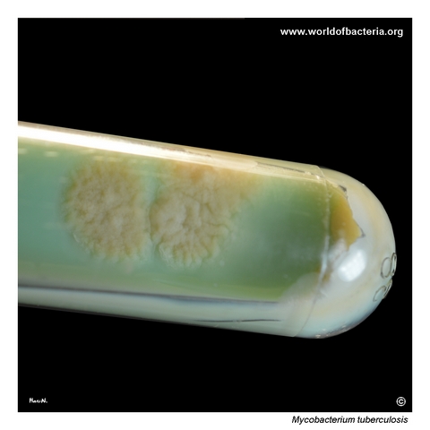
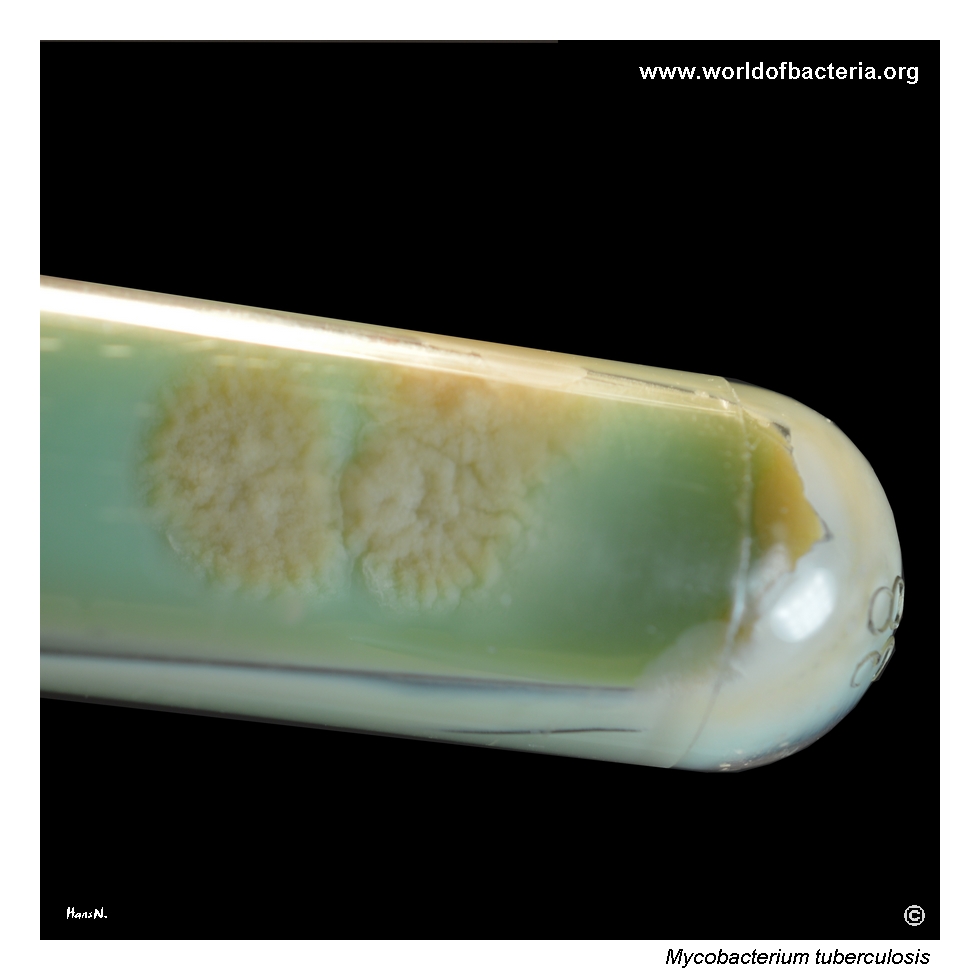
Mycobacterium tuberculosis on Ogawa medium after 9 weeks of incubation at 36 °C in ambient air. The culture forms rough, light yellow-brown colonies with irregular margins. Broader culture tubes or similar closed systems are used to reduce water evaporation during prolonged incubation and at the same time improve safety for laboratory staff when handling the cultures. The image illustrates the slow growth and characteristically dry, uneven colonial appearance of this species.
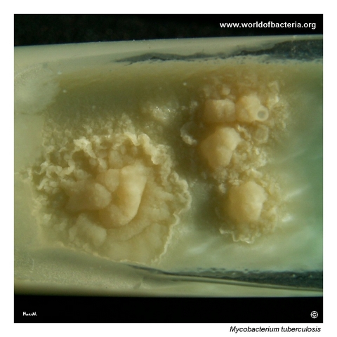
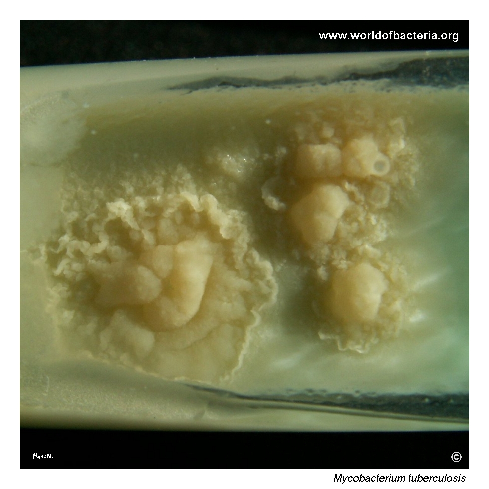
Colonies of Mycobacterium tuberculosis on Löwenstein–Jensen medium after 9 weeks of incubation at 36 °C in ambient air. The colonies are light yellow-brown, with a rough irregular surface and a wavy, uneven margin. The green colour of the medium is caused by the addition of malachite green. This image shows the classic appearance of tubercle bacilli on egg-based solid medium, where growth remains slow but develops a very characteristic rough colonial texture.
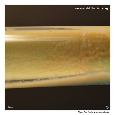
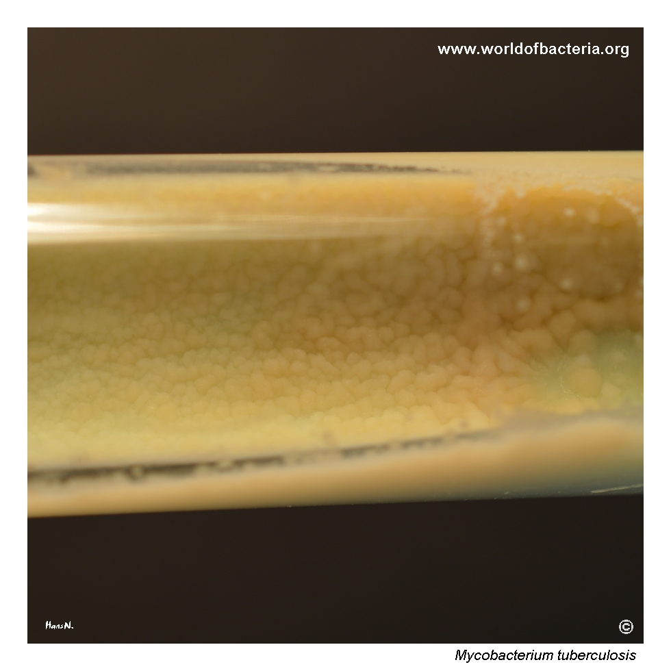
Close-up view of confluent growth of Mycobacterium tuberculosis on Ogawa medium. The grown culture has an uneven, rough surface, and older cultures gradually acquire a darker brownish shade. Culture conditions: 9 weeks at 36 °C in ambient air followed by a further 7 days at room temperature. Both Ogawa and Löwenstein–Jensen media are examples of egg-based media, which remain standard solid media for the cultivation of M. tuberculosis.
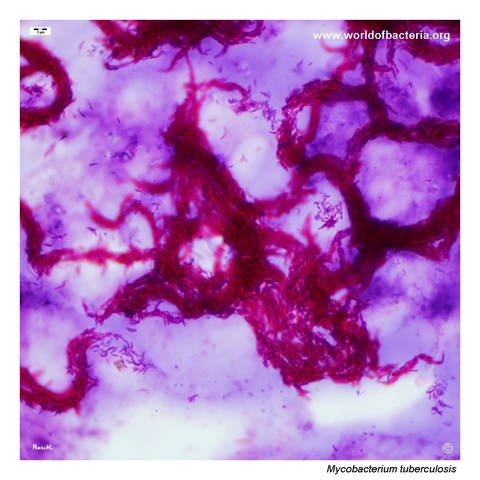
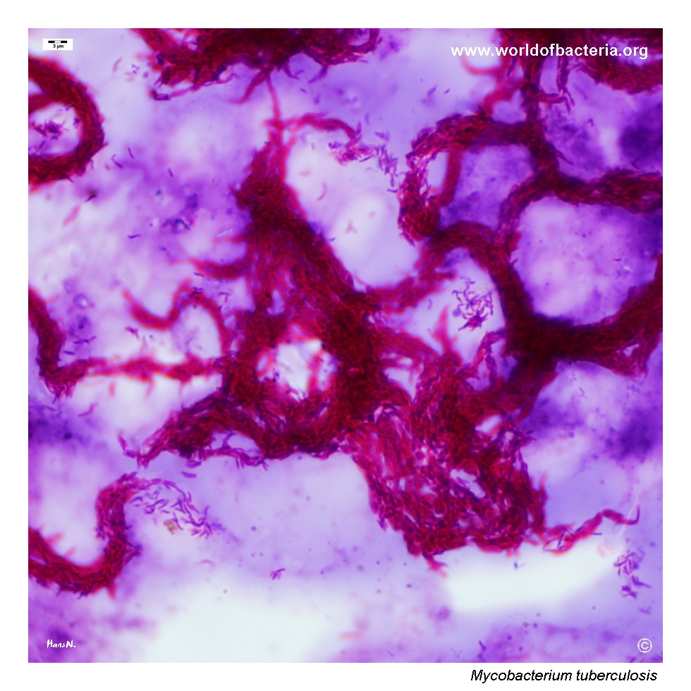
Smear prepared from liquid Middlebrook 7H9 medium and stained for acid-fast bacilli by a cold method. In addition to acid-fastness, virulent strains of Mycobacterium tuberculosis characteristically show so-called cording, meaning that daughter cells remain attached after division and form structures resembling threads or cords of varying thickness. This microscopic arrangement is one of the classic phenotypic features associated with virulent tubercle bacilli.
Diagnostic and Clinical Notes
This page set is didactically valuable because it documents the very different laboratory tempo of tubercle bacilli compared with routine clinical bacteria. Visible culture required many weeks rather than one or two days, and the resulting colonies developed a dry, rough, irregular appearance that is strongly characteristic on egg-based media.
The comparison between Ogawa and Löwenstein–Jensen medium is especially useful. Both media support the typical rough colonial morphology of Mycobacterium tuberculosis, while the malachite-green colour of Löwenstein–Jensen medium provides a familiar visual laboratory context for mycobacterial culture.
The close-up of confluent growth also demonstrates an important practical point: older cultures become darker and more uneven in appearance, so reading the plate or tube at different time points can materially change the visual impression of the isolate.
The acid-fast smear from liquid Middlebrook medium adds an equally important microscopic correlate. Cording is not, by itself, a complete identification method, but it remains one of the classic visual phenotypes historically associated with virulent tubercle bacilli.
Overall, the page illustrates why mycobacterial work requires dedicated methods, prolonged incubation, and cautious interpretation that differs substantially from the workflow used for common pyogenic pathogens.
Laboratory Identification
Colony Morphology
On egg-based solid media, Mycobacterium tuberculosis typically forms dry, rough, uneven colonies with irregular or wavy margins. In this page set the colonies are light yellow-brown to darker brownish with age, and the overall surface remains distinctly coarse rather than smooth.
Microscopy
Microscopy is best performed with acid-fast staining rather than with routine Gram staining. In the illustrated smear from liquid Middlebrook 7H9 medium, acid-fast bacilli show cording, a classic arrangement in which daughter cells remain attached and form elongated aggregates.
Key Identification Clues
Very slow growth compared with routine bacterial pathogens
Dry, rough colonies on egg-based media such as Ogawa and Löwenstein–Jensen
Acid-fast bacilli on special staining
Characteristic cording in virulent cultured isolates
Need for dedicated laboratory identification and susceptibility methods
Modern Identification Methods
In modern tuberculosis laboratories, definitive identification and resistance assessment depend on validated molecular assays, reference culture procedures, and formal susceptibility testing. Colony morphology and cording remain valuable teaching features, but they are supportive rather than sufficient as stand-alone identifiers in contemporary diagnostic practice.
Antibiotic Characteristics
For Mycobacterium tuberculosis, antimicrobial interpretation must never be inferred from colony appearance. Drug susceptibility depends on formal tuberculosis-specific testing performed in an appropriate reference laboratory framework.
The principal value of the cultural phenotype shown on this page is rapid visual recognition of a tubercle-bacillus-like isolate and appreciation of the unusually slow, rough growth pattern on specialized media.
Note: Because tuberculosis management is tightly linked to resistance detection and public-health procedures, species confirmation and susceptibility work-up are more important than any presumptive impression based on morphology alone.