Bacillus anthracis is a large spore-forming bacillus and the classical agent of anthrax, notable in culture for rough white non-haemolytic colonies and a close visual relationship to other members of the Bacillus cereus group.
Basic Characteristics
Taxonomy
Domain: Bacteria
Phylum: Bacillota
Class: Bacilli
Order: Bacillales
Family: Bacillaceae
Genus:Bacillus
Species:Bacillus anthracis
Microscopy & Gram Stain
Large Gram-positive rods, typically arranged in long chains
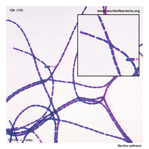
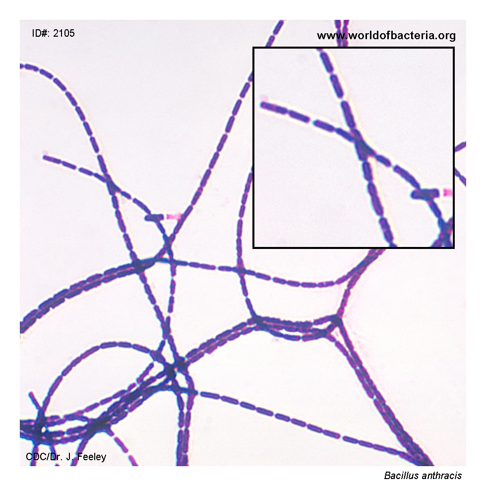
The illustrated smear shows the classic appearance of broad bacilli with a chained arrangement that fits well with the species
In routine culture, this morphology should always be interpreted together with colony appearance, because several large bacilli may look superficially similar on microscopy alone
Endospore formation is a defining property of the species, although spores are not the focus of the current image set
Oxygen Relationship
Aerobic to facultatively anaerobic spore-forming bacillus
Grows well on routine blood agar and develops a visually distinctive rough colonial texture within 24 hours
The species is not fastidious in culture, but its clinical importance means that recognition must go beyond routine plate reading
Rapid Identification Tests
Haemolysis on blood agar: characteristically absent
Motility: classically non-motile
Colony surface: rough, dry-looking, opaque white colonies with irregular margins
Classical supportive test: susceptibility to gamma phage in reference identification settings
Modern diagnostics: MALDI-TOF MS and confirmatory reference methods are preferred when a clinically suspicious isolate is encountered
Ecology and Clinical Relevance
Natural Habitat
Bacillus anthracis is an environmental spore-forming bacterium whose spores may persist for long periods in soil
Its epidemiological importance is strongly linked to herbivorous animals and to zoonotic transmission
Unlike many other environmental bacilli, this species is of major medical significance because it is the classical cause of anthrax
Common Clinical Specimens
Relevant clinical material depends on the form of anthrax and may include blood, lesion material, tissue, or respiratory specimens
The present page, however, is intended primarily as a morphology-focused atlas entry rather than as a diagnostic workflow page
Any suspected clinically significant isolate requires careful interpretation within the full clinical and laboratory context
Clinical Significance
Bacillus anthracis is one of the most important pathogenic species within the genus Bacillus
Human disease is uncommon in many routine settings, but when the organism is genuinely involved the clinical significance is high
For atlas purposes, the main didactic value of this page lies in the combination of rough colony texture, lack of haemolysis, long chains on Gram stain, and its differential relationship to Bacillus cereus
Differential Considerations
Bacillus cereus, especially because colonies can be visually similar at first glance
Other members of the Bacillus cereus group
Large Gram-positive rod-forming bacilli recovered from culture
Any rough white colony-forming aerobic spore-former where haemolysis and motility help guide differentiation
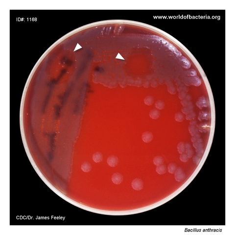
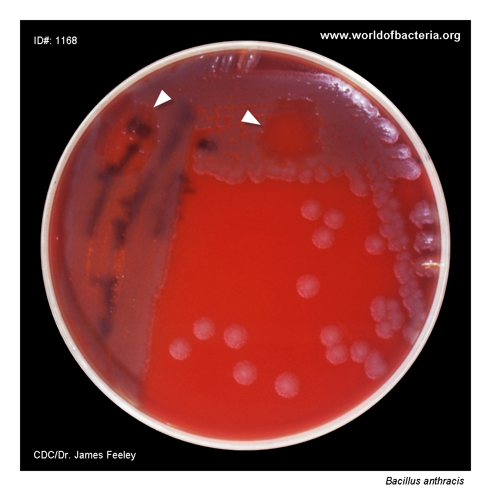
Bacillus anthracis on blood agar. B. anthracis belongs to the bacteria that do not produce haemolysis on blood agar. The white arrows indicate the area where a suspension of gamma phage was applied to the culture, resulting in lysis of the grown test organism in the phage test. This image highlights one of the classical laboratory features associated with the species.
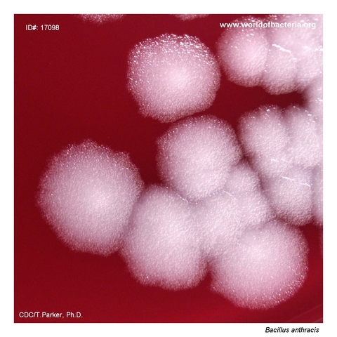
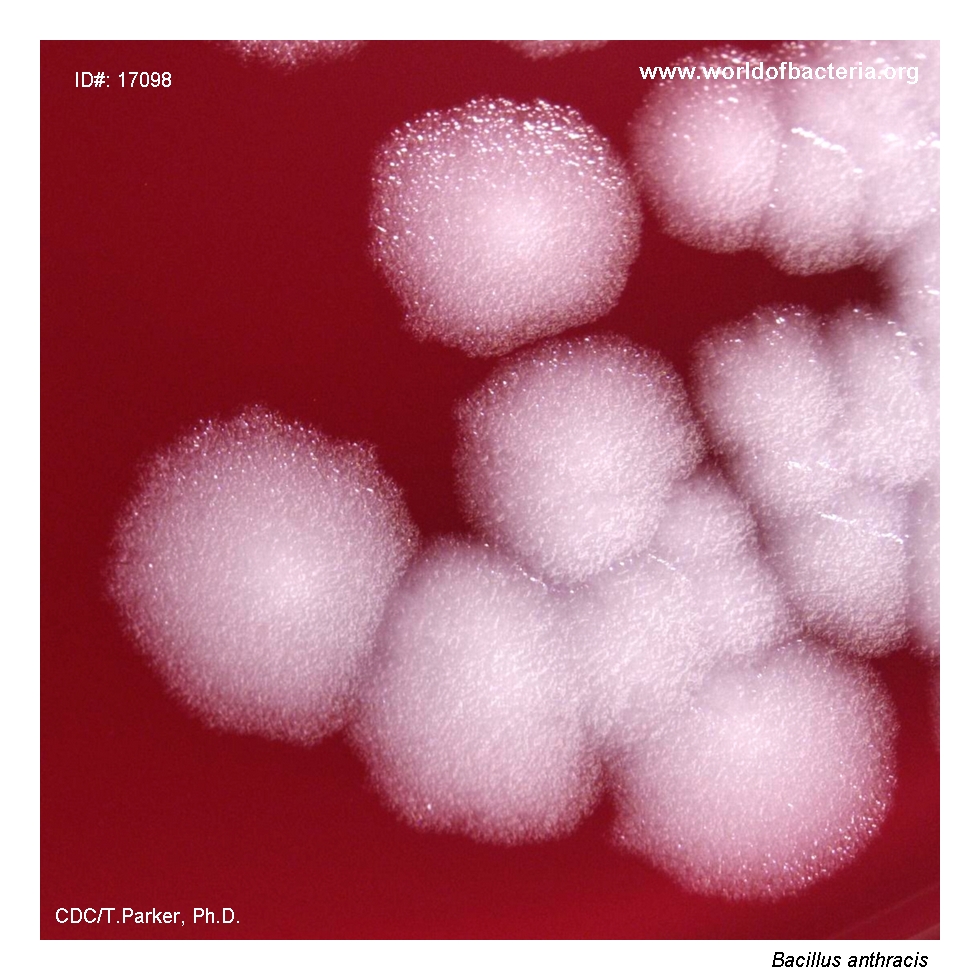
Colonies of Bacillus anthracis on blood agar after 24 hours of incubation at 37 °C. The colonies are large, opaque, white, and have a very rough surface with an irregular edge. Characteristically, anthrax colonies are non-haemolytic. Even at first glance, the combination of rough colony texture and lack of haemolysis gives the culture a distinctive appearance on routine solid medium.
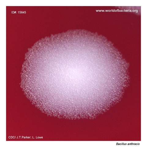
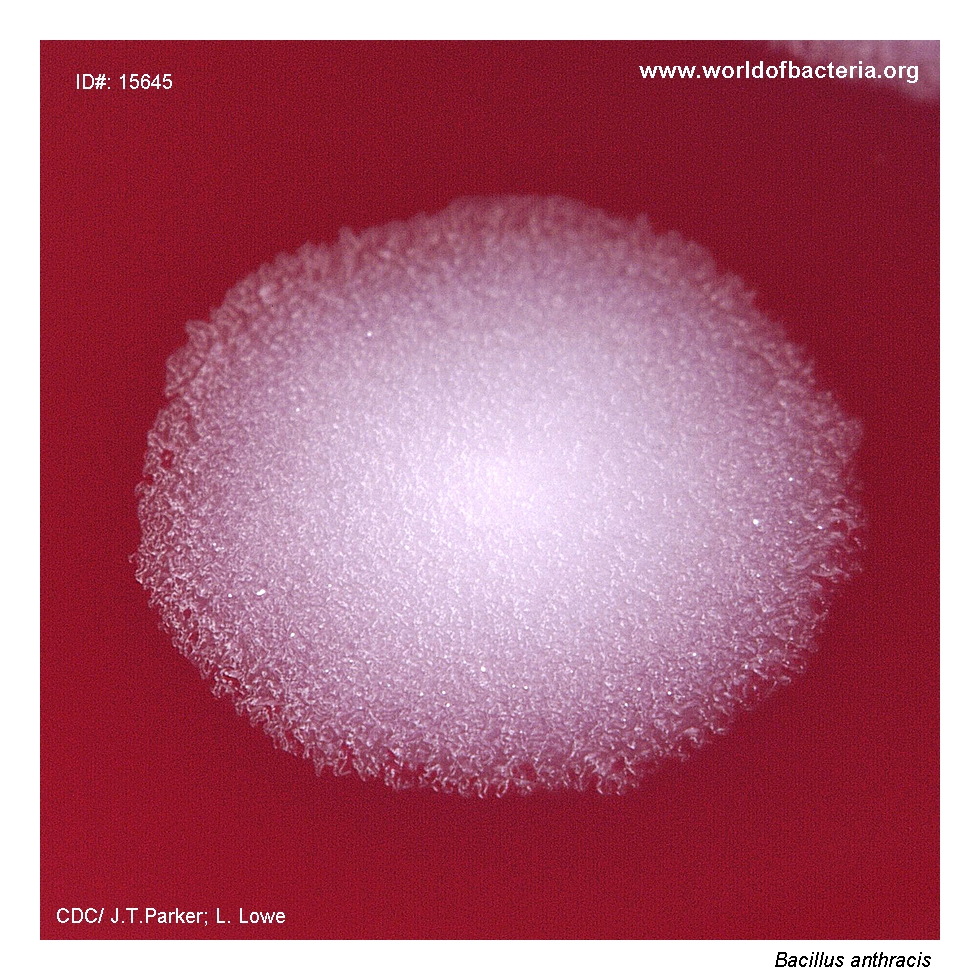
Close-up view of Bacillus anthracis colonies after 24 hours of incubation at 37 °C. Colonies of B. anthracis and Bacillus cereus can appear similar, although those of the former are generally smaller and non-haemolytic. This comparison is useful in routine plate reading, because visual differentiation within the genus Bacillus may depend on subtle but important colony features.
Gram-stained smear prepared probably from a liquid culture of Bacillus anthracis. The organism appears as Gram-positive rods arranged in long chains. This microscopic pattern is typical of large bacilli and fits well with the characteristic morphology of the species seen in laboratory preparations.
Diagnostic and Clinical Notes
This page is especially instructive because it brings together several of the classical visual features historically associated with Bacillus anthracis: rough white colony morphology, absence of haemolysis on blood agar, long chained Gram-positive rods, and visible lysis in the gamma phage test area.
The colony close-ups also demonstrate an important diagnostic caution. At first glance, colonies of B. anthracis may resemble those of Bacillus cereus or related bacilli, so the species should never be judged from colony roughness alone. The combination of non-haemolysis, microscopy, and confirmatory identification methods is much more informative than any single plate feature.
The first image highlights a classical identification concept rather than a routine appearance only: the culture shows lysis in the area marked by gamma phage application. In modern practice, however, species confirmation should be aligned with contemporary reference methods and laboratory biosafety expectations rather than relying on a single historic test result.
The Gram-stained smear is equally useful for atlas purposes. Long chains of broad Gram-positive rods fit the species well, but the image also reminds us that large bacilli are a morphological group, not a definitive identification by themselves.
Overall, the page is valuable less because it shows a common routine isolate and more because it documents one of the most recognizable classical plate phenotypes in medical bacteriology.
Laboratory Identification
Colony Morphology
On blood agar, Bacillus anthracis typically forms large, opaque, white colonies with a very rough, dry-looking surface and irregular margins. In the material shown here, the colonies are conspicuously non-haemolytic, a feature that remains one of the most practical clues when differentiating the species from haemolytic bacilli such as many isolates of Bacillus cereus.
Microscopy
The Gram-stained smear shows large Gram-positive rods arranged in long chains. This appearance is highly compatible with B. anthracis, but it should still be interpreted together with the colony phenotype and confirmatory identification methods, because other large bacilli may also produce chained rods.
Key Identification Clues
Rough opaque white colonies on blood agar
Absence of haemolysis
Long chained Gram-positive rods
Close differential relationship to Bacillus cereus and other members of the Bacillus cereus group
Classical susceptibility to gamma phage as a historical supportive feature
Need for confirmatory species identification by modern reference methods
Modern Identification Methods
Reliable identification is best based on modern laboratory methods interpreted within the appropriate clinical and biosafety framework. MALDI-TOF MS, molecular confirmation, and reference laboratory support are more appropriate for suspected clinically significant isolates than dependence on morphology alone. The image set remains most useful as a morphology teaching reference.
Antibiotic Characteristics
Antimicrobial interpretation should never be inferred from morphology alone. With a species of this importance, treatment decisions depend on formal clinical guidance, species confirmation, and appropriate susceptibility assessment where relevant.
For this atlas page, the educational value lies mainly in recognition of the classical phenotype rather than in therapeutic detail.
Note: Because Bacillus anthracis is a major pathogen, any real clinical interpretation belongs within formal diagnostic, infectious disease, and public health workflows rather than in a morphology-focused summary.