Staphylococcus pettenkoferi is an uncommon coagulase-negative staphylococcal species that may be overlooked in mixed clinical cultures because its colonies often resemble those of many other non-pigmented CoNS on routine aerobic blood agar.
Basic Characteristics
Taxonomy
Domain: Bacteria
Phylum: Bacillota (Firmicutes)
Class: Bacilli
Order: Bacillales
Family: Staphylococcaceae
Genus:Staphylococcus
Species:Staphylococcus pettenkoferi
Microscopy & Gram Stain
Gram-positive cocci
Cells occur predominantly in clusters, but pairs and small aggregates may also be seen
Microscopic morphology is consistent with staphylococci but is not species-specific
Oxygen Relationship
Facultatively anaerobic
Grows under routine aerobic blood agar conditions
May show altered colony size under anaerobic culture, as illustrated in this page set
Rapid Identification Tests
Catalase: positive
Coagulase: negative
Colony pigmentation: usually absent
Haemolysis on blood agar: typically absent
Routine recognition: often difficult because colonies resemble many other CoNS
Ecology and Clinical Relevance
Natural Habitat
Described from human clinical specimens
Probably associated with human skin or skin-related microbiological niches
Best regarded as an uncommon member of the coagulase-negative staphylococci encountered in clinical material
Common Clinical Specimens
Wound swabs and skin-associated specimens
Occasional blood culture isolates reported in the literature
Material from device-associated or postoperative settings
Mixed cultures in which the organism may be present only in low quantity
Clinical Significance
Rare opportunistic human pathogen
Clinical interpretation is often difficult because the species may occur in small quantity and alongside more familiar CoNS
Has been reported from invasive settings, but overall clinical significance is still being clarified
May be underrecognized when routine identification systems or plate reading rely mainly on typical phenotypic impressions
Differential Considerations
Staphylococcus epidermidis, because both may appear as non-pigmented coagulase-negative staphylococci in mixed clinical cultures
Other visually inconspicuous CoNS recovered from wounds or device-associated specimens
Species-level identification cannot be based reliably on colony appearance alone
Anaerobic growth characteristics may add interest but do not replace definitive identification methods
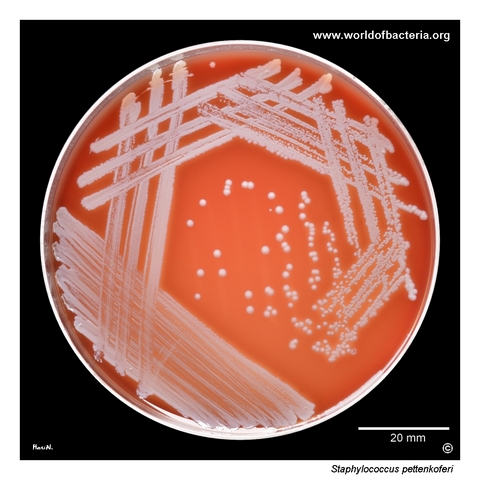
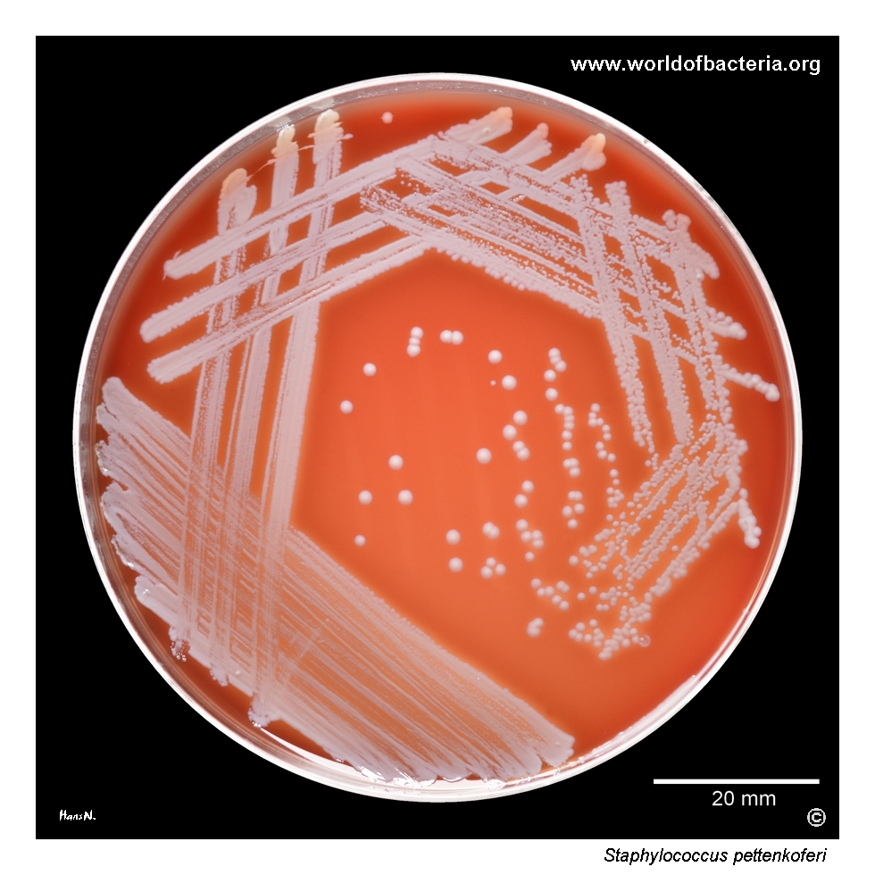
Staphylococcus pettenkoferi on blood agar after 24 hours of incubation at 36 °C in ambient air. The isolate originated from a wound swab from an 88-year-old woman with a complication following total joint replacement. In the specimen, the strain was present in low quantity together with Staphylococcus epidermidis. In mixed cultures of this kind, careful comparison of colony morphology is essential to avoid overlooking less prominent accompanying species.
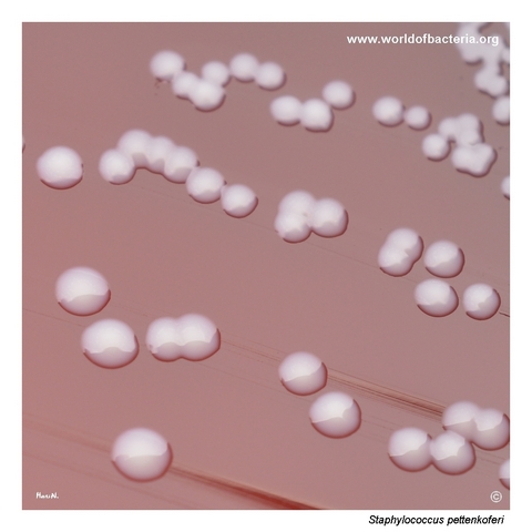
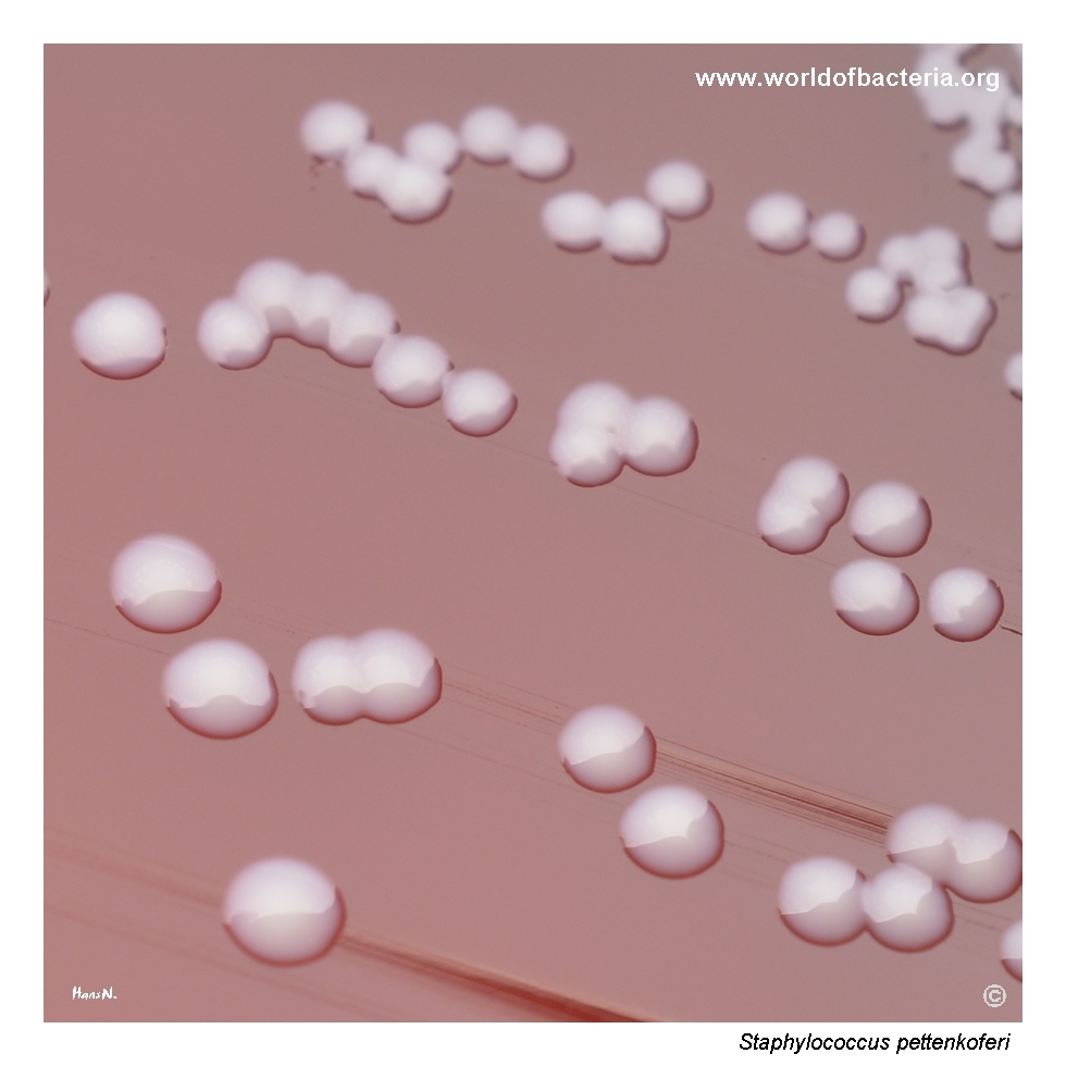
Colonies of Staphylococcus pettenkoferi on blood agar after 24 hours at 36 °C in ambient air. The colonies are smooth, convex, non-pigmented, and round with entire margins. On aerobic culture they appear fairly typical and do not immediately stand out from many other coagulase-negative staphylococci, which is why this species may easily remain unrecognized without careful observation and reliable identification methods.
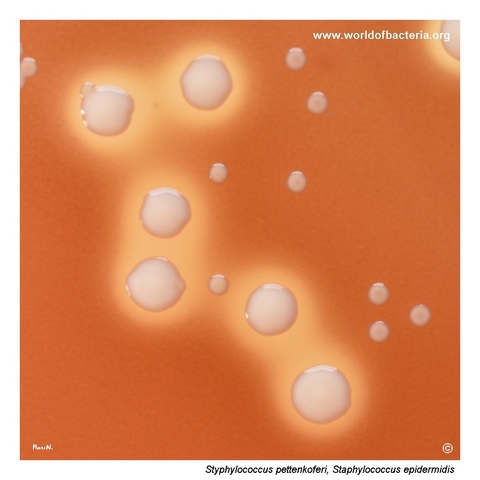
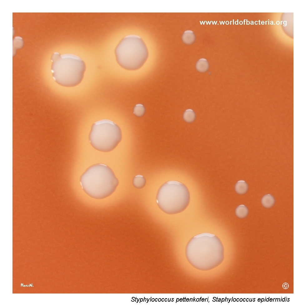
Close-up view of colonies of Staphylococcus pettenkoferi and Staphylococcus epidermidis on Schaedler agar after 48 hours of anaerobic incubation. In this setting, the colonies of S. pettenkoferi remain conspicuously small, although the same isolate formed ordinary colonies during aerobic cultivation. Around the S. epidermidis colonies, a striking zone of beta-haemolysis is visible, which is unusual for that species and illustrates the variability seen in clinical isolates.
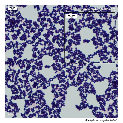
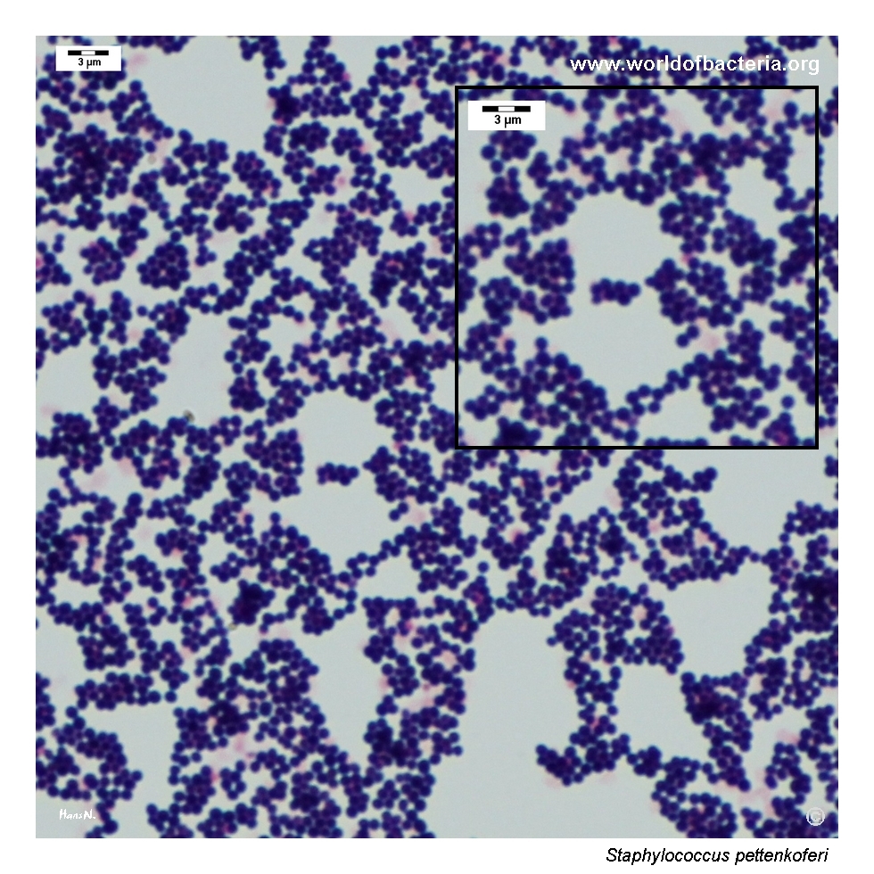
Gram-stained smear prepared from a colony of Staphylococcus pettenkoferi grown on blood agar. The organism appears as Gram-positive cocci occurring predominantly in clusters. As with other members of the genus Staphylococcus, this morphology supports generic classification but is not sufficient for species determination, which must be based on the overall culture picture and further laboratory identification.
Diagnostic and Clinical Notes
Staphylococcus pettenkoferi is diagnostically instructive mainly because it can be so easy to miss. On routine aerobic blood agar, the colonies are smooth, non-pigmented, and visually fairly ordinary for a coagulase-negative staphylococcus, which means that the isolate may disappear into the background of a mixed clinical culture.
The wound specimen shown here came from a patient with a complication following total joint replacement, and the organism was present only in low quantity together with Staphylococcus epidermidis. This type of setting is exactly where less prominent CoNS may be overlooked unless plate reading is careful and comparative.
The anaerobic image adds an additional practical lesson. On Schaedler agar after 48 hours, colonies of S. pettenkoferi remain conspicuously small, even though the same isolate formed ordinary colonies aerobically. Such behaviour is useful descriptively, but not sufficiently distinctive for species assignment on its own.
The comparison with Staphylococcus epidermidis is particularly relevant. Both species may appear as rather typical, non-pigmented CoNS, yet their clinical role in a specimen can differ and neither should be interpreted solely from first visual impression.
Because S. pettenkoferi is an uncommon species and may be underrecognized in routine diagnostics, reliable identification depends heavily on modern methods rather than on colony morphology alone.
Laboratory Identification
Colony Morphology
On aerobic blood agar, Staphylococcus pettenkoferi forms smooth, convex, round colonies with entire margins and without obvious pigmentation. This morphology is rather typical of many coagulase-negative staphylococci and therefore provides only limited discriminatory value. In the anaerobic setting shown here, the colonies remain much smaller, which is visually interesting but not by itself diagnostic.
Microscopy
Gram staining demonstrates Gram-positive cocci occurring predominantly in clusters. This appearance is fully consistent with the genus Staphylococcus, but it does not allow reliable distinction of S. pettenkoferi from Staphylococcus epidermidis or other coagulase-negative staphylococci encountered in clinical microbiology.
Key Identification Clues
Gram-positive cocci in clusters
Catalase positive
Coagulase negative
Smooth, convex, non-pigmented colonies on aerobic blood agar
Often visually inconspicuous among other CoNS
May show notably small colonies under anaerobic cultivation in some settings
Definitive identification requires modern instrumental or molecular methods
Modern Identification Methods
Species-level identification is best achieved by MALDI-TOF mass spectrometry when the reference database is sufficiently current, or by molecular methods when necessary. This is especially important for Staphylococcus pettenkoferi because the organism is uncommon, often phenotypically unremarkable, and therefore easy to miss or misclassify during routine work-up of coagulase-negative staphylococci.
Antibiotic Characteristics
No simple species-level susceptibility rule should be assumed for Staphylococcus pettenkoferi. As with other coagulase-negative staphylococci, antimicrobial susceptibility may vary between isolates and must be determined individually.
This is particularly important in postoperative or device-associated settings, where therapeutic interpretation depends not only on the susceptibility profile but also on whether the isolate is judged to represent contamination, colonization, or a true pathogen.
The low quantity of growth in mixed material should not automatically lead to dismissal, but neither should species identification alone be overinterpreted. Microbiological relevance must always be considered together with specimen type, culture context, and the patient’s clinical picture.
Note: Antimicrobial therapy should always be guided by formal laboratory susceptibility testing and overall clinical assessment.