Staphylococcus capitis is a human skin-associated coagulase-negative staphylococcal species, especially linked with the scalp and head region, usually forming non-pigmented and non-haemolytic colonies on blood agar.
Basic Characteristics
Taxonomy
Domain: Bacteria
Phylum: Bacillota (Firmicutes)
Class: Bacilli
Order: Bacillales
Family: Staphylococcaceae
Genus:Staphylococcus
Species:Staphylococcus capitis
Microscopy & Gram Stain
Gram-positive cocci
Cells occur singly, in pairs, and in clusters
Microscopic morphology is typical of staphylococci but not species-specific
Oxygen Relationship
Facultatively anaerobic
Grows readily under routine aerobic blood agar conditions
Behaves as a conventional coagulase-negative staphylococcal isolate in everyday diagnostic culture
Rapid Identification Tests
Catalase: positive
Coagulase: negative
Colony pigmentation: usually absent
Haemolysis on blood agar: usually absent
Practical note: visually rather inconspicuous on primary culture
Ecology and Clinical Relevance
Natural Habitat
Member of the normal human skin microbiota
Classically associated with the scalp, forehead, and other head skin sites
May be recovered from skin before invasive procedures such as blood culture collection
Common Clinical Specimens
Skin swabs
Blood cultures, especially when contamination is possible
Device-associated or hospital-associated material in selected patients
Mixed surface specimens in which interpretation depends on context
Clinical Significance
Often of low apparent virulence and may represent skin flora rather than a primary pathogen
Can nevertheless act as an opportunistic pathogen in the appropriate clinical setting
Particularly relevant when recovered from blood cultures or indwelling-device-associated material
Interpretation depends strongly on specimen type, sampling technique, and the overall clinical picture
Differential Considerations
Staphylococcus epidermidis, as another very common non-pigmented coagulase-negative staphylococcus
Staphylococcus hominis and Staphylococcus warneri, which may show a similarly bland colony phenotype on blood agar
Staphylococcus aureus, mainly as a practical comparator rather than because of close phenotypic similarity
The key difficulty is often not dramatic morphology but the ease with which this species may be overlooked or dismissed as background skin flora
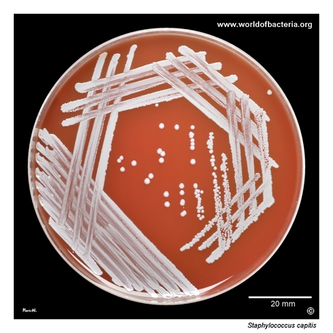
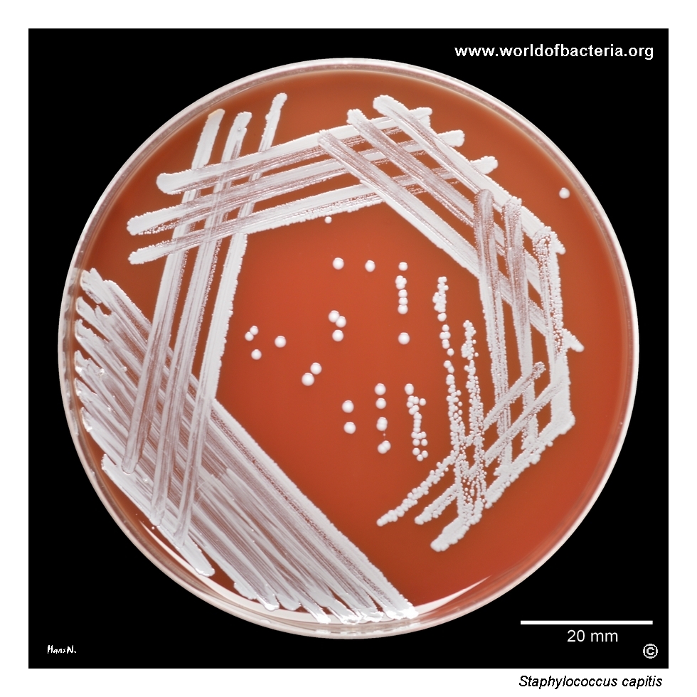
Staphylococcus capitis on blood agar. The strain was isolated from a skin swab taken before blood culture collection. Strains of S. capitis are non-pigmented and usually non-haemolytic. Incubation: 24 hours at 36 °C in ambient atmosphere.
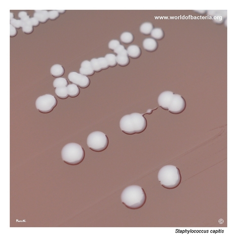
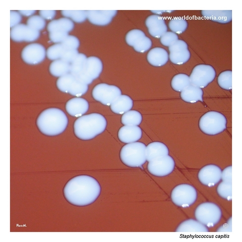
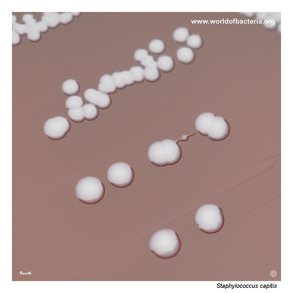
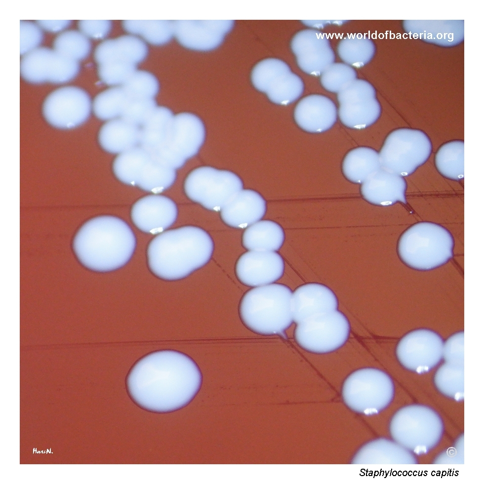
Close-up view of the colonies. Incubation: 24 hours at 36 °C in ambient atmosphere. The colonies are smooth, convex, and round with entire margins. Strains of S. capitis do not produce yellow pigment.
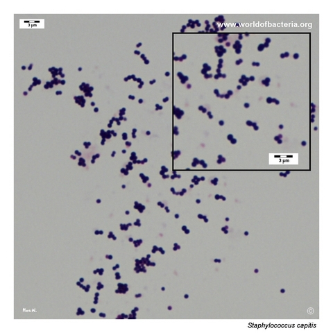
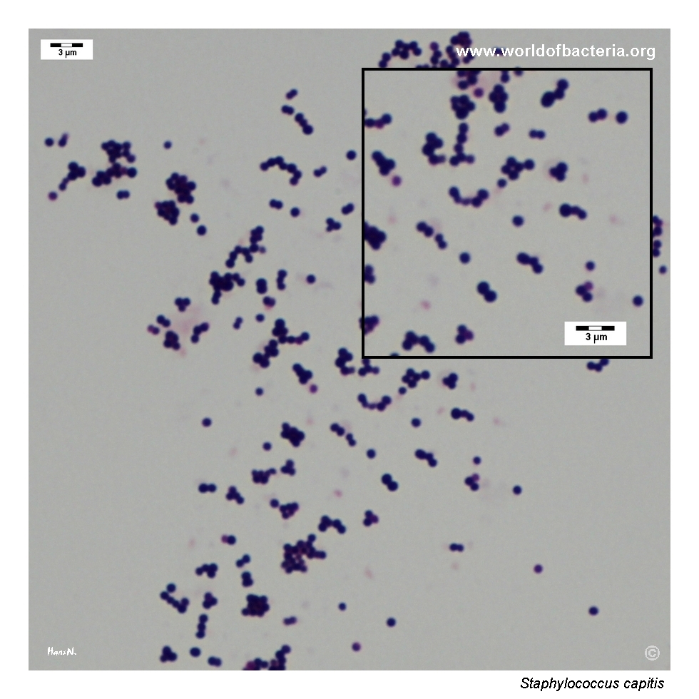
Smear prepared from a colony of Staphylococcus capitis grown on blood agar and stained by the Gram method. Gram-positive cocci are seen singly, in pairs, and in clusters.
Diagnostic and Clinical Notes
Staphylococcus capitis is a useful teaching example of a coagulase-negative staphylococcus that is microbiologically typical yet visually quite unobtrusive. On blood agar it usually appears as a non-pigmented, non-haemolytic isolate that can easily blend into the broader background of common skin flora.
That background matters diagnostically. This species is part of the human skin microbiota and may be recovered from skin swabs or introduced into blood cultures when skin disinfection before venepuncture is inadequate. Interpretation therefore depends not only on identification but also on sampling quality and specimen context.
The photographs on this page illustrate a classic low-profile colony phenotype: smooth convex colonies, no yellow pigment, and no haemolysis. Such isolates may receive less visual attention than more strongly pigmented or haemolytic staphylococci, even though careful recognition of the full mixed culture remains important in routine diagnostics.
For clinical interpretation, the key point is that S. capitis should not be judged by appearance alone. In many situations it may represent colonizing skin flora or contamination, but in predisposed patients it may also act as an opportunistic pathogen, especially when recovered repeatedly or from clinically significant material.
Laboratory Identification
Colony Morphology
On blood agar, Staphylococcus capitis typically forms smooth, convex, round colonies with entire margins. Pigmentation is usually absent and haemolysis is generally not seen. This results in a rather bland visual phenotype that resembles many other non-pigmented coagulase-negative staphylococci.
Microscopy
Gram staining shows Gram-positive cocci occurring singly, in pairs, and in clusters. This pattern is entirely compatible with the genus Staphylococcus, but—as with other coagulase-negative species—it does not provide definitive species-level identification.
Key Identification Clues
Gram-positive cocci in small groups and clusters
Catalase positive
Coagulase negative
Usually non-pigmented colonies
Usually non-haemolytic on blood agar
Commonly associated with human skin flora, especially of the head region
Reliable species-level confirmation requires instrumental or molecular identification methods
Modern Identification Methods
Definitive identification is best achieved by MALDI-TOF mass spectrometry when the database is sufficiently comprehensive, or by molecular methods where necessary. This is important because the routine phenotype of S. capitis overlaps broadly with several other coagulase-negative staphylococci commonly encountered in clinical laboratories.
Antibiotic Characteristics
No simple species-level susceptibility rule should be assumed for Staphylococcus capitis. Antimicrobial susceptibility must be determined on the individual isolate.
This is especially important when the organism is recovered from blood cultures, hospital-associated material, or device-related specimens in which the distinction between contamination and true infection is clinically relevant.
As with other coagulase-negative staphylococci, the therapeutic question is not only which agents are active in vitro, but also whether the isolate truly represents infection in the specific specimen context.
Note: Antimicrobial therapy should always be guided by formal laboratory susceptibility testing and the overall clinical picture.