Mycobacterium smegmatis is a rapidly growing non-tuberculous mycobacterial species whose waxy, irregular colonies on routine medium provide a useful contrast to the very slow culture phenotype of Mycobacterium tuberculosis.
Basic Characteristics
Taxonomy
Domain: Bacteria
Phylum: Actinomycetota
Class: Actinomycetes
Order: Mycobacteriales
Family: Mycobacteriaceae
Genus:Mycobacterium
Species:Mycobacterium smegmatis
Microscopy & Special Staining
Slender to short acid-fast bacilli; mycobacteria characteristically stain poorly by the Gram method because of their lipid-rich cell envelope
In this page set the illustrated smear is Gram stained, which usefully demonstrates the limited performance of routine Gram staining for mycobacteria
Specific methods for acid-fast bacilli remain more informative than Gram staining when mycobacteria are suspected
The colonies shown here were grown on blood agar, where the organism already displays the waxy, dry-looking character typical of the genus
Oxygen Relationship
Aerobic mycobacterium that grows well under routine laboratory oxygen conditions
Unlike Mycobacterium tuberculosis, this species belongs to the rapidly growing mycobacteria and produced visible growth within about 5 days
Its relatively fast culture kinetics make it useful as a teaching contrast to slow-growing tubercle bacilli
Rapid Identification Tests
Acid-fast stain: positive
Growth rate: rapidly growing mycobacterium
Routine blood agar: visible growth after several days, with waxy, irregular colonies
Gram stain: often weak or misleading for genus recognition
Modern diagnostics: species confirmation is best supported by MALDI-TOF MS or molecular methods when clinically relevant isolates are encountered
Ecology and Clinical Relevance
Natural Habitat
Mycobacterium smegmatis is primarily regarded as an environmental, non-tuberculous mycobacterium
It is associated with soil, water, and other external habitats rather than with obligate human infection
In laboratory teaching and research it is also widely known as a convenient model mycobacterium because of its comparatively rapid growth
Common Clinical Specimens
This page shows a collection strain rather than a clinical isolate
When clinically encountered, rapidly growing mycobacteria are more likely to appear in wound, soft-tissue, device-associated, or environmental contamination contexts than in classical tuberculosis specimens
Culture material and laboratory teaching strains are also common sources for practical microbiology demonstration
Clinical Significance
Mycobacterium smegmatis is usually of far lower clinical importance than M. tuberculosis, but occasional opportunistic infection is possible
Its greatest didactic value lies in demonstrating the mycobacterial phenotype in a species that grows much faster than tubercle bacilli
The contrast between rapid growth and typical mycobacterial colony texture makes it especially useful for teaching core features of the genus
Differential Considerations
Other rapidly growing non-tuberculous mycobacteria
Mycobacterium tuberculosis, mainly as a contrast in growth rate, colony texture, and biosafety significance
Dry, irregular colonies of non-mycobacterial organisms when interpretation is based only on gross plate morphology
Gram-positive organisms that stain poorly or appear unusual on routine Gram stain
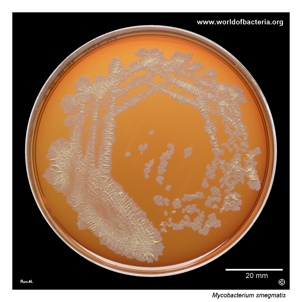
Mycobacterium smegmatis, collection strain DSM 43058, grown on blood agar after 5 days of incubation at 36 °C in ambient air. In contrast to Mycobacterium tuberculosis, the relatively rapid growth of M. smegmatis places it among the rapidly growing mycobacteria. This difference is diagnostically important, because growth rate remains one of the classic practical features used to distinguish groups of mycobacteria in routine culture work.
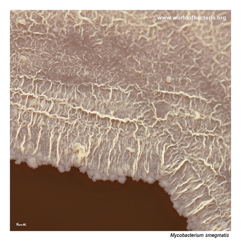
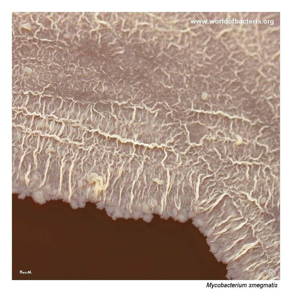
Appearance of Mycobacterium smegmatis culture in the area of confluent growth. Particularly striking is the formation of paler rib-like structures on the surface of the grown culture. Overall, the bacterial mass has a distinctly waxy appearance, which becomes especially evident during manipulation with an inoculating loop. This waxy character reflects the unusual composition of the mycobacterial cell envelope and is one of the classic visual features of the genus.
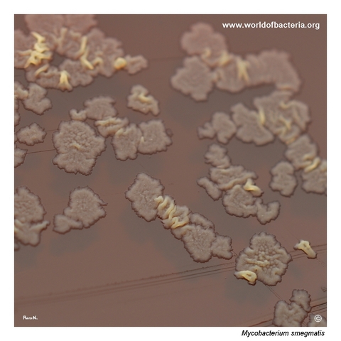
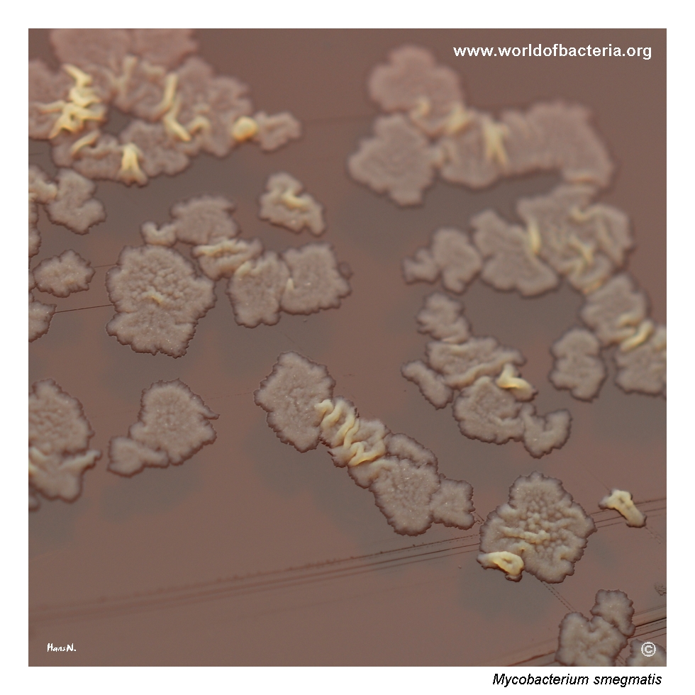
Close-up view of Mycobacterium smegmatis colonies on blood agar. The colonies are flat, irregular in shape, and have an uneven surface and margins. Their consistency is waxy and crumbly. Culture conditions: 5 days at 36 °C in ambient air. Even on routine medium, the colonies differ clearly from the smooth moist appearance of many other bacteria, which makes them visually distinctive once sufficient growth has developed.
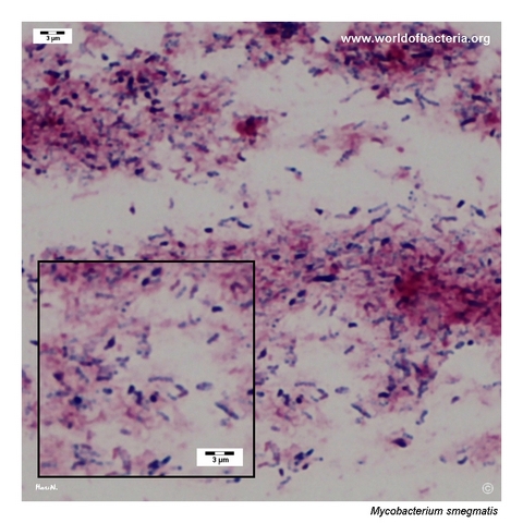
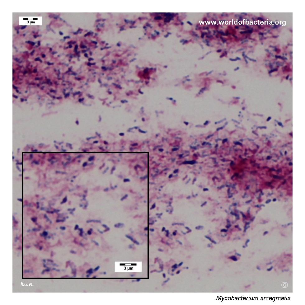
Gram-stained smear prepared from a colony of Mycobacterium smegmatis grown on blood agar. Mycobacteria generally stain poorly by the Gram method, which is a consequence of the composition of their cell wall and the related property of acid-fastness. For this reason, Gram staining is of only limited value for mycobacteria, and specific staining methods for acid-fast bacilli remain much more informative in routine laboratory practice.
Diagnostic and Clinical Notes
This page set is useful mainly because it documents a mycobacterial phenotype on ordinary blood agar within a practical laboratory timeframe. In contrast to the many weeks required for Mycobacterium tuberculosis, visible growth of M. smegmatis developed after only about 5 days, immediately placing the isolate among the rapidly growing mycobacteria.
The plate images also show an important tactile and visual teaching point: mycobacterial colonies are often waxy, dry-looking, and structurally uneven. In the confluent growth area, pale rib-like surface structures further emphasize that the culture mass behaves differently from the smooth, moist growth of many routine bacteria.
The close-up colony photograph reinforces this impression. The colonies are flat to irregular, with a crumbly and waxy character rather than a glossy surface, making them visually distinctive once enough growth has developed.
The Gram-stained smear is equally instructive because it demonstrates a limitation rather than a strength of the method. Mycobacteria often Gram stain poorly, so apparent weak staining or an unusual microscopic impression should prompt consideration of acid-fast staining rather than overinterpretation of the Gram result.
Overall, the page provides a convenient teaching bridge between routine culture microbiology and the more specialized diagnostic logic required for mycobacteria.
Laboratory Identification
Colony Morphology
On blood agar, Mycobacterium smegmatis forms flat to irregular colonies with an uneven surface and margins. The growth has a distinctly waxy, sometimes crumbly character, and confluent areas may show paler rib-like structures that make the culture mass look architecturally different from the moist confluent growth of many non-mycobacterial species.
Microscopy
Routine Gram staining is only of limited value because mycobacteria stain weakly and may be difficult to interpret confidently by this method alone. The more informative microscopic approach for genus recognition is acid-fast staining, which reflects the unusual lipid-rich cell envelope.
Key Identification Clues
Rapid growth for a mycobacterium, with visible colonies in about 5 days
Waxy, dry, irregular colony texture on routine solid medium
Poor or unreliable Gram staining compared with ordinary Gram-positive bacteria
Acid-fastness on specific staining methods
Clear laboratory contrast with the very slow growth of Mycobacterium tuberculosis
Modern Identification Methods
When clinically relevant isolates are encountered, reliable species identification is best supported by MALDI-TOF MS or molecular methods interpreted within the framework used for non-tuberculous mycobacteria. Colony morphology remains highly useful for teaching and for raising suspicion, but it is not sufficient on its own for definitive identification.
Antibiotic Characteristics
No simple species-level susceptibility rule should be inferred from colony appearance. As with other non-tuberculous mycobacteria, antimicrobial interpretation depends on formal testing rather than on morphology.
For educational purposes, the major value of this page lies less in therapy than in recognition of the rapidly growing mycobacterial phenotype and in comparison with much slower tubercle bacilli.
Note: Even when a mycobacterial isolate looks visually distinctive, definitive clinical interpretation still depends on species confirmation and, where relevant, dedicated susceptibility testing.